Abstract
Background. Research on the etiology of excessive strain at the implant neck, which may be associated with technical and biological complications, is still lacking.
Objectives. This in vitro study aimed to compare strain development at the implant neck between cement-retained and screw-retained implant-supported fixed restorations, as well as among 3 prosthesis designs, using strain gauge analysis.
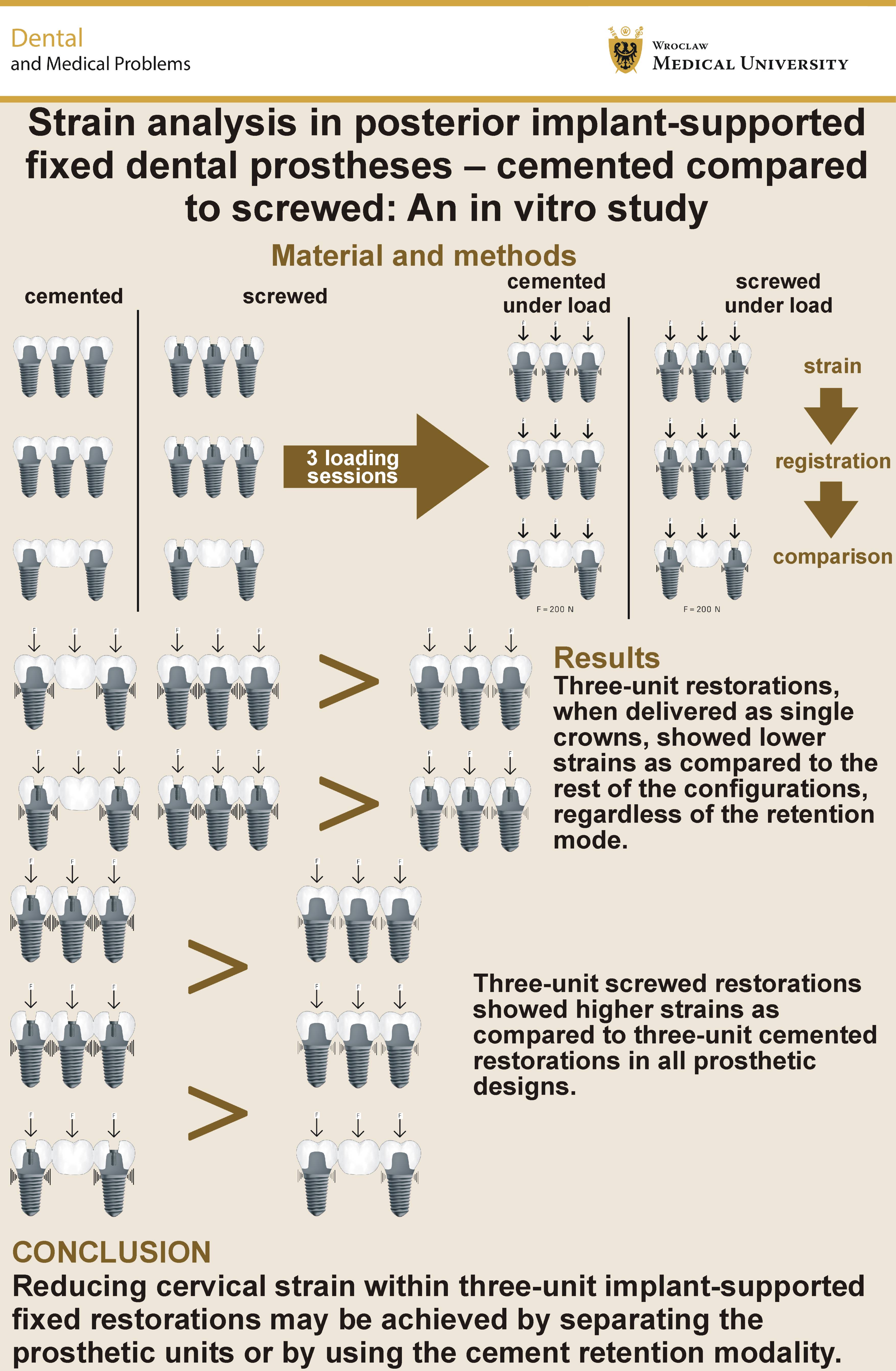
Material and methods. Six prosthetic configurations were fabricated and divided according to their retention mechanism: single crowns – cement-retained and screw-retained (SC-c and SC-s, respectively); splinted restorations – cement-retained and screw-retained (SP-c and SP-s, respectively); and simple three-unit bridges – cement-retained and screw-retained (SB-c and SB-s, respectively). Two strain gauges were attached to the implant necks. A vertical load of 200 N was applied simultaneously at 3 identical loading points on each restoration. Strain measurements were analyzed statistically using ANOVA and Scheffé’s post hoc contrast analysis.
Results. Cement-retained restorations transferred significantly less cervical strain to the implant neck than screw-retained restorations in all 3 prosthetic configurations tested (p < 0.001; SC-c < SC-s; SP-c < SP-s; and SB-c < SB-s). Single crowns transferred significantly less strain to the implant neck than the other restoration designs in both cement-retained and screw-retained groups (p < 0.001; SC-c < SP-c and SB-c; SC-s < SP-s and SB-s).
Conclusions. The use of cement-retained implant-supported fixed prostheses results in less strain development at the implant neck when subjected to high occlusal loads as compared to screw-retained prostheses. Splinting the adjacent implant-supported restorations does not necessarily reduce strain at the implant neck.
Keywords: cement retention, screw retention, posterior partial edentulism, strain analysis, three-unit prosthesis
Introduction
The consistently favorable long-term survival and success rates of posterior implant-supported fixed prostheses underscore the preference for this highly reliable treatment modality, even when narrow-diameter implants are used.1, 2
Technical complications and failures continue to drive research into their possible causes, which are generally attributed to biomechanical factors. These imply that the balance between occlusal forces and the durability of the prosthesis, its components, or its interface with the implant has been exceeded.3, 4, 5, 6, 7, 8 This phenomenon is referred to as overload.5, 8
As occlusal forces increase toward the posterior dentition,9 more than 70% of occlusal contacts and occlusal forces occur in these regions.10 Possible overload in the posterior dentition may result even from physiological occlusal forces because of the lower resilience of the implant–bone interface as compared to the tooth–periodontal ligament–bone interface. Stress generated during the masticatory function with implant-supported prostheses is therefore transmitted more directly to the implant and the surrounding bone.11 Furthermore, the markedly reduced (approx. 8-fold) tactile sensitivity associated with posterior implant-supported fixed prostheses may further contribute to this effect, since neuromuscular biofeedback is absent.12, 13 As previously reported, these 2 additive effects of occlusal loading are concentrated primarily within the cervical region of the loaded implant.14, 15
Excessive occlusal forces that may lead to overload can occur during parafunctional activities, such as bruxism and clenching.16 Tooth clenching commonly accompanies bruxism, which affects approx. 28–36% of the adult population.5, 17 During static clenching, the mandible is held against the maxillary dentition in the intercuspal position. In contrast, normal functions involving multiple intercuspal contacts include mastication (at the end of each chewing cycle) and deglutition. During mastication, the occlusal contact lasts for approx. 200 ms, amounting to less than 10 min per day.18 During swallowing, the mandible is also stabilized in the intercuspal position for an average of approx. ⅔ of a second while generating forces of about 300 N.18, 19 Swallowing occurs around 590 times over a 24-hour period.20
During parafunctional activity, individual clenching episodes may last 20–40 s,21 amounting to nearly 40 min over a single night’s sleep and generating considerable loads through multiple simultaneous occlusal contacts.22 Moreover, although tooth grinding predominates during sleep, clenching is more common during wakefulness, further increasing the cumulative duration of clenching.23, 24 These 3 clinical situations – mastication, swallowing and clenching – all involve multiple contacts in the intercuspal position, yet they are infrequently simulated in laboratory studies.
Prosthetic design may modify stress distribution within the prosthetic complex, as demonstrated by previous laboratory studies.25, 26, 27 One of the key components of prosthetic design is the choice between cement and screw retention. Each retention modality has its own advantages and limitations, and the selection between the two depends largely on clinician preference. Regarding implant and restoration survival, no significant differences between the two retention methods have been reported.28 The prevailing belief regarding the maintenance of fixed prostheses is that screw-retained restorations are primarily associated with technical complications, whereas cement-retained crowns are more prone to biological complications.29 The limited retrievability of cement-retained restorations as compared to screw-retained restorations has been a concern; however, current evidence suggests that newer cement-retention designs offer improved retrieval techniques.30
Recently, screw-retained restorations have gained popularity due to their inherent advantages, including retrievability, the elimination of cementation procedures, and the widespread adoption of digital workflows that simplify impression procedures.31, 32 However, recent clinical studies have reported increased biological complications, such as crestal bone loss around these restorations, in both short- and long-term follow-up periods.33, 34 This is in addition to the previously reported higher rate of technical complications, primarily related to screw loosening and porcelain chipping.29, 35 In contrast, in vitro studies have reported different findings regarding the crestal bone region surrounding implants, with outcomes associated with parameters such as the welding technique, the framework material and the engagement modality, which may reduce stress development in the crestal bone area.36
Since the primary cause of technical complications is occlusal overload, direct stress and strain measurements within implants should be obtained using strain gauge analysis. This approach enables the assessment of the actual strain generated within implants and the surrounding tissues under loading conditions, without relying on intermediate or theoretical modeling.37
Considering the above, when exposed to comparable occlusal loads, the stress distribution patterns within implants supporting cement-retained and screw-retained posterior three-unit fixed partial dentures (FPDs) with different prosthetic configurations should be investigated.
The purpose of this study was to compare implant-supported cement-retained restorations with their screw-retained counterparts, using strain gauge analysis across 3 common partial prosthesis designs. The 1st working hypothesis was that there would be differences among the tested configurations with regard to strain registration. The 2nd hypothesis was that the retention mechanism would not influence strain development in the cervical region of the implant.
Material and methods
This in vitro strain gauge study was performed in one of the laboratories of Vishay Precision Group Corporation (Vishay Measurements Group, Raleigh, USA) to compare 3 posterior designs of cement-retained restorations with their screw-retained counterparts under simulated occlusal loading. Owing to its in vitro design, ethical approval was not required.
Test model preparation
A resin block (PLM-4B; Vishay Measurements Group, Raleigh, USA), measuring 15.8 × 35.4 × 32 mm (Cat. No. S-116H) and exhibiting an elastic modulus similar to that of human bone (2.8 GPa), was used. Three screw-shaped, SLA (sand-blasted, large-grit, acid-etched)-coated, internal-hex implants (MIS Seven; M.I.S. Implant Technology, Bar Lev Industrial Area, Israel), each measuring 3.75 mm in diameter and 13 mm in length, were inserted into the block. The implants were fabricated from Grade V titanium (Ti-6Al-4V; density: 4.4 g/cm3; elastic modulus: 113 GPa). The implant necks protruded 2 mm above the model surface and were positioned 6 mm apart in a straight line. Open-tray impression copings (Grade V titanium: Ti-6Al-4V; density: 4.4 g/cm3; elastic modulus: 113 GPa) were secured to the implants and splinted with acrylic resin (DuraLay; Reliance Dental Manufacturing Co., Alsip, USA) to enhance stability and impression accuracy. An implant-level impression was made using a polyether-based impression material (Impregum; 3M ESPE, Seefeld, Germany) and a custom-made acrylic resin tray, after which a working cast was poured. Three cement-retained abutments and three conical screw-retained abutments, all fabricated from Grade V titanium (Ti-6Al-4V; density: 4.4 g/cm3; elastic modulus: 113 GPa), were connected to the implant analogs, using a controlled tightening torque of 20 N·m.
Fabrication of prostheses
Six cast base-metal fixed prostheses, including cement-retained and screw-retained designs, were fabricated from a Co-Cr-Mo alloy (Remanium GM380; Dentaurum, Ispringen, Germany; composition: Co – 64.5%; Cr – 29.0%; Mo – 4.5%; Mn, C and Si – <2%; density: 8.2 g/cm3; elastic modulus: 220 GPa). All frameworks were fabricated to identical vertical, buccolingual and mesiodistal dimensions, using a silicone index for standardization. Investment, casting, spruing, and framework finishing procedures were standardized and performed in a single dental laboratory, which fabricated all 6 framework configurations. Framework fit was verified using a low-viscosity silicone material by a single operator (O.R.).
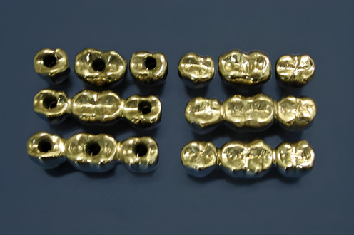
The prosthetic configurations were divided into 3 groups (Figure 1):
– 3 separate cement-retained single crowns and 3 separate screw-retained single crowns (SC-c and SC-s, respectively);
– 2 three-unit FPDs with a central pontic supported by the 2 adjacent abutments, fabricated as either cement-retained or screw-retained restorations (SB-c and SB-s, respectively); and
– 3 splinted cement-retained crowns and 3 splinted screw-retained crowns (SP-c and SP-s, respectively).
After the fabrication of all 6 restorations (3 cement-retained: SC-c, SB-c and SP-c; and 3 screw-retained: SC-s, SB-s and SP-s), the corresponding abutments were connected to the implants according to the planned prosthetic configuration, using a tightening torque of 20 N·m. Ten minutes after initial tightening, all abutment screws were retightened at 20 N·m. For the SB configurations, the central abutment was removed to accommodate the pontic design.
Temporary cement (TempBond NE; Kerr Dental, Brea, USA) was used for all cement-retained restorations. All abutments and screw-retained restorations were installed, and all cement-retained restorations were cemented by a single investigator (G.B.I.). The cement-retained restorations were initially seated with light finger pressure, followed by the application of a pressure of 5 kg/cm3 for 4 min, using a Lutron FG-20KG device (Lutron Electronics, Coppersburg, USA).
Strain registration
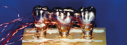
Two strain gauges (EA-06-015EH-120; Vishay Measurements Group) were attached horizontally to the neck of each implant on opposite surfaces (buccal and lingual) before abutment and crown placement. All strain gauges were bonded using a designated adhesive (M-Bond 200; Vishay Measurements Group), as shown in Figure 2. During load application to the restorations, the changes in electrical resistance recorded by the strain gauges were used to determine the absolute strain values generated within the implant necks. This location was selected, as it has been shown to experience the highest strain concentrations during loading of implant-supported restorations.14, 15
The strain gauge terminals were connected to a wiring adapter interface via soldered leads (134 DFV; Vishay Measurements Group). The interface was then connected to an electronic data scanner (Strain Gauge Scanner, model 5100; Instruments Division, Vishay Measurements Group), which communicated with a notebook computer through a designated interface card mounted in the docking station.
Load application
The 3 common posterior three-unit restoration designs, in both cement-retained and screw-retained configurations, were evaluated under 3 simultaneous occlusal loading points, and the resulting strain distribution within the implant necks was recorded. This experimental setup was designed to simulate oral conditions, in which full posterior intercuspation occurs during clenching, mastication and deglutition.
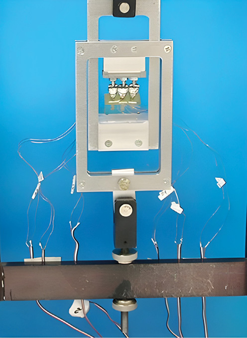
A custom-made loading device was fabricated to apply a vertical load of 200 N to each of the 6 restoration assemblies through 3 individual loading pins positioned in the central fossae of the restorations. The loading points were identical for all prosthetic configurations (Figure 3). Each loading pin was individually adjusted using a tightening screw and 8-µm-thick articulating paper until the paper was ruptured. The loading device was then calibrated to zero, after which the load was gradually increased to 200 N. Each loading cycle lasted 20 s, followed by a 10-second unloading interval. Each prosthetic configuration was tested 3 times. After each testing session and exchange of the prosthetic configuration, the loading pins were recalibrated before the subsequent measurements.
For each loading session, strain measurements were recorded. The deformation detected by the strain gauges was expressed in microstrain (με) according to the following equation:
where:
ε – strain;
ΔL – change in the strain gauge length [mm];
L – initial strain gauge length [mm];
ΔR – change in the electrical resistance of the strain gauge [Ω]; and
R – initial electrical resistance specified by the manufacturer [Ω].
Although strain measurements may represent either compression or tension, the primary outcome measure was the absolute strain value, calculated as the sum of both components and expressed in microstrain (µε) values for statistical analysis. All strain measurements were recorded by a single investigator (A.S.).
Statistical analysis
The groups were compared using one-way parametric analysis of variance (ANOVA). A p-value <0.05 was considered statistically significant. Specifically, variance analysis was performed to evaluate differences in strain distribution among the different prosthetic configurations under loading conditions. Additionally, differences among the results obtained from loading the various prosthetic configurations were assessed using Scheffé’s contrast analysis. Statistical analyses were performed using IBM SPSS Statistics for Windows, v. 24 (IBM Corp., Armonk, USA).
Results
Comparing retention mechanisms
The ANOVA was performed separately for each of the 3 restoration types – single crowns (SC), splinted restorations (SP) and the three-unit bridge design (SB) – to ascertain differences between the 2 retention mechanisms used (cement and screw retention). Strain gauge recordings were obtained from all 6 registration points located on the implants. The means (M), standard deviations (SD), and F-values of these recordings are presented in Table 1.
Significant differences were found between cement- and screw-retained restorations in all 3 restoration types, with cemented restorations generating less strain. In the SC configuration, all but one strain gauge exhibited less strain with cemented restorations (strain gauge no. 3). This trend was repeated in the SP assembly, where all strain gauges except for no. 5 exhibited significantly less strain with cemented restorations.
When the strain gauge recordings were combined at the implant level, as presented in Table 2, the overall strain pattern became even more apparent. Each implant exhibited greater strain under screw-retained restorations than under cemented restorations. These differences were well beyond the accepted level of significance for all implants and across all prosthetic configurations (p < 0.001). Therefore, the 2nd hypothesis was rejected, as substantially less strain was recorded for cemented restorations than for screw-retained restorations, regardless of the restoration design.
Comparing restoration types
Significant overall differences were observed among the 3 restoration types for both cement- and screw-retained designs (p < 0.001). The strain gauge data showed that single restorations generated the least strain as compared to the bridge and splinted crown configurations, regardless of the retention mechanism (Table 1 and Table 2). Therefore, the 1st hypothesis was accepted, corroborating the findings of previous studies.25, 27
Discussion
Parallel laboratory reports using multiple simultaneous occlusal contacts to simulate static clenching, chewing and swallowing are rare.18, 38 As noted in previous scoping reviews,37, 39 most in vitro studies evaluate simulated occlusal loading using a single loading point. Consequently, there are few comparable studies against which to evaluate our findings, apart from related publications from our own research group, owing to our unique methodological design.
The present study confirmed the 1st working hypothesis. Our findings revealed that 3 separate crowns yielded the most favorable strain distribution in both cement- and screw-retained configurations. These results are supported by previous theoretical models.7, 40 A previous finite element analysis (FEA) reached similar conclusions, demonstrating that 3 separate screw-retained crowns generated less strain than a three-unit bridge; at the level of the implant neck, 3 separate crowns produced the least strain, as did the abutment and the prosthetic screw.41
Disregarding the differences in our use of screw-retained prostheses and the additional restorative designs evaluated, more comprehensive reports have documented comparable findings. A recent in vitro study examined stress distribution among various cement-retained prosthetic configurations, using strain gauge analysis and the same loading protocol as the present study.25 Regarding three-unit prostheses, the lowest strain was observed with 3 separate crowns, consistent with our findings. A simple bridge and a three-unit splint also registered significantly higher strain, as observed in the present study.25 Similar strain gauge results for cement-retained prostheses were reported by Nissan et al. in an in vitro study, which also favored 3 separate units over a three-unit splint.27 Another strain gauge analysis employed three-point loading on 3 screw-retained prosthetic designs.42 Unlike the screw-retained assemblies in the present study, a simple 3-unit bridge generated the least strain at both the prosthesis level and the bone crest model as compared to a three-unit cantilever bridge. No strain gauges were placed at the implant neck, and a proximal contact point was present, both of which may have contributed to the differences between those findings and the results reported here.42
The 2nd working hypothesis was rejected, as the comparison of the 2 retention modes demonstrated that the cement-retained modality was significantly more favorable, with less cervical strain recorded at the implant level. Guichet et al. reported that cement-retained restorations distributed occlusal loads more evenly to the abutments because of the cushioning effect provided by the cement layer.43 A comparative in vitro study evaluating the probability of surviving cyclic loading for cement-retained vs. screw-retained three-unit bridges found that cement-retained prostheses had a higher probability of survival than screw-retained prostheses, regardless of the implant–abutment interface.44 Another study, using FEA, compared the biomechanical performance of the 2 retention designs.45 The results indicated that screw-retained prostheses exhibited a higher biomechanical risk of failure at the abutment fixation screw, which was more prone to loosening or fracture.45
Fracture resistance has also been compared in vitro between cement- and screw-retained prostheses. Two studies demonstrated the superiority of cement retention for both five-unit and three-unit prostheses.46, 47 Gaur, comparing the strain patterns of cement-retained and screw-retained prostheses, found that under vertical loading, cement-retained prostheses generated less strain in the cervical region of the implants.48 In that study, 2 point loads were applied to the pontics of a five-unit bridge supported by a pier abutment. Complementing our findings, the bridges were supported by 3 implants, each instrumented with 2 strain gauges placed on the mesial and distal aspects, and, as in the present study, exhibited lower overall strain with cement retention.48
Strain concentration within the implant neck warrants the analysis of the adjacent bone, which intimately surrounds this region. Two clinical reviews and a leading textbook in periodontology reported evidence linking cement retention with reduced crestal bone loss as compared to screw retention.29, 33, 34 The structural superiority of cement-retained prostheses is well accepted.29, 42 Establishing a mechanical etiology that may contribute to peri-implant crestal bone loss is of primary importance for both understanding and managing peri-implantitis. Although this research is laboratory-based, it attempts to provide a theoretical adjunctive mechanism contributing to crestal bone loss.
An experimental setting that directly reflects strain development within the cervical region of a bone-level implant is not feasible through strain gauge analysis, as there is no space for strain gauge placement when implants are submerged within a bone substitute. The resorption of the bone surrounding implants in the crestal region is primarily considered a biofilm-mediated infectious and inflammatory process, sharing considerable similarities with the etiology and clinical characteristics of periodontitis. This premise has been described in several reviews and textbooks.49, 50, 51 According to some authors, occlusal overload is not considered an initiating factor in peri-implantitis.52, 53 Others have described it as an “alternative hypothesis for the cause of peri-implantitis”,50 implying a limited cause-and-effect relationship between occlusal overload and disease development. Conversely, other researchers maintain that, in addition to microbial involvement in the initiation of peri-implantitis, occlusal overload may contribute to disease progression and the exacerbation of crestal bone loss.8, 54, 55 The present laboratory model may, theoretically, reveal additional mechanical factors that contribute to the initiation and possible progression of crestal bone loss in conjunction with bacterial colonization.
Limitations
Limitations of this in vitro research include the assessment of strain only along the buccolingual axis and the acquisition of data from areas in close proximity to the strain gauges, without information from more distant regions. Anteroposterior strains remain unknown, and their inclusion in the total strain dataset might influence the primary outcome parameters.
We may assume that anteroposterior strains are considerably smaller than buccolingual strains, as previously reported56; however, this cannot be regarded as evidence until this relationship has been thoroughly examined. The forces applied in our experiment were purely vertical and do not represent the average direction of physiological occlusal forces, which are predominantly three-dimensionally oblique.57 Our study evaluated the various prosthetic assemblies in a “stand-alone” setting, without interproximal contacts on either side, and therefore does not fully represent clinical stress dissipation in the presence of the adjacent units. The sample size was small, and the insertion torque during the installation of abutments and screw-retained crowns remained constant, and was not compared with higher torque values exceeding 20 N·m.
For future research, a more clinically relevant setting should be considered, including the presence of neighboring teeth with interproximal contacts, thereby influencing strain magnitude through the integrity of the dental arch. Higher insertion torque values should also be examined to evaluate their impact on the same primary outcome parameters. The use of contemporary materials, such as all-ceramic prostheses and abutments fabricated from high-performance polymers, should be considered due to their reported potential influence in reducing peri-implant bone strain.58
Conclusions
Within the limitations of this in vitro study and based on its results, cementing posterior implant-supported prostheses may be considered as a modality for reducing strain at the implant neck in comparison with screw-retained restorations. Prosthetic design influences stress distribution within posterior three-unit implant-supported restorations, with 3 separate crowns demonstrating the lowest inherent strain.
Ethics approval and consent to participate
Not applicable.
Data availability
The datasets supporting the findings of the current study are available from the corresponding author on reasonable request.
Consent for publication
Not applicable.
Use of AI and AI-assisted technologies
Not applicable.