Abstract
Background. More than 90% of transgender patients undergoing gender-affirming procedures are individuals transitioning from male to female. Forehead feminization is one of the most commonly performed procedures in this patient population. Frontal sinus and fronto-orbital anatomy are crucial considerations in determining the surgical approach and technique.
Objectives. The present study aimed to analyze the anatomy of the frontal sinus and fronto-orbital region in transgender women undergoing forehead feminization surgery.
Material and methods. A retrospective analysis of computed tomography (CT) scans of 32 patients of European ethnicity, obtained between June 2022 and June 2024, was performed. Cross-sectional facial CT images were acquired using the Carestream CS 9600 system with the following scanning parameters: 120 kV; 5 mAs; and a voxel size of 300 × 300 × 300 µm. The modeling and superimposition of the frontal sinuses were performed. The data were analyzed using IBM SPSS Statistics for Windows, v. 22.0.
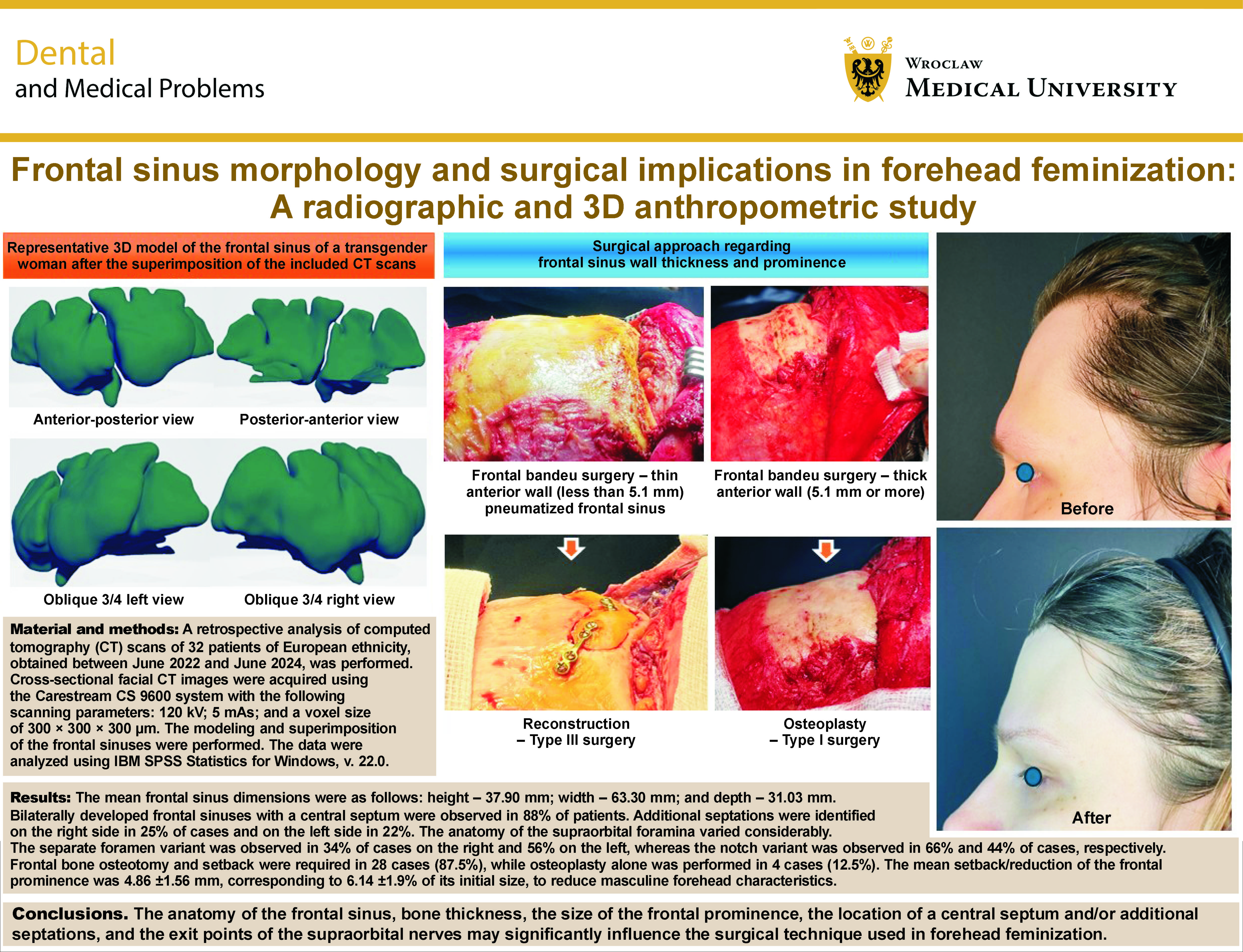
Results. The mean frontal sinus dimensions were as follows: height – 37.90 mm; width – 63.30 mm; and depth – 31.03 mm. Bilaterally developed frontal sinuses with a central septum were observed in 88% of patients. Additional septations were identified on the right side in 25% of cases and on the left side in 22%. The anatomy of the supraorbital foramina varied considerably. The separate foramen variant was observed in 34% of cases on the right and 56% on the left, whereas the notch variant was observed in 66% and 44% of cases, respectively. Frontal bone osteotomy and setback were required in 28 cases (87.5%), while osteoplasty alone was performed in 4 cases (12.5%). The mean setback/reduction of the frontal prominence was 4.86 ±1.56 mm, corresponding to 6.14 ±1.9% of its initial size, to reduce masculine forehead characteristics.
Conclusions. The anatomy of the frontal sinus, bone thickness, the size of the frontal prominence, the location of a central septum and/or additional septations, and the exit points of the supraorbital nerves may significantly influence the surgical technique used in forehead feminization.
Keywords: surgery, computed tomography, craniofacial, facial feminization
Introduction
More than 90% of transgender patients seeking gender-affirming care are individuals transitioning from male to female.1 Forehead reduction is one of the most frequently performed procedures in transgender women, as the upper third of the face accounts for approximately 35% of facial height, and its anatomical features play an important role in the perception and psychological categorization of an individual’s appearance.2
Some anatomical differences between male and female faces are subtle, whereas others are more distinctive. Sexual dimorphism in cranial morphology, in addition to differences in the overall body size and proportions, is an important factor contributing to differences in appearance between the sexes. The overall size and shape of male and female faces differ, particularly in the upper third of the face and the mandibular region.3 Female faces are generally smaller, with a more prominent forehead, more pronounced frontal eminences, and higher, more arched eyebrows. Females also tend to have relatively larger eyes with a slightly more oblique orientation.4, 5 Male skulls are generally characterized by more pronounced cranial contours. The male forehead is relatively lower, the eyebrows are positioned lower, and the eyeballs are more deeply set. The glabellar region and the supraorbital ridges are also more strongly pronounced compared with female skulls.5 Physicians performing surgery in this region must be aware of the anthropological diversity and anatomical variations of the craniofacial region.6, 7
Typically masculine features, such as prominent brow ridges and frontal bossing, are reduced and reshaped during forehead feminization surgery. The surgical approaches to forehead reduction were first described by Sir D. Ousterhout in his work on facial feminization surgery (FFS).8, 9 Type I surgery involves the reduction of the frontal cortical bone through burring. This is the most basic technique and is generally appropriate when the frontal sinus is underdeveloped and/or the anterior table of the frontal sinus is sufficiently thick to achieve the desired esthetic outcome while maintaining bone wall integrity. It is also considered the safest approach, since the frontal sinus is not entered during the procedure.9, 10 Type II surgery is performed when no frontal bone setback is required, but the augmentation of the forehead is necessary to achieve a more feminine forehead slope and nasofrontal angle (the angle formed between the facial line and the vertical line of the nasal dorsum, which is generally greater in females than in males). Type III surgery, however, is a considerably more advanced technique and involves the complete reconstruction of the forehead through frontal bone osteotomy, followed by the remodeling and setback of the frontal bone segment, usually stabilized with plates and screws or wires.1, 9 Nowadays, Type III surgery is performed in more than 90% of transgender patients requiring forehead feminization.11, 12, 13, 14
The surgical procedure must be performed in a manner that avoids the disruption of the nasofrontal drainage pathway, injury to vital structures such as the supraorbital and olfactory nerves, and cerebrospinal fluid leakage. The extent of reduction should be determined according to the individual patient’s facial anatomy and esthetic expectations. Limiting the extent of reduction solely out of concern about entering the frontal sinus is a common surgical mistake. This may result in patient dissatisfaction with the esthetic outcome and the need for revision surgery, which is frequently associated with complications related to re-entry, such as ischemia, hair loss, scarring, and an increased risk of injury to nerves and blood vessels.2 The undercorrection of frontal bossing is, therefore, one of the most common reasons for revision surgery, highlighting the importance of meticulous preoperative assessment.7, 15
The anatomy of a central septum and additional septations may influence the surgical technique used for the osteotomy, removal and stabilization of the frontal bone segment. Therefore, the precise localization of the septa is important when planning osteotomy and bone resection, which may be performed as a single-piece or two-piece procedure. Bone thickness along the borders of the repositioned anterior wall determines the appropriate length of the screws and the type of plate system required for bone fixation. Inadequate stabilization may lead to non-union, inflammation, sinusitis, and mucocele formation.11, 14
From this perspective, it is particularly important to consider modern approaches, such as digital surgical planning. Wide-field cone-beam computed tomography (CBCT) can be used to plan bone resections and anticipate the final surgical outcome, as described by Perrotti et al.16 Recent advances in three-dimensional (3D) surgical planning also provide detailed insight into frontal sinus anatomy and have been shown to be beneficial in facial feminization procedures.17, 18
The aim of this study was to evaluate the anatomical landmarks of the frontal sinus in transgender women (individuals transitioning from male to female) to identify features that may be helpful in surgical planning and decision-making.19, 20, 21 Through this work, we aspired to contribute to broader scientific discourse on craniofacial anatomy and its application in surgical interventions tailored to individual facial anthropometry. To the best of the authors’ knowledge, this is the first study to propose an approach to surgical feminization of the upper face based on frontal sinus anatomy using 3D modeling, and to compare frontal sinus anatomy between cisgender and transgender women.
Material and methods
The study was approved by the Bioethics Committee of the Medical University of Warsaw, Poland (approval No. AKBE/21/2025).
A retrospective case–control analysis of computed tomography (CT) scans from 32 patients of European ethnicity who underwent forehead feminization surgery between June 2022 and June 2024 was performed (case group). The facial CT images were acquired using the Carestream CS 9600 system with the following scanning parameters: 120 kV; 5 mAs; and a voxel size of 300 × 300 × 300 µm.
The inclusion criteria were as follows: patients undergoing gender-affirming facial surgery involving the orbitofrontal region; at least 6 months of hormone replacement therapy (HRT); and ongoing care under the supervision of a psychologist and sexologist. The exclusion criteria comprised incomplete medical records, facial surgery not involving the upper third of the face, and age below 18 years.
For comparison, the CT scans of the skulls of 32 adult cisgender women were retrieved and analyzed as the control group.
3D modeling and superimposition of the frontal sinuses
The modeling and superimposition of the frontal sinuses of transgender women (n = 32) and cisgender women (n = 32) were performed. For each participant, a 3D model of the frontal sinus was generated by the segmentation of the corresponding Digital Imaging and Communications in Medicine (DICOM) data.
The raw imaging data were stored in the DICOM format and imported into 3D Slicer software (v. 4.1.20; GitHub). Before segmentation, all image data were analyzed using identical parameters (window level: 485 HU; window width: 2,730 HU). The coronal slices of the outer compact layer of the frontal bone were obtained perpendicular to the hard palate, extending posteriorly to the posterior wall of the sphenoid bone. In contrast, axial slices were restricted to the soft tissue region.
The Segment Editor module was used to isolate the frontal sinus from the surrounding tissues. Three-dimensional models were generated using a semi-automatic segmentation procedure, which involved automatic segmentation followed by manual inspection and the correction of segment boundaries and cross-sectional contours. The intensity threshold for the segmentation of the frontal sinus in the 3D reconstructed models ranged from −1,023 to −350 HU. The axial views of the cross-sectional CT images were primarily used for 3D modeling, while coronal and sagittal views were used to identify and correct unwanted or incorrectly segmented areas, using the Segment Editor tool (3D Slicer, v. 4.1.20, stable release). The final 3D models were subsequently generated and exported according to the software’s model-exporting procedure.
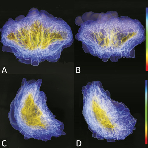
Subsequently, the reconstructed frontal sinus models were superimposed to evaluate anatomical similarities and differences between the groups. The CT-derived 3D models were aligned and registered using the ImageJ software (v. 1.42; National Institutes of Health (NIH), Bethesda, USA). All images were registered and stacked using identical spatial resolution and inter-slice distances with the Z Project function (ImageJ, v 1.42). The degree of overlap between the models was then evaluated using root mean square (RMS) values. This process generated a color-coded map illustrating the accuracy of superimposition.
The resulting color scale ranged from blue to red, with red indicating areas of substantial overlap between the models and therefore a high degree of congruence, while blue represented regions with minimal or no overlap, indicating anatomical discrepancies. Green represented an intermediate degree of alignment. This color-coded representation enabled the visualization and comparison of anatomical variations and alignment between the groups, providing insight into the structural variability of the frontal sinuses in transgender and cisgender women.
Anatomical measurements and clinical parameters
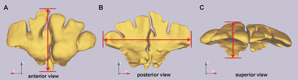
Using the 3D reconstructed models, the following anatomical parameters were assessed and measured in cisgender and transgender women: frontal sinus morphology (unilateral or bilateral development); the location and number of septa; and sinus width, height and depth (Figure 1). Based on these measurements, the volume of each frontal sinus was calculated. To assess intra-rater reliability, all measurements were repeated after a 15-day interval, and the mean values of the two measurements were used for subsequent analysis.
Frontal bone thickness was measured at the midpoint between the coronal suture and the nasion, and was referred to as anterior wall thickness. The distance from the outer table of the frontal sinus to the midsellar point was also measured and defined as the frontal prominence-to-midsella distance (hereafter referred to as frontal prominence). These parameters were assessed both before and after surgery in transgender women. Additionally, the type of surgery required for forehead feminization and the extent of anterior wall reduction/setback were evaluated.
Statistical analysis
Before conducting the analyses, sample size calculations were performed to ensure sufficient statistical power. Cohen’s effect size (d) was estimated at 1.98. Assuming a significance level of α = 0.01 and a statistical power of 0.95, the required sample size was 22 individuals (11 in each of the 2 study groups). With a total sample size of 64 individuals, the achieved statistical power was estimated to exceed 99.99%.
Descriptive statistics, including the mean (M), standard deviation (SD), and minimum–maximum (min–max)values, were calculated for all frontal sinus measurements. Differences in frontal sinus measurements between cisgender and transgender women were assessed using the Mann–Whitney U test. Changes in frontal prominence before and after surgery were evaluated using the Wilcoxon signed-rank test. Differences in frontal prominence between preoperative and postoperative measurements in transgender women and measurements in cisgender women were assessed using Tukey’s post hoc test.
Additionally, the frequencies of individuals with or without a central septum and with additional septations on the right and/or left sides were calculated. The frequencies and percentages of supraorbital foramina and notches, as well as their distribution between the right and left sides, were also determined. The χ2 test was used to assess the statistical significance of differences in categorical variables. Pearson’s correlation coefficient was used to evaluate the relationship between preoperative frontal prominence and the extent of the required setback/reduction.
All statistical analyses were performed using IBM SPSS Statistics for Windows, v. 22.0 (IBM Corp., Armonk, USA), and Statistica, v. 13.3 (StatSoft. Krakow, Poland). A p-value of less than 0.05 was considered statistically significant (supplementary material: Statistical analysis, available on request from the corresponding author)
Results
A total of 32 adult transgender women who underwent FFS involving the upper third of the face were included in the study. Patients ranged in age from 18 to 51 years, with a mean age of 30.91 ±9.45 years. The mean height of the frontal sinuses was 37.90 mm, while the mean width and depth were 63.30 mm and 31.03 mm, respectively. The mean frontal prominence was 79.09 ±3.30 mm, and the mean anterior wall thickness was 2.63 ±1.73 mm. In the control cohort, consisting of adult cisgender women within the same age range, the mean frontal sinus dimensions were as follows: height – 29.30 mm; width – 51.10 mm; and depth – 23.9 mm (Table 1). One individual in the cisgender cohort had an absent frontal sinus. Three-dimensional superimposition enabled the generation of a model-based visualization of frontal sinus anatomy in transgender women before feminization surgery, compared with the anatomy of cisgender women (Figure 2, supplementary material: Video 1).
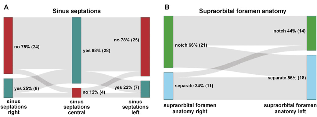
Transgender women exhibited bilaterally developed frontal sinuses. A central septum was observed in 88% of cases, whereas it was absent in the remaining 12%. Additional septations were identified on the right side in 25% of cases and on the left side in 22%, with no statistically significant difference in their occurrence between sides (Figure 3A). The anatomy of the supraorbital foramina also varied. The notch variant was observed in 66% of cases on the right and 44% on the left, whereas the separate foramen variant was observed in 34% on the right and 56% on the left (Figure 3B). A statistically significant difference in supraorbital anatomy between the right and left sides was observed in the transgender cohort (p = 0.035).
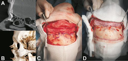
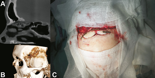
Type III surgery was required in 28 cases (87.5%), whereas osteoplasty (Type I) was performed in 4 cases (12.5%) (Figure 4 and Figure 5). No indications for Type II or Type IV surgery were identified in this study.
The mean required setback of the frontal bone was 4.86 ±1.56 mm, while the mean desired frontal prominence after surgery was 74.22 ±3.08 mm (Table 2).
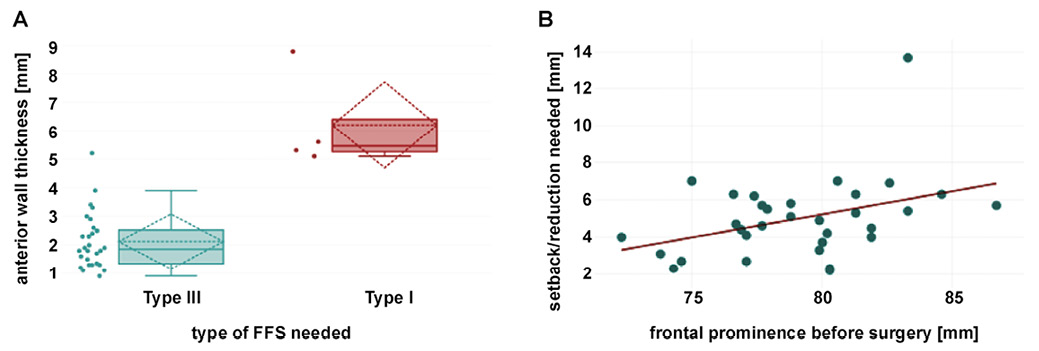
Patients with thinner anterior frontal sinus walls more frequently required bone reconstruction (Type III surgery) rather than bone burring (Type I surgery) (p = 0.002). An anterior wall thickness of 5.1 mm or greater was associated with a 95% probability of achieving satisfactory results using Type I surgery (Figure 6A).
A statistically significant correlation was also observed between preoperative frontal prominence and the extent of forehead reduction required, with greater frontal prominence being associated with an increased likelihood of requiring Type III surgery (p = 0.005) (Figure 6B). Each 1-mm increase in preoperative frontal prominence was associated with a 0.25-mm increase in the required setback/reduction.
In patients undergoing Type III surgery in whom a thick central septum was identified preoperatively on CT scans, a two-piece segmental frontal bone ostectomy was performed, followed by the reduction of the septum using a surgical burr.
Discussion
Recently, a growing body of literature has focused on quantitative assessments in reconstructive oral and maxillofacial surgery, particularly regarding facial characteristics and the roles of both hard and soft tissues.22, 23, 24 Volumetric assessments of the frontal sinuses and maxillofacial region are increasingly performed using 3D reconstruction techniques based on high-resolution imaging technologies, such as CT.25, 26, 27, 28
Studies by Hamed et al.29 and Mrożek et al.30 demonstrated that the measurements of frontal sinus dimensions, particularly the anteroposterior length, are valuable in investigating sexual dimorphism between biological males and females. Similarly, our study revealed significant differences in anteroposterior sinus length, height and width between transgender and cisgender women. Additionally, sex-related differences were observed in other dimensions of the fronto-orbital region: cisgender women had significantly smaller frontal prominences, greater anterior wall thicknesses, and, consequently, smaller frontal sinus volumes. The magnitude and statistical significance of these differences highlight the importance of detailed anatomical assessment for surgeons performing facial feminization surgery in transgender women.
Forehead feminization is one of the most commonly performed procedures in transgender women. Therefore, a precise and individualized surgical plan is essential to avoid undercorrection, patient dissatisfaction, and eventually the need for revision surgery. In a recent study by Rochlin et al., undercorrection and patient dissatisfaction were identified as the main reasons for revision surgery, which was required in 25% of cases.7 Similar observations were reported in our previous study.2 These outcomes may result from inadequate preoperative diagnostics and surgical planning, a conservative surgical approach, insufficient experience or equipment, fear of entering the frontal sinus, or other contributing factors. Computed tomography and 3D surgical planning should include an assessment of frontal prominence (defined as the distance from the anterior table of the frontal sinus to the midsellar point), the degree of frontal sinus pneumatization, and frontal bone thickness.18 By evaluating these anatomical parameters, the surgical technique can be safely tailored to each individual case.
In our study, forehead feminization involved a mean anterior wall setback of 4.86 ±1.56 mm to achieve the desired esthetic outcome, corresponding to 6.14 ±1.9% of preoperative frontal prominence. The mean postoperative frontal prominence was 74.22 mm, which is comparable to the results reported by other authors.12 Importantly, the range of postoperative frontal prominence values in transgender women was not significantly different from the range observed in cisgender women. Frontal sinus aplasia has been reported in approx. 3–4% of individuals. In our transgender cohort, no cases of frontal sinus aplasia were observed. However, one individual in the cisgender cohort lacked a frontal sinus (3.1%), which is consistent with previously published literature.
In our study, no patients required Type II surgery, which is relatively uncommon and reportedly accounts for less than 10% of forehead feminization procedures in transgender women. Particular care should be taken when determining the indications for Type II surgery, as some patients may actually require Type III reconstruction. Surgical access to the orbital region requires the elevation of a subperiosteal flap through a coronal incision or its modifications, depending on the shape and position of the hairline.2, 31 During dissection, the surgeon must avoid injury to the supraorbital nerves, which provide sensory innervation to the forehead. Therefore, the position and morphology of the supraorbital foramina should be carefully evaluated preoperatively. The extracranial passage of these nerves may occur through a notch continuous with the supraorbital margin or through a distinct foramen separated from the margin. When a supraorbital notch is present, the supraorbital neurovascular bundles are released and protected intraoperatively. When a distinct foramen is present, osteotomy may be required to release and protect the neurovascular bundle.18, 32
Regarding flap preparation and nerve preservation, the supraorbital notch variant may provide a more predictable anatomical landmark during flap elevation than multiple foramina. This is particularly relevant, as orbital feminization requires wide exposure of the orbital aperture and the reduction of its upper and lateral borders. The extent of frontal bone reduction and setback may also be influenced by the presence and morphology of a central septum. In cases involving a thick central septum, the frontal bone may need to be resected in 2 separate segments. Forceful removal of the bone segment should be avoided, as this may result in the fractures of the frontal bone or the septum, and potentially damage the adjacent structures, including the ethmoidal cribriform plate, crista galli, the nasal cavity, and the orbital roof or its superomedial portion. Such complications may require additional surgical management similar to that used in the treatment of frontal bone fractures. To the best of the authors’ knowledge, this is also the first study to discuss the relevance of frontal sinus septations in planning upper-face feminization surgery.
Limitations
We acknowledge several limitations of this study. First, the study design did not account for differences in body measurements between the 2 groups (transgender and cisgender women). Body size exhibits biological sexual dimorphism, with significant differences between biological sexes not only in the overall body height ,but also in skull length, width and height. These differences may have influenced frontal sinus dimensions and anterior wall thickness before surgery, as well as the dimensions of the frontal sinus and the frontal region after surgery. Nevertheless, the postoperative frontal prominence values in transgender women were comparable to those observed in the cisgender control group, which was one of the primary goals of the surgical interventions. Second, no patients in the surgical cohort met the criteria for Type II or Type IV surgery, which limited the possibility of evaluating the applicability of these surgical techniques in our study population.
Conclusions
Within the limitations of this retrospective study, the frontal sinus was present in almost all transgender women undergoing the feminization surgery of the upper third of the face. The frontal sinuses were significantly more pneumatized than those in the cisgender control group, although considerable anatomical variability was observed among individual patients. The application of 3D virtual surgical planning is therefore crucial for ensuring safe and individualized surgical treatment, as the considerable variability in frontal sinus anatomy requires meticulous preoperative evaluation. Precise preoperative imaging enables the surgical plan to be tailored to each patient’s individual anatomy, potentially resulting in more predictable outcomes, shorter operative times, and a reduced risk of complications, while helping to achieve satisfactory esthetic effects and improve patient satisfaction.
Ethics approval and consent to participate
The study was approved by the Bioethics Committee of the Medical University of Warsaw, Poland (approval no. AKBE/21/2025), and was conducted in accordance with the Declaration of Helsinki. Written informed consent was obtained from all study participants.
Data availability
The datasets supporting the findings of the current study, including all the supplementary materials, are available from the corresponding author on reasonable request.
Consent for publication
Written informed consent was obtained from individuals whose photographs are presented in this article.
Use of AI and AI-assisted technologies
Not applicable.