Abstract
Background. Glucose-regulated protein 78 (GRP78), also known as binding immunoglobulin protein (BiP), is an endoplasmic reticulum (ER) chaperone involved in protein folding and processing, particularly during the unfolded protein response (UPR). It has become an important subject of research in cancer biology and cell proliferation.
Objectives. The aim of the present study was to investigate the relationship between GRP78 expression and temporomandibular disorders (TMDs). Given that alterations in the structure and morphology of the articular disc have been implicated in the pathogenesis of temporomandibular joint (TMJ) symptoms, investigating the role of GRP78 in chondrocyte proliferation may provide further insight into the underlying mechanisms of TMDs.
Material and methods. A total of 22 human TMJ disc samples underwent immunohistochemical staining for GRP78, followed by an assessment of staining intensity. The sample comprised 6 specimens from patients with anterior disc displacement with reduction (ADDwR), 12 from patients with anterior disc displacement without reduction (ADDwoR), and 4 control specimens.
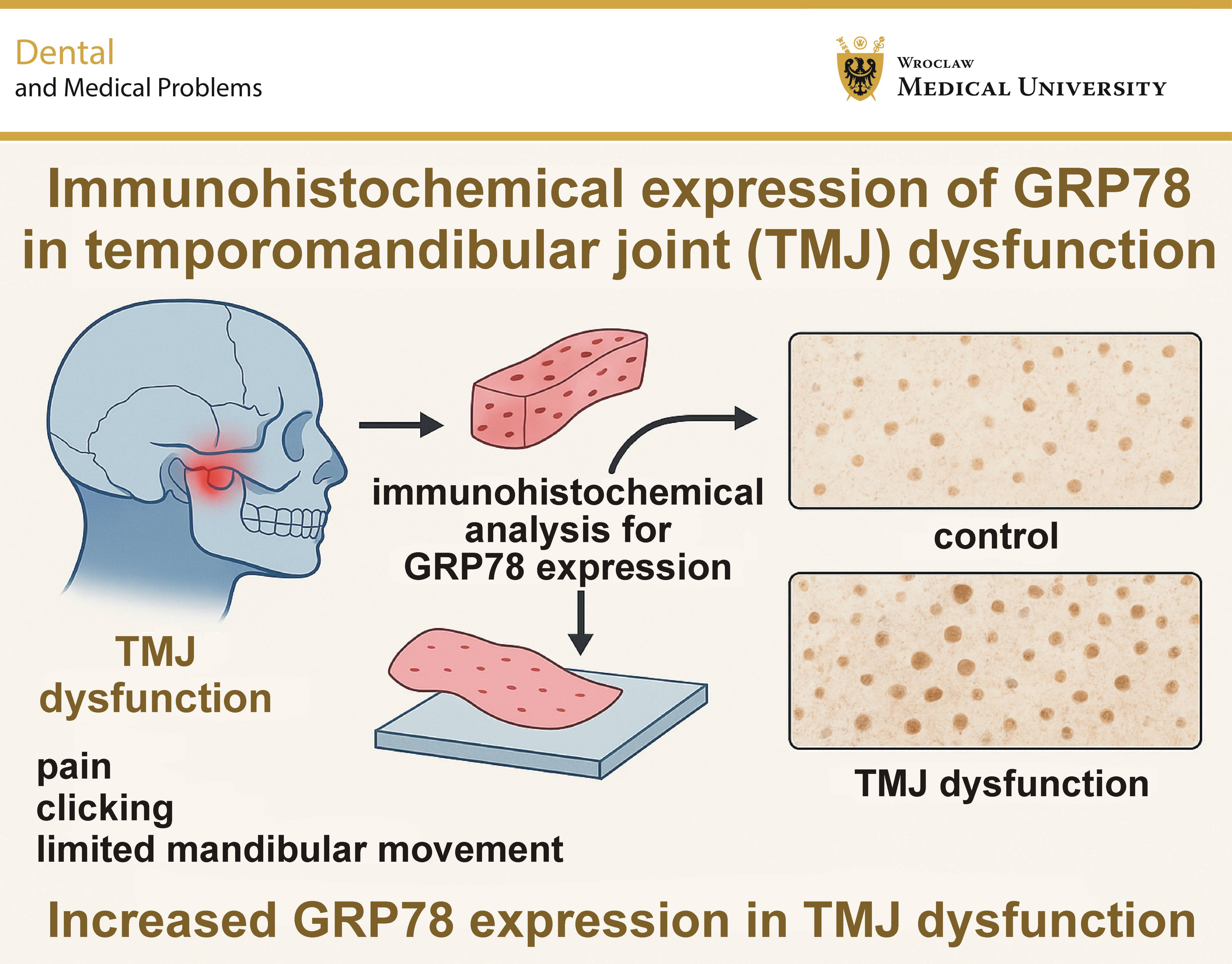
Results. A statistically significant difference in GRP78 staining intensity was observed between the control and experimental groups. GRP78 expression was significantly higher in both experimental groups than in the control group (p < 0.0001). These findings suggest that GRP78 may play an important role in the pathogenesis of TMDs.
Conclusions. GRP78 expression may be involved in the pathogenesis of TMDs, as its expression differed significantly between TMJ discs affected by TMDs and those from unaffected controls.
Keywords: inflammation, temporomandibular joint, protein expression, immunohistochemical analysis, GRP-78
Introduction
The temporomandibular joint (TMJ), which articulates the mandibular condyle with the glenoid fossa of the temporal bone, is considered one of the most complex joints in the human body. Classified as a ginglymoarthrodial joint, TMJ allows both hinge and gliding movements.1 Due to its anatomical and functional complexity, temporomandibular disorders (TMDs) are relatively common in the general population, with affected individuals frequently reporting jaw pain and restricted mouth opening.2 Limited knowledge of the underlying pathophysiology of TMDs makes it challenging for clinicians to develop effective treatment strategies and restore normal joint function and mobility. Consequently, current management approaches remain largely symptomatic rather than targeting the underlying causes of the disorders.
Recent studies have highlighted molecular mechanisms involved in TMJ pathology, including the association between mandibular condylar hyperplasia and vascular endothelial growth factor (VEGF), emphasizing the potential role of molecular markers in joint remodeling.3 Furthermore, the influence of health literacy on conditions such as bruxism illustrates how biological mechanisms and patient-related factors intersect in clinical management.4
Temporomandibular disorders are frequently associated with the anterior disc displacement (ADD) of the temporomandibular joint disc. Anterior disc displacement is characterized by an abnormal disc position relative to the mandibular condyle during jaw movement and the protrusion of the mandible, and is classified as either with reduction (ADDwR), when the disc returns to its normal position during mouth opening or closing, or without reduction (ADDwoR), when the displaced disc fails to return to its normal physiological position during mandibular movement. Since TMD management in the USA is largely palliative, patients may remain dissatisfied with treatment outcomes, and TMD-related symptoms continue to be a common reason for seeking dental care.5
The aim of the present study is to investigate the relationship between TMDs and binding immunoglobulin protein (BiP), also known as glucose-regulated protein 78 (GRP78). BiP/GRP78 is an endoplasmic reticulum (ER) chaperone that plays a central role in protein folding and quality control, particularly during the unfolded protein response (UPR).6, 7 Specifically, GRP78 facilitates the proper folding and refolding of unfolded or misfolded proteins to maintain cellular homeostasis. When these processes are insufficient to restore protein homeostasis, misfolded proteins may be targeted for ER-associated degradation (ERAD), a quality control pathway responsible for removing aberrant proteins.7 According to Walter and Ron, impaired ERAD can result in the accumulation of misfolded proteins within the ER lumen and membrane, contributing to ER stress,11 a phenomenon implicated in several diseases.6, 7, 8, 9, 10, 11 Under normal physiological conditions, GRP78 binds to the ER stress sensors – activating transcription factor 6 (ATF6), protein kinase R-like endoplasmic reticulum kinase (PERK) and inositol-requiring enzyme 1 (IRE1), maintaining them in an inactive state. In response to ER stress, GRP78 dissociates from these sensors, allowing UPR signaling pathways to become activated.11
GRP78 was selected as the primary marker in this study because of its central role in the ER stress response and its potential relevance to cartilage biology. Its expression may therefore provide valuable insight into the cellular stress mechanisms associated with TMJ disc pathology. Compared with other UPR-related markers, such as C/EBP homologous protein (CHOP) and spliced X-box binding protein 1 (XBP1s), GRP78 offers an opportunity to investigate an upstream component of the ER stress response.
Various pathological conditions can induce ER stress, and consequently activate UPR, including viral infections, mutations affecting secretory proteins, and nutrient or electrolyte imbalances.12 When the UPR mechanisms fail to adequately resolve ER stress and restore cellular homeostasis, apoptotic pathways may be activated, ultimately leading to cell death.13 Given that many pathological conditions involve dysregulated cell proliferation and/or apoptosis, GRP78 has attracted increasing research interest because of growing evidence of its involvement in a wide range of diseases.6 Its broad involvement in cellular stress responses raises the question of whether GRP78 also contributes to the pathogenesis of TMDs. Although its role in chondrogenesis remains poorly understood, GRP78 is believed to participate in cartilage formation, remodeling, cellular proliferation, and apoptosis.13 This perspective is consistent with emerging research highlighting the contribution of genetic factors and cellular stress-response pathways to disease pathogenesis.14, 15
It is important to note that GRP78 is expressed in the growth plates of chondrocytes, and its overexpression has been shown to promote chondrocyte proliferation by accelerating cell-cycle progression through the G1 phase.13 Given that alterations in the structure and morphology of the TMJ articular disc have been implicated in the pathogenesis of TMDs, investigating the role of GRP78 in chondrocyte proliferation may provide further insight into the underlying mechanisms of these disorders.
This study investigates the association between GRP78 expression and disc displacement in TMDs, acknowledging that the findings reflect a correlation rather than an etiological relationship.
Material and methods
A total of 22 articular disc samples were collected from patients treated at Hospital Universitário Evangélico de Curitiba (Mackenzie Evangelical University Hospital in Curitiba), Brazil, between 2002 and 2009. Of these, 4 samples consisted of unaffected human articular discs and served as the control group. The mean age of the patients was 31 years (range: 19–45 years). Control samples were obtained from patients undergoing surgery for condylar fractures who had no history of TMDs.
All participants completed a medical history questionnaire and provided written informed consent. The exclusion criteria for the control group comprised a history of orthodontic treatment, systemic diseases (e.g., diabetes, hepatitis, or human immunodeficiency virus (HIV) infection), immunosuppressive therapy, pregnancy, immune dysfunction, severe jaw trauma, dentofacial deformities, previous TMJ surgery, or other TMD-related interventions.
All patients who met the preliminary eligibility criteria were asked to complete a pain questionnaire and underwent clinical examinations performed by an experienced oral and maxillofacial (OMF) surgeon. The examination included the palpation of the TMJ region, auscultation for joint sounds, including crepitus, and a functional assessment. Any signs of pain during mandibular movements were recorded. Following the initial evaluation, patients whose pain was attributed exclusively to muscular spasms were excluded from the experimental group.
After inclusion in the study, all participants underwent magnetic resonance imaging (MRI) and panoramic radiography. Based on the MRI findings, patients were classified into 2 experimental subgroups according to their disc displacement status: anterior disc displacement with reduction (ADDwR; n = 6) and anterior disc displacement without reduction (ADDwoR; n = 12). The control group consisted of 4 articular disc samples obtained from patients undergoing surgery for condylar fractures who had no history of TMDs.
The staining protocol was adapted from the Thermo Fisher Scientific Ultra-Sensitive ABC Peroxidase Standard Staining Kit, with all steps modified as appropriate. The articular disc samples were deparaffinized in xylene, and subsequently rehydrated using graded ethanol solutions. This process involved 9 sequential solvent baths in Coplin jars, comprising 3 xylene baths, 2 baths of 100% ethanol, 2 baths of 95% ethanol and 5% deionized water, and a final two-step rinse with 100% deionized water.
Antigen retrieval was then performed to unmask potential antigenic sites. The samples were immersed in 10 mM sodium citrate buffer and heated to boiling for 14 min, followed by a 20-minute cooling period. Subsequently, the samples were treated with a peroxidase suppressor to inhibit endogenous peroxidase activity and incubated with a 5% bovine serum albumin (BSA) blocking solution in phosphate-buffered saline (PBS) for 15 min.
All samples were incubated with the GRP78 primary antibody (1:200 dilution; Thermo Fisher Scientific, Rockford, USA) for 30 min at 16°C. Subsequently, the samples were incubated with a biotinylated secondary antibody (1:400 dilution) for an additional 30 min at the same temperature. All antibodies were diluted in PBS.
The avidin–biotin complex (ABC) was applied to the samples to amplify the immunohistochemical signal. The samples were incubated with the ABC reagent for 30 min, followed by a 5-minute application of 3,3'-diaminobenzidine (DAB) chromogen, prepared at a 1:9 dilution from the 10× concentrate. The slides were subsequently dehydrated using deionized water, ethanol and xylene. The same sequence of solvent baths used for deparaffinization was followed in reverse order, with each bath lasting 18 s. Following dehydration, the articular disc sections were systematically examined under a light microscope, and staining intensity and the number of stained fibroblasts were recorded.
Evaluation protocol
Staining intensity was evaluated using a 5-point scale ranging from 0 to 4, where 0 indicated no staining, 1 – light staining, 2 – moderate staining, 3 – strong staining, and 4 – very strong staining.
This study was reported in accordance with the Strengthening the Reporting of Observational Studies in Epidemiology (STROBE) .
Statistical analysis
Statistical analysis was performed using the SPSS software, v. 12.0 (SPSS Inc., Chicago, USA). For the analysis of staining area and density, the average of the measurements obtained from all available samples for each patient was calculated. Group means were compared using the one-way analysis of variance (ANOVA), followed by the Bonferroni post hoc test for multiple comparisons. A p-value of less than 0.05 was considered statistically significant.
Results
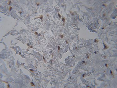
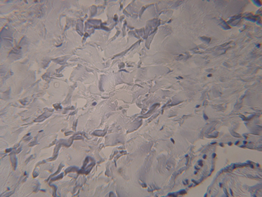
Following immunohistochemical staining, GRP78 expression was found to be higher in both experimental groups than in the control group. Five areas of each sample were subjectively evaluated for staining intensity using the 0–4 scale. The resulting scores were analyzed using ANOVA. A marked increase in GRP78 immunoreactivity was observed in both the ADDwR and ADDwoR groups (Figure 1) compared with the control group (Figure 2). This difference was statistically significant (p < 0.0001). However, no statistically significant difference was observed between the ADDwR and ADDwoR groups (p = 0.198). The mean staining intensity scores are presented in Table 1: 3.58 ±0.31 for ADDwR, 3.27 ±0.35 for ADDwoR, and 0.28 ± 0.22 for the control group.
Discussion
The human skeleton is in a constant state of remodeling, and a healthy TMJ is no exception.16, 17, 18, 19, 20, 21, 22, 23, 24, 25 Optimal function and mobility are maintained throughout life through extensive adaptation, healing and inherent regenerative potential.20 When an inflammatory process develops and the joint no longer functions at an optimal level, the disc often undergoes a well-documented displacement from its normal functional position.18, 21 Symptoms often begin with clicking or popping during mandibular opening, and may progress to muscle tenderness, limited maximum mouth opening, headaches, and other symptoms.20, 21
The results of this study clearly demonstrate an increased prevalence of GRP78 in pathological joints. It has been shown that GRP78 overexpression enhances cell proliferation during chondrocyte development.13, 17, 18 Increased expression of GRP78 within the joint space may alter the remodeling processes that occur in TMJ. Increased cartilage deposition could affect proper disc positioning and contribute to the development of TMD symptoms, which may originate at the microscopic level.17, 18, 19, 20, 21
In line with our findings, recent reports have expanded the biomarker-based approach to oral and maxillofacial research. For instance, machine-learning models, such as Light Gradient Boosting Machine (LightGBM), have been employed to predict quality of life (QoL) among oral cancer survivors.22 Similarly, imaging-based reviews have evaluated the comparative effectiveness of different palatal expansion techniques using cone-beam computed tomography (CBCT).23 These complementary efforts highlight the growing convergence of molecular biology, computational analysis and advanced imaging in shaping contemporary oral health research.
Digital workflows are increasingly shaping orthodontic and prosthetic practice, offering more efficient and precise approaches to treatment.24, 25 Preliminary implant studies and systematic reviews further highlight the potential of translational research approaches to improve our understanding of TMJ pathology and inform rehabilitation strategies.26, 27
Following the same line of thinking, recent studies have further expanded our understanding of TMDs and related conditions from both clinical and molecular perspectives. Conservative approaches, such as Kinesio Taping, have demonstrated measurable improvement in mandibular mobility and pain reduction in patients with TMDs, supporting the role of non-invasive therapies in symptom management.28 At the same time, experimental studies investigating agents such as penehyclidine hydrochloride have demonstrated protective effects against inflammation and oxidative stress through the activation of the ROS/Nrf2/HO-1 pathway, highlighting the important role of cellular stress responses in musculoskeletal disorders.29 Research on stress-related disorders further suggests that biological, mechanical and psychosocial factors interact in the development and progression of TMDs.30 These findings reinforce the rationale for investigating molecular markers such as GRP78 to better understand the contribution of cellular stress pathways to the pathogenesis of TMDs.
Our findings demonstrate elevated GRP78 expression in pathological discs; however, this observation remains correlative in nature. Therefore, no causal relationship can be inferred, and GRP78 should currently be regarded as a potential biomarker rather than an established therapeutic target.
Conclusions
In this preliminary study, GRP78 expression was significantly elevated in discs from patients with TMDs compared with those from control subjects. These findings suggest that GRP78 may have potential as a biomarker of disc displacement. Further studies are needed to validate this association and establish the clinical relevance of GRP78 expression in TMDs.
Ethics approval and consent to participate
This study was conducted in accordance with the Declaration of Helsinki. The study was approved by the Research Ethics Committee at the Pontifical Catholic University of Paraná (PUCPR), Curitiba, Brazil (CAAE: 55535716.8.0000.0100; approval No. 1.533.139). The subsequent analysis of the de-identified specimens at Marquette University, Milwaukee, USA, was reviewed by the Marquette University Institutional Review Board (protocol No. HR-2859), which determined that it did not constitute human subjects research; therefore, further IRB approval was not required. All participants provided written informed consent prior to participation in the study.
Data availability
The datasets supporting the findings of the current study are available from the corresponding author on reasonable request.
Consent for publication
Not applicable.
Use of AI and AI-assisted technologies
During the preparation of this manuscript, the authors used DeepL Translate for language checking and grammar correction. Following its use, the authors thoroughly reviewed, verified and revised all content to ensure accuracy and originality.