Abstract
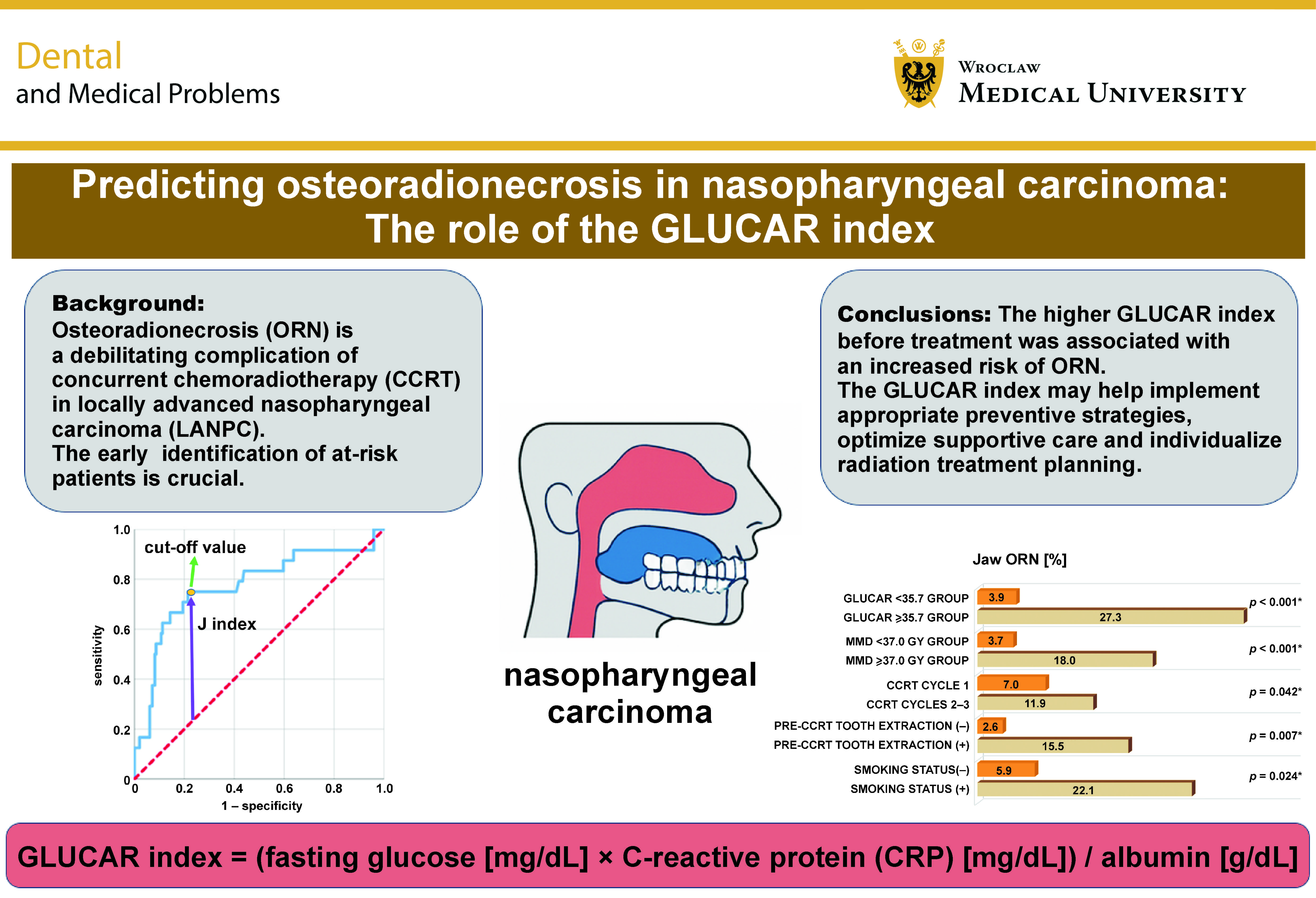
Background. Osteoradionecrosis (ORN) is a debilitating complication of concurrent chemoradiotherapy (CCRT) in patients with locally advanced nasopharyngeal carcinoma (LANPC). The early identification of patients at risk is crucial.
Objectives. The aim of the present study was to evaluate the predictive value of the GLUCAR index, which integrates fasting glucose and the C-reactive protein (CRP)-to-albumin ratio (CAR), for assessing the risk of ORN in patients with LANPC undergoing CCRT.
Material and methods. A retrospective analysis was conducted in a consecutive cohort of 220 patients with LANPC who underwent definitive CCRT. The patients were stratified into high- and low-GLUCAR groups according to their pre-treatment GLUCAR index levels, using the optimal cut-off value determined by the receiver operating characteristic (ROC) curve analysis. Osteoradionecrosis was defined as the event of interest. Multivariate Cox proportional hazards regression analysis was performed to identify independent predictors of ORN development.
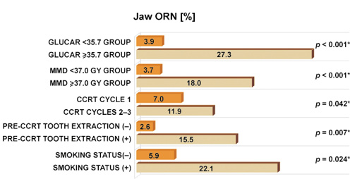
Results. The overall incidence of ORN was 10.9%. The optimal cut-off value of the GLUCAR index for predicting ORN was 35.7 (area under the curve (AUC): 0.773; sensitivity: 75.0%, and specificity: 76.5%). Patients with a GLUCAR index ≥35.7 had a significantly higher incidence of ORN than those with a GLUCAR index <35.7 (27.3% vs. 3.9%; p < 0.001). The multivariate analysis identified a GLUCAR index ≥35.7 as an independent predictor of ORN development (odds ratio (OR): 9.09; 95% confidence interval (CI): 2.48–25.02; p < 0.001). Other independent predictors included smoking, pre-CCRT tooth extractions, a higher number of chemotherapy cycles, and a higher mandibular radiation dose (p < 0.05 for all).
Conclusions. The results of this study suggest that a pre-treatment GLUCAR index ≥35.7 is independently associated with an increased risk of ORN in patients with LANPC undergoing definitive CCRT.
Keywords: nasopharyngeal carcinoma, osteoradionecrosis, predictive biomarker, GLUCAR index, chemoradiotherapy
Introduction
Nasopharyngeal carcinoma (NPC) is a distinct malignancy within the spectrum of head and neck cancers, characterized by unique epidemiological features and marked sensitivity to both radiotherapy and chemotherapy. In patients with locally advanced nasopharyngeal carcinoma (LANPC), concurrent chemoradiotherapy (CCRT) remains the cornerstone of curative treatment, administered either as definitive therapy or following induction chemotherapy, and is associated with high rates of locoregional control and improved survival.1, 2 However, with the increasing intensity of treatment protocols, the risk of serious late radiation-induced complications, particularly osteoradionecrosis (ORN) of the jaw, has become an important clinical concern.
Although the primary tumor site in NPC is anatomically distant from the mandible, cumulative radiation exposure, particularly in patients with advanced primary tumors or extensive nodal disease requiring larger radiation fields, may still compromise mandibular structures and lead to ORN. Although the incidence of ORN in NPC is generally lower than that reported in oral cavity or oropharyngeal cancers, the reported rates in LANPC populations remain substantial, reaching 7–10%.3, 4, 5, 6 Given the debilitating nature of ORN and its profound impact on patients’ quality of life (QoL), the early identification of individuals at high risk remains an important unmet clinical need.
Osteoradionecrosis (ORN) is defined as exposed irradiated bone that fails to heal within 3 months, in the absence of tumor recurrence or metastatic involvement of the mandible.7 Its development involves a multifactorial pathogenesis characterized by radiation-induced endarteritis, tissue hypoxia, hypocellularity, and progressive fibrosis, which collectively impair the regenerative capacity of bone.8 While traditional risk assessment has primarily focused on local factors, such as the radiation dose, dental status and oral hygiene, increasing attention is being paid to systemic factors that may influence tissue healing and host susceptibility to ORN.9
The GLUCAR index, an emerging composite biomarker integrating the fasting blood glucose levels with the C-reactive protein (CRP)-to-albumin ratio (CAR), has been proposed as a potential indicator of systemic metabolic stress and inflammation.10 Both components of the GLUCAR index are associated with biological mechanisms that can compromise wound healing. Hyperglycemia disrupts angiogenesis and impairs immune function, whereas an elevated CAR reflects systemic inflammation, immune dysregulation and malnutrition, all of which may synergistically impair tissue recovery following radiation therapy.11, 12, 13 Elevated glucose levels promote the accumulation of advanced glycation end-products (AGEs), induce endothelial dysfunction and interfere with bone remodeling.14 Similarly, an elevated CAR reflects a catabolic state characterized by depleted protein reserves, potentially limiting the availability of essential substrates for cellular repair and collagen synthesis.13
Clinically, the first pivotal study by Somay et al. investigated the prognostic value of the GLUCAR index for tooth loss following CCRT in patients with LANPC.8 The authors demonstrated that patients with a pre-treatment GLUCAR index ≥31.8 had a significantly higher risk of tooth loss than those with lower values (84.4% vs. 47.4%; p < 0.001).8 These findings suggest that hyperglycemia, systemic inflammation and hypoalbuminemia may collectively reflect an inflammatory-catabolic state that predisposes patients to radiation-induced bone complications.
Beyond NPC, the GLUCAR index has also demonstrated prognostic significance in other malignancies characterized by systemic inflammation and metabolic dysregulation. For instance, a recent analysis showed that a GLUCAR index ≥42.8 independently predicted poorer survival in patients with locally advanced pancreatic cancer.15 Despite its potential biological relevance, no previous studies have investigated the predictive value of the GLUCAR index for ORN in patients with LANPC undergoing definitive CCRT.
Given the established associations between metabolic-inflammatory dysregulation and impaired tissue healing following radiation therapy, we hypothesized that an elevated pre-treatment GLUCAR index could serve as a systemic marker of ORN risk.16 Accordingly, this study aimed to investigate the association between pre-treatment GLUCAR index levels and the subsequent ORN development, with the ultimate goal of refining the current risk stratification models and informing individualized pre-radiation management strategies.
Patients and methods
Ethical considerations
This retrospective study was conducted in collaboration with the Department of Radiation Oncology and the Dentistry Clinics at Baskent University Faculty of Medicine, Adana, Turkey. Before data collection, the study protocol received formal approval from the Institutional Review Board of Baskent University Faculty of Medicine (project no. DKA:19/39), ensuring compliance with applicable ethical standards. Eligible participants were selected according to the predefined inclusion criteria, and provided written informed consent before undergoing comprehensive oral and dental assessments and CCRT. The consent covered the collection and subsequent analysis of relevant clinical data, including blood samples, sociodemographic characteristics, medical history, and dental radiographs, as well as the use of anonymized data for academic presentations and publications. The present study was reported in accordance with the Strengthening the Reporting of Observational Studies in Epidemiology (STROBE) guidelines.17
Patient selection
The Dentistry Clinics at Baskent University Adana Research and Treatment Center reviewed the medical records of patients with LANPC who underwent CCRT, including pre- and post-CCRT oral and dental examinations performed between February 2010 and January 2025. Patients were eligible for inclusion if they met the following criteria: age ≥18 years; histopathologically confirmed squamous cell carcinoma (SCC); locally advanced disease according to the 8th edition of the American Joint Committee on Cancer (AJCC) staging system; and no history of previous malignancy, systemic chemotherapy, or head and neck radiotherapy. Additionally, patients were required to have available complete blood count (CBC) and biochemical test results obtained before CCRT initiation, as well as documented pre- and post-CCRT dental examination findings and panoramic radiographs.
Patients with mandibular invasion by the primary tumor or metastatic lymph nodes, a previous diagnosis of jaw ORN, a history of jaw surgery, the current use of corticosteroids or other immunomodulatory agents, or a history of blood transfusion within 30 days before CCRT initiation were excluded from the study. Additionally, patients with active systemic inflammatory or immunological conditions, including rheumatological, renal and respiratory diseases, viral hepatitis, immunosuppressive conditions, collagen vascular diseases, chronic inflammatory disorders, and glycogen storage diseases, were considered ineligible.
These restrictions were applied to minimize potential confounding related to pre-existing systemic, metabolic, inflammatory, and immunological conditions, as well as medication use. To further reduce potential bias, patients with periodontitis, cardiovascular or other vascular diseases, a history of stroke, metabolic syndrome, or diabetes mellitus were also excluded, as these conditions may independently increase the risk of tooth loss.
Baseline oral examination
All patients underwent a comprehensive dental assessment performed by an experienced oral and maxillofacial surgeon (E.S.) before initiating CCRT. The assessment followed the established protocols of the American Dental Association (ADA) and the relevant regulations of the US Food and Drug Administration (FDA).18 Panoramic radiographic examinations were performed using the J. Morita Veraviewepocs 2D system (Kyoto, Japan) to facilitate a thorough evaluation of each patient’s dental status. These examinations formed part of the routine dental assessment protocol for all patients diagnosed with head and neck cancer.
Each tooth was carefully examined for dental caries in accordance with the World Health Organization (WHO) criteria.19 Clinical examinations were performed using illuminated dental mirrors and explorers to detect even subtle signs of caries. Teeth with inadequate periodontal support, extensive structural deterioration rendering them non-restorable, or apical lesions considered unsuitable for successful endodontic treatment were scheduled for extraction. Superficial carious lesions were treated with appropriate dental restorations to prevent further progression.
To optimize oral health before and during oncological treatment, all patients received individualized instructions on appropriate oral hygiene practices. Professional dental scaling was also performed to remove plaque and calculus, thereby improving oral hygiene and helping maintain dental health throughout the treatment period.
GLUCAR index calculation and measurement
The GLUCAR index, previously developed by our research group as a novel prognostic biomarker, was calculated using the following formula: GLUCAR index = (fasting glucose [mg/dL] × CRP [mg/dL]) / albumin [g/dL].8 This index integrates pre-treatment systemic glucose metabolism, inflammation, immune status, and nutritional status using routinely available clinical parameters. Fasting glucose, CRP and serum albumin levels were measured in the blood samples collected on the first day of CCRT. All biochemical analyses were performed using the Abbott ARCHITECT c8000 biochemistry autoanalyzer (Chicago, USA), in accordance with the manufacturer’s instructions and standard quality assurance protocols.20
Chemoradiotherapy protocol
All patients received simultaneous integrated boost intensity-modulated radiotherapy (SIB-IMRT), as previously described.21 Target volumes were delineated using co-registered imaging modalities, including computed tomography (CT), 18-F-fluorodeoxyglucose positron emission tomography/CT (18-F-FDG PET/CT) and magnetic resonance imaging (MRI). The prescribed radiation doses were 70 Gy, 59.4 Gy, and 54 Gy for the high-, intermediate-, and low-risk planning target volumes (PTVs), respectively, delivered in 33 daily fractions, with no treatment sessions scheduled on weekends.22 Concurrent cisplatin chemotherapy was administered every 21 days for 3 cycles during radiotherapy. Following CCRT, all patients were advised to receive 2 additional cycles of adjuvant chemotherapy consisting of cisplatin and 5-fluorouracil. Antiemetic medication, hydration and nutritional support were provided as clinically indicated.
Follow-up examination
The protocol described in the “Baseline oral examination” section was consistently applied to ensure a comprehensive assessment of each patient’s oral health. Additional oral and dental evaluations were performed according to a predefined follow-up schedule, including routine examinations at 1, 3, 6, 9, and 12 months after CCRT, followed by assessments every 6 months thereafter or more frequently, as clinically indicated. At each follow-up visit, clinical findings and radiographic data were systematically recorded to monitor changes in oral health and facilitate the early detection of potential complications. Using the same assessment criteria described in the “Baseline oral examination” section, each patient’s ongoing dental care needs were evaluated, taking into consideration oral hygiene status, periodontal health, and any signs of treatment-related adverse effects. All findings were thoroughly documented to provide a comprehensive record for guiding subsequent dental management and therapeutic interventions.
Diagnosis of osteoradionecrosis of the jaw
The diagnosis of ORN was established based on clinical and radiological findings, including radiographic evidence of ORN in the presence of intact overlying mucosa.23 Clinically, ORN was defined as necrotic irradiated bone that remained unhealed for at least 3 months, in the absence of evidence of tumor recurrence or metastatic disease. To facilitate early detection, all patients underwent systematic clinical and radiological assessments at 1, 3, 6, 9, 12, 18, and 24 months after the completion of CCRT. Thereafter, the same follow-up protocol was continued at 6-month intervals or more frequently if ORN was detected. The severity of ORN was classified according to the Notani classification system, which takes into account the anatomical extent of the lesions and the associated osseous changes.24
Statistical analysis
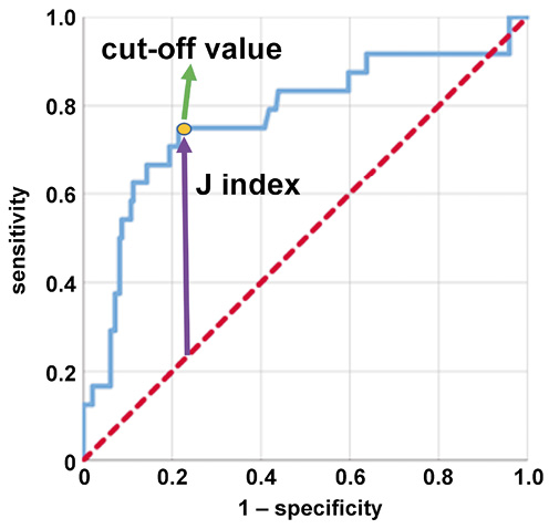
The primary objective of this study was to evaluate the association between pre-treatment GLUCAR index values and the subsequent incidence of ORN during the follow-up period. Continuous variables were summarized using medians (Me) and interquartile ranges (IQR), while categorical variables were presented as frequencies and percentages. Between-group comparisons were performed using appropriate statistical methods, including Student’s t test for continuous variables and the χ2 test for categorical variables. The receiver operating characteristic (ROC) curve analysis was performed to determine the optimal cut-off value of the pre-treatment GLUCAR index for discriminating between patients with and without ORN. The optimal cut-off was identified using the maximum Youden index (J), which represents the difference between the true-positive rate (sensitivity) and the false-positive rate (1 − specificity), thereby optimizing the balance between sensitivity and specificity. Furthermore, logistic regression analysis was performed to identify independent predictors of ORN development following treatment. All statistical analyses were conducted using IBM SPSS Statistics for Windows, v. 26.0 (IBM Corp., Armonk, USA). All statistical tests were two-sided, and a p-value of less than 0.05 was considered statistically significant.
Results
This analysis involved 220 patients who met the inclusion criteria. The optimal cut-off value of the GLUCAR index for predicting the risk of ORN was determined using the ROC curve analysis, which identified an optimal threshold of 35.7 (area under the curve (AUC): 77.3%; sensitivity: 75.0%; specificity: 76.5%; Youden’s J index: 0.515). Based on this threshold, 154 patients (70.0%) were classified as having a GLUCAR index <35.7, while 66 patients (30.0%) had a GLUCAR index ≥35.7 (Figure 1).
Baseline characteristics
Baseline characteristics according to GLUCAR index group are summarized in Table 1. The median age of the overall cohort was 55.5 years (range: 18–78 years). Patients with a GLUCAR index ≥35.7 were significantly younger than those with a GLUCAR index <35.7, with median ages of 53.5 and 57.0 years, respectively (p = 0.015). The cohort comprised 145 males (65.9%) and 75 females (34.1%), with no significant differences in gender distribution between the GLUCAR index groups (p = 0.350). Smoking prevalence was higher in the GLUCAR index <35.7 group (35.1%) than in the GLUCAR index ≥35.7 group (21.2%) (p = 0.042). Additionally, a significantly larger proportion of patients in the GLUCAR index ≥35.7 group had diabetes mellitus (40.9%) than in the GLUCAR index <35.7 group (13.6%) (p = 0.001). The frequency of pre-CCRT tooth extractions was also significantly higher in the GLUCAR index ≥35.7 group (90.9%) than in the GLUCAR index <35.7 group (53.2%) (p < 0.001). No significant differences were observed between the groups regarding alcohol consumption, T-stage or N-stage. The median follow-up duration for the entire cohort was 36.0 months (range: 12–78 months).
Post-CCRT characteristics
The characteristics observed following CCRT are summarized in Table 2. The prevalence of tooth extractions after CCRT was significantly higher in the GLUCAR index ≥35.7 group (90.9%) than in the GLUCAR index <35.7 group (53.2%) (p < 0.001). Additionally, the incidence of ORN was significantly higher in the GLUCAR index ≥35.7 group (27.3%) than in the GLUCAR index <35.7 group (3.9%) (p < 0.001). No significant differences were found between the groups regarding the number of concurrent or adjuvant chemoradiotherapy cycles, the maximum mandibular dose (MMD), or the MMD group.
The overall incidence of ORN in the study cohort was 10.9%, with 24 of the 220 patients affected. Among these 24 patients, the distribution of the Notani stages was as follows: stage 0 in 1 patient (4.2%); stage 1 in 14 patients (58.3%); and stage 2 in 9 patients (37.5%). In the subgroup analysis, ORN was observed in 27.3% of patients with a GLUCAR index ≥35.7, compared with 3.9% of those with a GLUCAR index <35.7 (p < 0.001). No significant differences in Notani stage distribution were observed between the 2 GLUCAR index groups (p = 0.790)
Osteoradionecrosis predictors: Univariate and multivariate analysis
In the univariate analysis (Table 3), the following factors were significantly associated with the occurrence of ORN: smoking status (p = 0.011); pre-CCRT tooth extractions (p = 0.003); the number of CCRT cycles (p = 0.030); the MMD group (p < 0.001); and the GLUCAR index group (p < 0.001).
In the multivariate logistic regression analysis, the following factors were identified as independent predictors of ORN: smoking status (odds ratio (OR): 3.70; 95% confidence interval (CI): 1.85–8.33; p = 0.024); pre-CCRT tooth extractions (OR: 5.88; 95% CI: 2.70–7.14; p = 0.007); the number of CCRT cycles (OR: 1.69; 95% CI: 1.19–2.44; p = 0.042); the MMD group (OR: 4.76; 95% CI: 1.54–8.17; p < 0.001); and the GLUCAR index group (OR: 9.09; 95% CI: 2.44–25.67; p < 0.001). Patients with a GLUCAR index ≥35.7 had a substantially higher risk of developing ORN than those with a GLUCAR index <35.7 (27.3% vs. 3.9%; p < 0.001), independent of other clinical factors (Table 3, Figure 2).
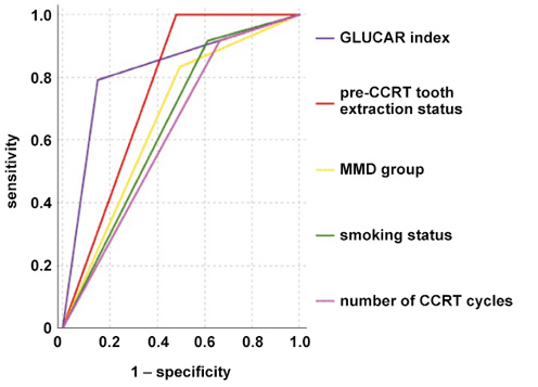
The ROC curve analysis evaluating the predictive performance of the 5 significant factors for ORN demonstrated that GLUCAR index grouping was the strongest predictor, with the highest AUC (82.2%), followed by pre-CCRT tooth extraction status (76.0%), the MMD group (66.9%), smoking status (65.2%), and the number of CCRT cycles (62.7%) (Figure 3).
Discussion
In this retrospective study of 220 patients with LANPC treated with CCRT, we found that an elevated pre-treatment GLUCAR index (≥35.7), a novel composite biomarker reflecting metabolic, immune, inflammatory, and nutritional status, was independently associated with a significantly increased risk of developing jaw ORN. To the best of our knowledge, this is the first study to investigate the predictive value of the GLUCAR index for radiation-induced bone complications in patients with NPC.
In addition to the GLUCAR index, our multivariate analysis identified several independent risk factors associated with an increased risk of ORN, including smoking, pre-CCRT tooth extractions and the number of CCRT cycles. Smoking is a well-established risk factor for impaired tissue health, as it promotes hypoxia, impairs angiogenesis and alters immune responses, all of which compromise soft tissue and bone healing following radiotherapy.25, 26 Moreover, the vasoconstrictive and pro-inflammatory effects of tobacco constituents may exacerbate radiotherapy-induced endothelial damage, further increasing the risk of ORN.27 Although pre-CCRT dental extractions are often necessary to eliminate potential sources of infection, these procedures may predispose patients to ORN, particularly when radiotherapy begins before adequate healing of the oral mucosa and the underlying bone has occurred.28 Our results also indicate that patients receiving more intensive CCRT regimens, as reflected by a higher number of CCRT cycles, may face an increased risk of ORN. This association may reflect cumulative systemic toxicity, which can impair bone remodeling, increase oxidative stress, deplete the essential nutrients required for tissue repair, and exacerbate radiation-induced endothelial dysfunction and hypoxia.29 Taken together, these factors may create an unfavorable environment for mandibular bone healing, particularly when compounded by high radiation doses.
The incidence of ORN in our cohort was 10.9%, which is consistent with previously reported rates of 7–10% in high-risk LANPC populations treated with modern intensity-modulated radiation therapy (IMRT) techniques.29, 30 The pathogenesis of ORN is multifactorial and involves radiation-induced vascular injury, chronic hypoxia, hypocellularity, and impaired bone remodeling.7, 29, 31 High-dose radiation damages endothelial cells in small blood vessels, leading to progressive obliterative endarteritis, reduced tissue perfusion and persistent hypoxia. This chronic ischemic state compromises osteoblast function and suppresses bone remodeling, both of which are essential for maintaining skeletal integrity and facilitating tissue repair.32 Moreover, radiation induces fibroatrophic changes in the periosteum and bone marrow, further diminishing the regenerative capacity of these tissues.33 Importantly, patients undergoing CCRT may be at an increased risk of ORN compared with those receiving radiotherapy alone, as chemotherapy may potentiate radiation-induced damage to bone and the surrounding soft tissues.34
In addition to the detrimental effects of radiation on bone healing, systemic pro-inflammatory and catabolic states, including hyperglycemia, increased systemic inflammation, immunosuppression, and malnutrition, as reflected by an elevated CAR, may further impair tissue repair mechanisms by promoting oxidative stress, dysregulated cytokine signaling and impaired cellular metabolism. In this context, the novel GLUCAR index, which integrates fasting glucose and CAR, may serve as a surrogate marker of host vulnerability to ORN. Hyperglycemia disrupts endothelial nitric oxide signaling, promotes the formation of AGEs and induces oxidative stress, all of which contribute to endothelial dysfunction, impaired angiogenesis and reduced collagen synthesis.8, 10 Furthermore, hyperglycemia adversely affects osteoblast differentiation and promotes osteoclast-mediated bone resorption, thereby compounding the detrimental effects of radiation on bone metabolism. Therefore, an elevated GLUCAR index may reflect an unfavorable systemic metabolic and inflammatory environment that facilitates the initiation and progression of ORN following CCRT.
Nonetheless, despite the compelling biological rationale supporting this hypothesis, it requires validation through well-designed basic and clinical research. Future studies should aim to elucidate the underlying biological mechanisms and provide a more comprehensive understanding of the predictive capacity of the GLUCAR index for ORN. Such investigations will help establish more robust evidence regarding the clinical utility of the GLUCAR index as a predictive tool.
From a translational perspective, the current findings highlight the potential of the GLUCAR index as a pre-treatment biomarker for stratifying the risk of ORN in patients with LANPC undergoing definitive CCRT. If validated in prospective multicenter studies, the routine assessment of the GLUCAR index could facilitate the early identification of patients at increased risk of radiation-induced mandibular injury. In these high-risk individuals, appropriate preventive strategies could be implemented proactively. These may include stricter glycemic control, individualized nutritional interventions aimed at optimizing protein and micronutrient intake, and intensified dental management protocols designed to minimize the need for tooth extractions during or after treatment.
Beyond systemic interventions, the GLUCAR index may also have implications for radiation treatment planning. In patients with elevated GLUCAR index values, clinicians could consider more aggressive mandibular-sparing approaches, including advanced IMRT techniques with stricter mandibular dose constraints or, where available, intensity-modulated proton therapy (IMPT), to further reduce radiation exposure to the mandible. Such individualized approaches may help reduce the incidence of ORN, preserve long-term oral function, and ultimately improve QoL in this vulnerable population.25, 26, 30, 35
Limitations
Several limitations of this study should be acknowledged. First, the retrospective design may have introduced inherent biases, including selection bias and unmeasured confounding. Although we attempted to address these issues through multivariate analysis incorporating key clinical factors, the inherent limitations of retrospective studies may still affect the robustness of our findings. Second, this study was conducted at a single institution, which may limit the generalizability of the results. Validation in large, independent, multicenter cohorts encompassing diverse clinical settings, patient demographics and radiotherapy techniques is therefore necessary to establish the broader applicability of our findings. Third, although the overall incidence of ORN in our cohort (10.9%) was consistent with previously reported rates, the absolute number of ORN cases (n = 24) was relatively small. This limitation may have reduced the statistical power of subgroup analyses, particularly those evaluating the relationship between the GLUCAR index and Notani stage distribution, which reflects ORN severity. Fourth, although the GLUCAR index incorporates important metabolic, immune, inflammatory, and nutritional markers, it does not account for other systemic factors that may significantly influence bone healing. These include vitamin D deficiency, sarcopenia, and alterations in the oral and gut microbiomes. Therefore, future prospective studies should consider incorporating these additional variables and evaluating changes in the GLUCAR index throughout treatment. Such approaches may provide a more comprehensive understanding of the pathophysiology of ORN and facilitate the development of more accurate predictive models for identifying patients at risk.
Conclusions
The findings of this study suggest that an elevated GLUCAR index, reflecting systemic disturbances in metabolic, immune, inflammatory, and nutritional status, is a significant independent predictor of ORN in patients with LANPC undergoing CCRT. If validated in future studies, incorporating GLUCAR index assessment into routine pre-CCRT evaluations could help clinicians identify patients at increased risk and implement appropriate preventive strategies, optimize supportive care and individualize radiation treatment planning. Such approaches may ultimately reduce the incidence of ORN and improve patient outcomes.
Ethics approval and consent to participate
The study was conducted in accordance with the Declaration of Helsinki. The study protocol received formal approval from the Institutional Review Board of Baskent University Faculty of Medicine, Andana, Turkey (project no. DKA:19/39), ensuring compliance with applicable ethical standards. Written informed consent was obtained from all the participants. The consent covered the collection and subsequent analysis of relevant clinical data, as well as the use of anonymized data for academic presentations and publications.
Data availability
Data are available from Baskent University Institutional Data Access Committee for researchers meeting criteria for confidential data access.
Consent for publication
Not applicable.
Use of AI and AI-assisted technologies
Not applicable.