Abstract
Background. Temporomandibular disorders (TMDs) are a group of musculoskeletal pathologies that are characterized by pain that occurs or worsens during function, temporomandibular joint (TMJ) noises, and limited jaw movements. The relationship between occlusion and TMDs has been the subject of extensive research, yet the findings have been contradictory.
Objectives. The aim of the present study was to examine the relationship between partial edentulism, prosthodontic rehabilitation, and a selection of TMD symptoms. Therefore, we re-examined data from a national screening examination from a novel perspective.
Material and methods. A retrospective analysis was conducted on data from a representative national epidemiological pathfinder survey for oral symptoms. In our analysis, we investigated the registered major TMD symptoms in 3 groups: patients with extensive tooth loss; patients with prosthodontic rehabilitation; and patients with normal dentition as control.
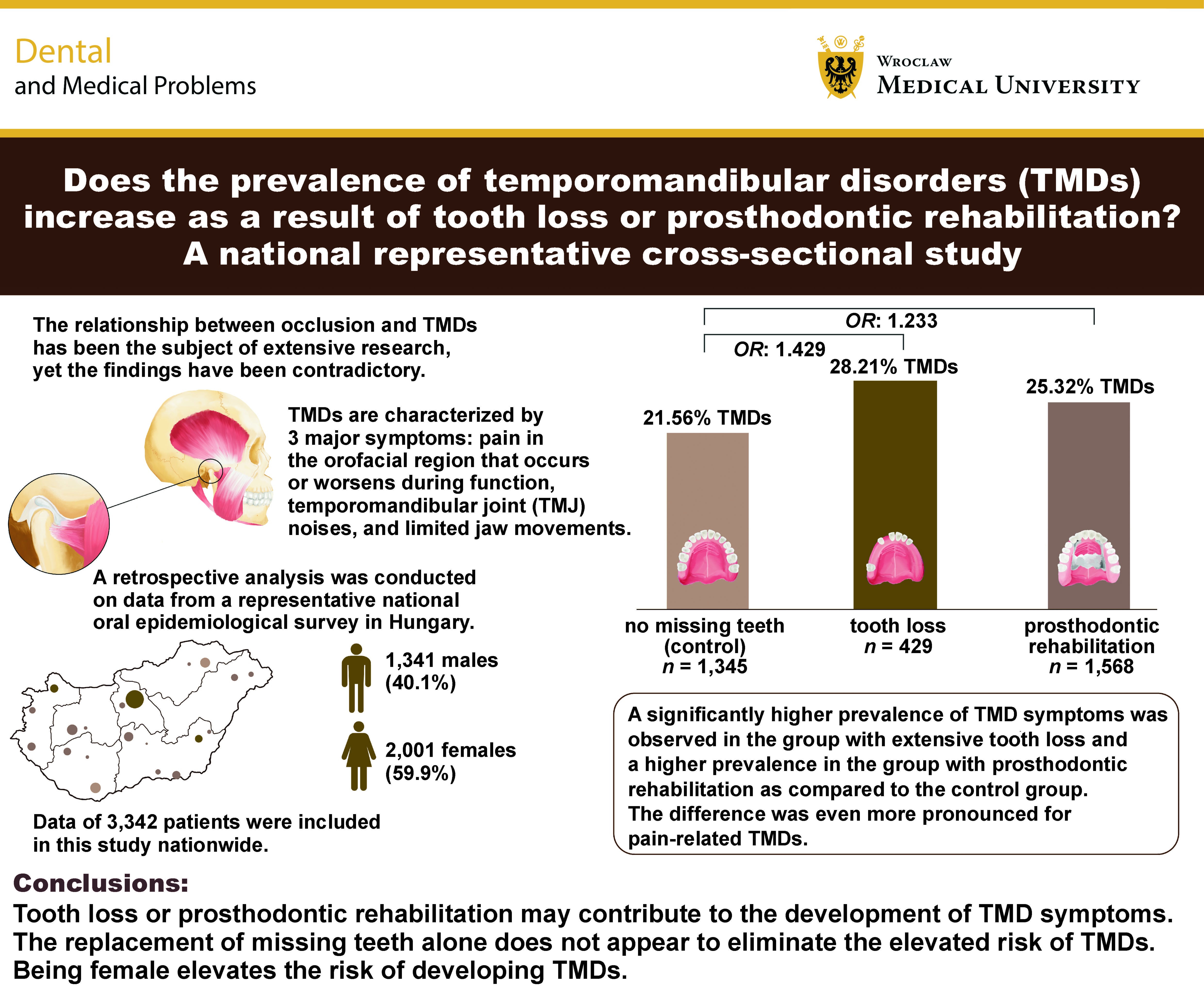
Results. The present study utilized data from a total of 3,342 patients. A significantly higher prevalence of TMD symptoms was observed in the group with extensive tooth loss (28.21%) as compared to the control group (21.56%) (p = 0.017; OR: 1.429). Similarly, a higher prevalence of TMD symptoms was observed in the group with prosthodontic rehabilitation (25.32%) (p = 0.057; OR: 1.233) as compared to the control group. The difference was even more pronounced for pain-related TMDs. The logistic regression analysis revealed that female gender was a significant predictor of pain, limited mouth opening and TMJ clicking.
Conclusions. Our findings suggest that tooth loss or prosthodontic rehabilitation may contribute to the development of TMD symptoms. They also show that the replacement of missing teeth alone does not appear to eliminate the elevated risk of TMDs. Consistent with the existing literature, being female elevates the risk of developing TMDs.
Keywords: temporomandibular disorders, dental occlusion, cross-sectional survey, prosthetic rehabilitation, tooth loss
Introduction
Temporomandibular disorders (TMDs) are a group of musculoskeletal pathologies that are characterized by 3 major symptoms in the orofacial region. These symptoms include pain that occurs or worsens during function, temporomandibular joint (TMJ) noises, and limited jaw movements, according to the Diagnostic Criteria for Temporomandibular Disorders (DC/TMD).1
Current estimates suggest that the prevalence of TMDs in the general population is substantially higher than previously thought, at approx. 34%.2 A wide range of psychosocial, behavioral, and physiological features have been described as etiological factors in TMDs. Depression and anxiety are well-documented psychological comorbidities,3 while behavioral factors,4 such as awake5, 6 and sleep bruxism,7, 8, 9 as well as sleep quality,10 have been identified as significant contributors. The relationship between bruxism and TMDs remains somewhat controversial. However, recent studies have reported a co-occurrence rate of 18%.2 In addition, individual biological characteristics, ranging from genetic factors to dentition-related features, have been shown to contribute to the development and persistence of TMDs.11
Further research is essential to advance the diagnosis of TMDs, patient pathways, and interprofessional collaboration. In view of the findings of recent research, it is worthwhile to examine the medications used to treat chronic conditions that coexist with TMDs.12 Information about changes in the tissues and the volume of the surrounding structures, such as the neck muscles, may also be important.13 New questionnaires may be of significant help in assessing the emotional state of patients.14 Furthermore, it may be worthwhile to consider evaluating therapeutic alternatives for bruxism,15 which is common among patients with TMDs.
The potential links between occlusal factors and TMDs have been the subject of investigation for decades, yet neither definitive proof nor comprehensive refutation has emerged.16 A number of studies have identified a correlation between occlusal factors and TMDs,17, 18, 19, 20 while others have not.21, 22, 23 Okeson provides a useful framework for interpreting the disparate results of previous studies: “One might conclude that if occlusion were the major etiologic factor in TMD, the profession would have confirmed this many years ago. On the other hand, if occlusion has nothing to do with TMD, the profession would have likewise already confirmed this conclusion.”24 Current knowledge suggests that TMDs may result from multiple etiological factors, thus indicating a biopsychosocial origin.25
In the field of TMD treatment, conservative therapy has been identified as the most effective approach in the majority of the existing literature.16 This approach encompasses counselling, self-management techniques, physical therapy, pharmacotherapy, and splint therapy, which are considered the preferred treatment modalities in most cases. Irreversible therapies include the provision of dental prostheses; however, the classification of dentures as a therapeutic intervention for TMDs remains debatable.
A review of the literature reveals numerous examples of studies investigating the relationship between tooth loss and TMDs. For instance, in 2023, Rawat et al. conducted a systematic review of 13 selected publications, which indicated that tooth loss affects TMJ function.26 The negative effect appears to increase with the number of missing teeth, and the absence of posterior teeth is associated with greater TMJ damage. However, due to the complexity of TMD development, it cannot be definitively concluded that tooth loss causes TMDs.26
In 2023, Manfredini et al. initiated an investigation to address 2 key questions – “Can prosthodontic rehabilitation be used as treatment for TMDs?” and “Can prosthodontic rehabilitation cause TMDs?”27 However, they were unable to include any articles in the planned systematic review due to the lack of high-quality clinical trials or randomized clinical trials (RCTs) on this topic.27 This suggests that very little evidence is available in the existing literature regarding the effects of prosthodontic rehabilitation on TMJ.
The aim of the present study was to examine the relationship between partial edentulism, the presence of prosthodontic restorations, and selected TMD symptoms. Therefore, we re-examined data from a national screening examination from a novel perspective. The null hypothesis was that tooth loss or prosthodontic rehabilitation would not be associated with an increased prevalence of TMDs.
Material and methods
A retrospective analysis was conducted using data from a representative national oral epidemiological pathfinder survey. The survey was carried out by calibrated physicians and assistants from the Department of Prosthodontics at Semmelweis University, Budapest, Hungary, and was based on the World Health Organization (WHO) criteria for screening surveys.28 Data were collected over a 2-year period, from 2003 to 2004. Ethical approval was obtained from the Regional and Institutional Committee for Scientific and Research Ethics of Semmelweis University (SE TUKEB 206/2003).
To determine the appropriate sample size for the study, the number of subjects required to detect small clinical effects was assessed. A sample size of more than 2,180 participants was considered sufficient to detect small clinical effects with 90% statistical power.29
Dental screening was conducted in conjunction with chest X-ray examinations. In Hungary, the chest X-ray screening program has a long history, having originally been introduced to detect tuberculosis. All Hungarian adults aged 18 years and older receive postal invitations every 5 years. Although this examination is no longer mandatory, high participation rates are observed among the population. A total of 304 locations were randomly selected from the comprehensive list of chest X-ray stations in Hungary, thereby ensuring the representation of all types of settlements. To determine the appropriate sample composition, data on gender and age groups at each location were used in accordance with data from the Hungarian Central Statistical Office. Socioeconomic characteristics were not directly taken into account during sample selection; however, as these characteristics correlate with a geographical region in Hungary, regional differences were also considered. All individuals who volunteered to participate and provided written informed consent underwent screening.
The inclusion criteria were as follows: participants had to be at least 18 years of age and provide written informed consent to participate in the study. The gender distribution and age of participants at each study site were determined as outlined above to ensure the representativeness of the survey. The exclusion criterion was refusal to participate in the study.
The survey team comprised 7 calibrated examiners and 5 data recorders, all affiliated with the Department of Prosthodontics at Semmelweis University. All team members had previously undergone theoretical and practical training and calibration. Inter-examiner reliability during examiner calibration demonstrated almost perfect agreement, with a Cohen’s kappa value of 0.85.
Dental screening examinations were conducted using a portable dental X-ray chair with a headrest, appropriate artificial lighting, and a range of diagnostic instruments, including a dental probe, a dental mirror, a dental forceps, and a WHO periodontal probe.
A questionnaire was used to collect the following information about the participants: gender; age; type of settlement; educational level; general health status; oral hygiene habits; frequency of dental visits; and smoking status.
The clinical data obtained during the dental examinations were analyzed. The condition of each tooth was recorded according to the following categories: intact; decayed; filled; prepared for fixed prostheses; or missing. Where applicable, the type of partial tooth loss and the existing prosthodontic appliances were recorded. Partially edentulous cases were classified according to the Fábián and Fejérdy classification of partial edentulism.
Additionally, selected TMD symptoms were assessed during the clinical examination. All pain triggered or aggravated by jaw movements was documented, irrespective of its origin, whether in the muscles involved in jaw movements or in TMJ. The degree of mouth opening was measured using a ruler, and any limitation was recorded. The presence of TMJ clicking was also documented.
Data collection was conducted before the development of validated diagnostic criteria; therefore, no such criteria were used.
Although the most widely recognized classification system for tooth loss is the Kennedy classification,30 a logical and straightforward classification system for partial edentulism was developed in Hungary at Semmelweis University: the Fábián and Fejérdy classification.31, 32, 33, 34 This classification is based on an analysis of the forces acting on the planned prosthodontic appliance. Each class is determined by the position of the remaining teeth, which may provide support against the chewing forces acting on the dental prosthesis. Consequently, the classification also provides recommendations regarding the most appropriate type of prosthodontic appliance. In the national representative screening survey, tooth loss was classified according to this system.
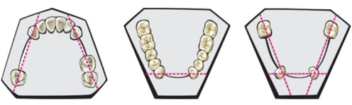
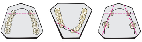
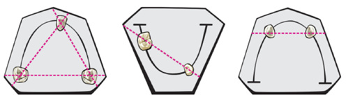
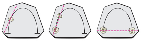
The Fábián and Fejérdy classification differentiates between 3 principal classes (Classes 1–3), 2 or 3 subclasses (1A, 1B, 2A, 2B, and 2A/1), and 2 categories: the completely edentulous situation (T); and the dentate situation without tooth loss (Class 0).
In Class 1, a fixed prosthetic appliance can be constructed, since no torque forces are present (Class 1A) (Figure 1), or because the torque forces can be compensated for by extending the bridge (Class 1B) (Figure 2). In Class 1A, a sufficient number of occlusal units are present in each quadrant, thereby maintaining stable occlusion. This class is analogous to Kennedy Class III, in which the teeth on either side of the edentulous area can be used as abutments.
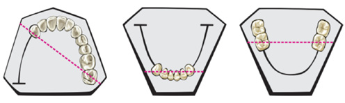
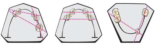
In Classes 2 (Figure 3 and Figure 4) and 3 (Figure 5), the extent of tooth loss necessitates the fabrication of either a removable partial denture or a complex prosthesis, constructed in accordance with the established prosthodontic principles. In these classes, the absence of stable occlusion is attributed to the extensive edentulous areas located posterior to the remaining teeth. Class 2 is analogous to Kennedy Classes I and II, whereas Class 2A/1 (Figure 6) encompasses subtotal edentulism with only 1 or 2 remaining teeth.
In our analysis, the recorded TMD symptoms were investigated in 3 distinct groups. Patients classified as prosthodontic Classes 1B or 2 according to the Fábián and Fejérdy classification were assigned to the tooth loss (L) group. These patients had several missing teeth, so occlusion was not stable in all quadrants. Patients with any type of prosthodontic restoration, either fixed or removable, regardless of whether the restorations were appropriately designed or occlusally sound, were assigned to the prosthodontic (P) group. Patients without extensive tooth loss, with stable occlusion in all quadrants (Classes 0 and 1A according to the Fábián and Fejérdy classification), and without prosthodontic appliances were assigned to the control (C) group. If the two dental arches belonged to different classes, the more extensive tooth loss was taken into account for group assignment. If a prosthodontic restoration was present in only one dental arch, even if it consisted of a single crown, the patient was assigned to the P group.
Statistical analysis
The data were processed using Microsoft Excel spreadsheets, and statistical analysis was performed using the GraphPad Prism software (v. 8.0.1). The χ2 test was used as an omnibus test, followed by Fisher’s exact test for post hoc pairwise comparisons. Multivariable logistic regression analysis was also performed, with effect sizes calculated where applicable.
Although the following values are commonly used to define effect sizes according to Cohen’s d – small: 0.2, medium: 0.5, and large: 0.8 – Zieliński and Gawda proposed lower effect size thresholds for TMD research, categorizing them as follows: 0.1 for small; 0.2 for medium; and 0.7 for large effects.29 As categorical variables were used in our study, the proposed thresholds were converted into odds ratios (ORs). The following values were adopted: OR = 1.2 for small effects; OR = 1.7 for medium effects; and OR = 3.6 for large effects.
Results
A total of 6,397 individuals were invited to participate in the screening process at the designated sites. Of these, 28% (1,791 individuals) did not respond. The majority of non-respondents cited a lack of time as the reason for their non-participation. Of the 4,606 individuals who underwent screening, those classified as Class 2A/1, Class 3 or Class T according to the Fábián and Fejérdy classification, indicating complete edentulism or the presence of no more than 3 remaining teeth, were excluded from the present study (n = 1,264; 27.4%). This exclusion was based on the study objective, which was to examine the relationship between tooth loss, the use of partial dentures, and TMDs.
The present study included data from 3,342 participants. The study population consisted of 1,341 males (40.1%) and 2,001 females (59.9%). The mean age of the participants was 43.9 ±15.0 years. The group with extensive tooth loss (L) comprised 429 individuals (12.8%), with a mean age of 50.4 ±13.0 years. The gender distribution in this group was 217 males (50.6%) and 212 females (49.4%). The group with prosthodontic rehabilitation (P) consisted of 1,568 individuals (46.9%), with a mean age of 51.1 ±12.6 years. Of these, 490 (31.3%) were male and 1,078 (68.8%) were female. A total of 1,345 participants (40.2%) were included in the control group (C), in which occlusion was stable in all quadrants. Their mean age was 33.5 ±11.9 years, and 634 (47.1%) were male and 711 (52.9%) were female (Table 1). The mean age of the control group appeared to be lower than that of the L and P groups. However, multivariable logistic regression analysis revealed no statistically significant association between age and group membership (OR: 0.99; 95% confidence interval (CI): 0.99–1.00).
A total of 808 participants (24.18%) exhibited symptoms consistent with TMDs. The most prevalent symptom was TMJ clicking, which was present in 786 individuals (23.52%). Pain triggered or aggravated by jaw movements was reported by 48 participants (1.44%), while limited jaw movement was observed in 12 participants (0.36%). In 36 participants (1.08%), 2 major TMD symptoms occurred simultaneously, while all 3 major symptoms were present in one participant (0.03%) (Table 1).
The gender distribution showed a notable difference, with males accounting for 31% of participants presenting with TMD symptoms and females accounting for the majority (69%). Among the 1,341 male participants examined, 248 (18.49%) exhibited TMD symptoms, whereas among the 2,001 female participants, 560 (27.99%) presented with TMD symptoms (p < 0.01). Multivariable logistic regression analysis further demonstrated that women had higher odds of presenting with TMD symptoms than men (OR: 1.71; 95% CI: 1.44–2.03). A greater proportion of female participants also reported limited mouth opening (OR: 3.79; 95% CI: 0.99–24.87), pain in TMJ or the orofacial region (OR: 4.85; 95% CI: 2.21–12.81), and joint clicking (OR: 1.66; 95% CI: 1.40–1.97) (Table 2). The distribution of individual symptoms was similar in both sexes, with joint clicking being overwhelmingly the most prevalent symptom. Among male participants presenting with TMD symptoms, joint clicking was reported in 98.79%, compared with 96.61% of female participants presenting with TMD symptoms.
Upon examining the occurrence of TMD symptoms across the 3 groups, differences in their prevalence were observed. A significantly higher prevalence of TMD symptoms was observed in the group with extensive tooth loss compared with the control group (p = 0.017; OR: 1.429; 95% CI: 1.116–1.830). Similarly, a higher prevalence of TMD symptoms was observed in the group with prosthodontic rehabilitation compared with the control group (p = 0.057; OR: 1.233; 95% CI: 1.038–1.466). Multivariable logistic regression analysis further demonstrated that the group with tooth loss had higher odds of presenting with TMD symptoms (OR: 1.55; 95% CI: 1.18–2.04). No significant difference was observed in the prevalence of TMD symptoms between the groups with tooth loss and prosthodontic rehabilitation. With regard to individual symptoms, a similar pattern was observed for joint clicking. The occurrence of this symptom was higher in both the L group (p = 0.016; OR: 1.437; 95% CI: 1.120–1.844) and the P group (p = 0.051; OR: 1.241; 95% CI: 1.042–1.477) compared with the control group (Table 3).
In the multivariable logistic regression analysis of pain triggered or aggravated by jaw movements, a significant association with a large effect size was found for tooth loss (OR: 3.60; 95% CI: 1.42–8.88), whereas prosthodontic rehabilitation was associated with a medium effect size (OR: 2.03; 95% CI: 0.93–4.60) (Table 4).
Discussion
The aim of this study was to examine the relationship between partial edentulism, the presence of prosthodontic appliances, and selected TMD symptoms. The null hypothesis was that tooth loss or prosthodontic rehabilitation would not increase the prevalence of TMD symptoms. The findings of the present study revealed statistically significant differences in the prevalence of TMD symptoms, particularly pain triggered or aggravated by jaw movements, between the control group and the groups with tooth loss (L) and prosthodontic rehabilitation (P). Therefore, the null hypothesis was rejected.
The results of the nationwide comprehensive oral epidemiological pathfinder survey conducted in Hungary have previously been evaluated from several different perspectives. These include general epidemiology,35 dental caries,36 periodontology,37 and prosthodontics.38, 39, 40 Previous studies have also addressed several other topics, including oral mucosal lesions,41 nutritional and oral hygiene habits,42 and the association between TMDs and certain orthodontic abnormalities.43
The association between occlusion and TMDs has also been investigated through the analysis of national surveys conducted in other countries. A study comparable to the present research was conducted in Finland as part of the nationally representative Health 2000 Survey, with data collection taking place between 2000 and 2001.19 In 2013, Sipilä et al. published their findings on the impact of tooth loss and the use of prosthodontic appliances on the clinical manifestations of TMDs. The authors concluded that complete edentulism, the use of complete dentures, and the wearing of poorly maintained removable dentures were associated with pain-related TMD symptoms in women. Furthermore, they found that these associations were influenced by psychosocial factors.19
In a study published in 2023, Liu et al. investigated the relationship between congenital tooth agenesis and the occurrence of TMDs.44 The study included 583 patients with congenital edentulism and 586 controls. The results indicated a significant association between congenital edentulism and both intra-articular (p = 0.017) and pain-related TMDs (p = 0.009). Additionally, the number of quadrants with missing teeth was found to be associated with the occurrence of TMDs.44
A comparable finding was reported by Jaber et al. in a radiological study examining TMJs in patients with TMDs.45 The study indicated that unilateral tooth loss was associated with a higher occurrence of TMDs.45
Tallents et al. investigated the potential association between the absence of posterior teeth and TMDs.46 They compared findings from magnetic resonance imaging (MRI) of the TMJs in patients with asymptomatic and pain-related TMDs with the number of missing premolars and molars. The results showed that TMD-related pain and MRI-confirmed disc displacement were significantly positively associated with the number of missing premolars and molars (p < 0.05).46
In their retrospective cross-sectional study, Chatzopoulos et al. examined the relationship between TMDs, parafunctional habits and missing teeth using patient questionnaires and radiographs.47 The results demonstrated statistically significant associations between the number of missing teeth, gender, age, and the occurrence of TMD symptoms (difficulty opening and closing the mouth, joint clicking, TMJ pain, and difficulty chewing). TMD symptoms were more prevalent among women and younger individuals. However, only chewing difficulty was found to be positively associated with the number of missing teeth, and this complaint is not necessarily directly related to TMDs.47
In a study examining the relationship between missing posterior teeth and TMDs, Wang et al. identified slight associations between the occurrence of TMDs and patients’ gender, age, and the number of missing teeth using multivariable logistic regression analysis.48 Furthermore, the study investigated the distribution of missing teeth according to the number of affected dental quadrants. This distribution was also found to be associated with the occurrence of TMDs. The findings suggest that the odds of presenting with TMDs are higher among individuals with missing posterior teeth in a greater number of quadrants (p < 0.01), particularly among young women.48
De Sousa et al. investigated potential associations between malocclusion, the presence of 5 or more missing teeth, and TMDs.49 Their findings suggest that these factors contribute only minimally to the development of TMDs.49
In a study published in 2016, Malheiros et al. found that TMD severity was higher among completely edentulous patients.50 However, they were unable to demonstrate any statistically significant differences between partially edentulous and dentate patients.50
A systematic literature review published in 2024 included 21 publications and aimed to examine the relationships between malocclusion and TMJ morphology, as well as the effects of tooth loss and bruxism on TMDs.51 Eight studies addressing the relationship between TMDs and tooth loss were selected for analysis. Of these, 3 examined morphometric features using radiographs, 2 identified TMD symptoms through self-reported questionnaires, and the remaining 3 performed and reported clinical examinations. These 3 studies concluded that pain and joint noises were associated with the number of missing teeth. These findings are consistent with those of our study, which demonstrated a higher prevalence of TMD symptoms in the group with extensive tooth loss compared with the control group. As outlined in the systematic review, the findings suggest that malocclusion is associated with alterations in TMJ morphology, tooth loss is associated with an increased prevalence and altered characteristics of TMDs, and bruxism is associated with TMD symptoms, including myofascial pain, disc displacement, joint pain, and muscle disorders.51
The results of the aforementioned studies are consistent with those obtained in our investigation. The present study revealed differences in the prevalence of selected TMD symptoms among the 3 examined groups: individuals with extensive tooth loss (L); those with prosthodontic rehabilitation (P); and the control group (C). A comparison between the control group and the group with extensive tooth loss revealed a significantly higher prevalence of TMD symptoms in the latter (Fisher’s exact test; p = 0.017). The corresponding effect size was OR: 1.429, which is slightly below the threshold for a medium effect. Logistic regression analysis further revealed that the odds of presenting with TMD symptoms were 1.55 times higher in the presence of tooth loss. The association between the control and prosthodontic rehabilitation groups was less pronounced, with a small effect size (OR: 1.233).
A further analysis focusing on pain triggered or aggravated by jaw movements revealed larger effect sizes in both the tooth loss and prosthodontic rehabilitation groups (OR: 3.60 and 2.03, respectively). These findings are consistent with the existing literature, which suggests that occlusal factors may play a modest role in the development of TMDs, particularly in cases involving pain that occurs or worsens with jaw movements.
The results of our study revealed a notably high prevalence of TMD symptoms, with nearly ¼ of the individuals examined exhibiting at least one of the major symptoms. Women accounted for approx. twice as many cases as men among participants presenting with TMD symptoms. A significantly higher proportion of women than men exhibited at least one of the major TMD symptoms. Furthermore, although the most prevalent finding in both sexes was joint clicking, pain triggered or aggravated by jaw movements and limited mouth opening were also observed more frequently among women. In the multivariable logistic regression analysis, female gender was identified as a significant predictor of pain triggered or aggravated by jaw movements and limited mouth opening, with large effect sizes (OR: 4.85 and 3.79, respectively). Female gender was also associated with a higher likelihood of joint clicking and the occurrence of any of the examined TMD symptoms. The effect sizes in these cases were medium (OR: 1.66 for joint clicking and OR: 1.71 for the occurrence of major TMD symptoms).
There are several potential explanations for the higher prevalence of TMD symptoms among women, and accumulating evidence suggests that pain sensitivity differs between males and females.52, 53 In addition, pain amplification resulting from the co-occurrence of multiple pain conditions has been identified as a contributing factor to the higher prevalence of TMDs in women. For instance, a study conducted by Marciniak et al. reported a significantly higher prevalence of TMDs (77.3%) among women diagnosed with endometriosis compared with the general population.54
Limitations
It is important to acknowledge the limitations of the present study. The data were collected between 2003 and 2004, which should be taken into consideration when interpreting the findings. Epidemiological data collected 2 decades ago may not be directly comparable with the current findings. Nevertheless, the results remain relevant, as they provide insights into the potential impact of tooth loss and prosthodontic rehabilitation on TMJ function and TMD symptoms.
Another limitation is that, at the time of data collection, the diagnostic criteria for TMDs were not as clearly established as they are today. The Diagnostic Criteria for Temporomandibular Disorders (DC/TMD) protocol was not published until 2014,1 and no Hungarian translation of the Research Diagnostic Criteria for Temporomandibular Disorders (RDC/TMD), originally published in 1992, was available.55 Consequently, the identification of TMD symptoms relied on the clinical expertise of the data collectors rather than on the standardized diagnostic criteria. Furthermore, no information was available regarding comorbidities, potential mental health conditions or previous TMD treatment.
Although our study, which was based on the results of a cross-sectional screening examination, would not meet the inclusion criteria established by Manfredini et al., it provides some insight into the 2 questions they posed – “Can prosthodontic rehabilitation be used as treatment for TMDs?” and “Can prosthodontic rehabilitation cause TMDs?”27 In other words, if prosthodontic rehabilitation were an effective therapeutic approach for TMDs, one might expect the group with the restored dentition to exhibit a lower prevalence of TMD symptoms. Conversely, if prosthodontic rehabilitation contributed to the development of TMDs, a higher prevalence of TMD symptoms might be expected in this group.
Conclusions
The relationship between TMDs and tooth loss has been the focus of extensive research. Despite mounting evidence suggesting that occlusal factors play only a limited role in the development of TMD symptoms, the findings of the present study suggest that tooth loss may be associated with a higher prevalence of TMD symptoms, particularly pain triggered or aggravated by jaw movements. Moreover, prosthodontic rehabilitation alone does not appear to eliminate this association. Female gender was also identified as a significant predictor of the occurrence of all the TMD symptoms examined.
Ethics approval and consent to participate
The study was conducted in accordance with the Declaration of Helsinki, and approved by the Regional and Institutional Committee for Scientific and Research Ethics of Semmelweis University, Budapest, Hungary (SE TUKEB 206/2003). Written informed consent was obtained from all subjects involved in the study.
Data availability
The datasets supporting the findings of the current study are available from the corresponding author on reasonable request.
Consent for publication
Not applicable.
Use of AI and AI-assisted technologies
Artificial intelligence technology was solely utilized to verify the linguistic accuracy of the text.