Abstract
Background. Oral health is one of the most important aspects of general well-being, and early tooth loss is associated with an increased risk of complications and mortality. These effects were even more pronounced in historical populations due to the limited effectiveness of the available treatment options.
Objectives. The primary aim of the present study was to investigate the prevalence of ante-mortem tooth loss (AMTL) and periapical lesions in a historical population from Gródek on the Bug River (Lublin Voivodeship), dating from the 11th to the 15th century, as valuable indicators of the population’s general health.
Material and methods. The study sample consisted of skulls of adult individuals whose permanent teeth had completed the eruption process (N = 156). A novel 6-stage classification system (0–5) for quantifying the severity of AMTL, accompanied by descriptive labels ranging from ‘none’ to ‘total’, was proposed to improve methodological consistency in paleoanthropological research. Periapical lesions were assessed macroscopically, with lesions smaller than 3 mm classified as granulomas.
Results. Out of the 156 examined skulls, only 9 (5.8%) retained their full dentition until death, while the remaining individuals had experienced tooth loss during their lifetimes. Nearly 2/3 of the examined skulls showed considerable AMTL. A considerable proportion (32%) of the individuals from the historical population of Gródek on the Bug River exhibited severe, potentially life-threatening periapical lesions resulting from oral health complications.
Conclusions. Ante-mortem tooth loss is a common consequence of poor oral hygiene and inadequate diet in historical populations. However, AMTL is a more complex phenomenon, as its consequences can have a substantial and long-term negative impact on general health. The observed patterns of AMTL may reflect difficulties in maintaining oral health, with potential implications for broader health and dietary conditions.
Keywords: periapical granuloma, periapical cyst, paleopathology, ante mortem tooth loss (AMTL)
Introduction
Oral health is one of the most important aspects of general well-being and depends on the proper functioning of all components of the stomatognathic system (SS). The SS comprises the teeth and periodontium (dentoalveolar system), the articulatory and occlusal systems of the upper and lower dental arches (dento-dental system), functionally interconnected temporomandibular joints (TMJs) (joint complex), as well as the facial bones, soft tissues, and the organs of the oral cavity and face. The proper structure and functioning of these components are essential for several critical processes, including the sucking reflex, articulation, mastication, and the initial stages of food digestion.1, 2, 3
The SS is structurally and functionally complex, and is therefore susceptible to various forms of damage and dysfunction. While modern dentistry can effectively treat or mitigate the consequences of many such conditions, diseases and disorders affecting the SS in historical populations often had serious health implications due to the limited medical knowledge and therapeutic options available at the time.
Ante-mortem tooth loss (AMTL) in historical populations most commonly resulted from the interplay of dietary habits, cultural practices and environmental factors. Dietary and nutritional factors played a significant role in AMTL. For instance, the consumption of tough and unprocessed foods led to increased dental wear, which was a major contributor to tooth loss.4 Conversely, a diet consisting of soft, highly processed foods rich in carbohydrates may have promoted the development of extensive carious lesions, leading to pulp exposure, abscess formation, and ultimately tooth loss.5, 6 Smith and Betsinger demonstrated that, in prehistoric Tennessee, the transition to maize agriculture was associated with changes in AMTL patterns, suggesting that dietary carbohydrates and nutritional deficiencies may have contributed to tooth loss.7 Poor oral hygiene also contributed significantly to AMTL in historical populations. The substantial accumulation of dental calculus could lead to the inflammation of the periodontal tissues, potentially resulting in tooth loosening and eventual loss. Cases of AMTL resulting from trauma are less frequently reported in the literature, although traumatic injuries could undoubtedly cause serious complications within the SS.4, 8 Finally, teeth could also be lost during an individual’s lifetime as a result of deliberate practices. Dental ablation for esthetic or cultural (ritual or symbolic) reasons was a recognized cause of AMTL in certain populations, including Hawaiians and prehistoric populations in Africa, China and Japan.9, 10, 11
One of the most serious dental pathologies observed in historical populations and contributing to AMTL was the development of periapical lesions and inflammatory apical cysts resulting from bacterial infections of the root canal system. The incidence of periapical lesions and the mortality associated with dental infections in historical populations were considerably higher than today, with dental diseases representing an important cause of death. Historical records indicate that, in the early 17th century, “teeth” were consistently listed as the 5th or 6th leading cause of death in London.12 This finding highlights the potentially severe consequences of untreated dental infections, which could lead not only to AMTL, but also to systemic complications and death. This is further supported by case studies of prehistoric populations, demonstrating that oral abscesses could result in life-threatening conditions.13
The primary aim of the present study was to investigate the prevalence of AMTL in a historical population from Gródek on the Bug River (Lublin Voivodeship), dating from the 11th to the 15th century, in order to characterize oral health patterns within this medieval community. The study also examined the occurrence of periapical bone destruction in this population, assessing the frequency and severity of these pathological lesions as indicators of dental health status. From a methodological perspective, we propose a comprehensive 6-stage classification system (0–5) for AMTL, accompanied by descriptive labels, to provide a standardized approach to quantifying the severity of tooth loss in paleoanthropological studies.
Material and methods
The stronghold at Gródek on the Bug River (Polish: Gródek nad Bugiem) forms part of a large medieval settlement complex situated near the confluence of the Huczwa and Bug Rivers, close to the present-day eastern border of Poland. Initially, the settlement at Gródek on the Bug River was dated to the 12th–15th centuries. However, recent research using radiocarbon (14C) dating has shown that the cemetery is older, establishing the 11th–15th centuries as a more reliable chronological framework for its use.14
The skeletal material was excavated during the 1952–1955 field seasons, and the preliminary results of anthropological analyses involving 303 individuals (170 males and 133 females) were published by Belniak et al.15 Currently, the osteological collection from Gródek on the Bug River is less numerous than it was in the 1950s, owing to the damage caused by the flood that affected Wroclaw in 1997. The remains of 197 individuals are currently housed at the Department of Anthropology, Ludwik Hirszfeld Institute of Immunology and Experimental Therapy, Polish Academy of Sciences, in Wroclaw, Poland. Skulls in relatively good condition constitute the largest part of this osteological collection (191 out of 197 individuals).16
Long-term archaeological research conducted at this site has revealed numerous artifacts providing valuable insights into the everyday lives of the inhabitants of medieval Gródek on the Bug River. Clusters of graves containing similar grave goods, such as buttons and fabric fragments, sometimes interwoven with a gold thread, have been identified in the eastern part of the cemetery. The concentration of graves, both with and without grave goods, in this area is particularly intriguing and may suggest that this part of the cemetery was established earlier than the others. It is possible that this area once constituted the central part of the cemetery, where individuals from the wealthiest and most prominent families were buried.17 These findings may indicate social stratification within the medieval community of Gródek on the Bug River, particularly given that some graves contained silk textiles and fabrics incorporating gold or gilded silver threads, which would have been affordable only to the most affluent members of society.18
The study sample consisted of 156 adult skulls from individuals whose permanent teeth had completed the eruption process. An essential criterion for inclusion in the study was the good state of preservation of both the skulls and the anatomical structures of the SS. Sex and age at death were estimated based on cranial features.19, 20, 21 The biological age categories followed the classification proposed by Martin and Saller.22 The sample was divided into 2 age groups: younger adults (juvenis, including only individuals with their third molars erupted, and adultus) and older adults (maturus and senilis).
For each individual in the examined sample, AMTL was documented by identifying the specific tooth positions (incisors, canines, premolars, and molars) in both the maxilla and the mandible, based on visible evidence of healing in the alveolar sockets. The 6-point classification scale developed for this study was used to assess the severity of AMTL, taking into account various functional aspects of tooth loss (Table 1).
Kennedy’s classification served as the initial methodological framework for this study.23 However, this system focuses on patterns of partial edentulism relevant to prosthetic rehabilitation and does not account for individual tooth losses. Our approach differs fundamentally, as it examines all patterns of tooth loss in the Gródek population as indicators of the oral health challenges faced by this historical community. Consequently, documenting single-tooth losses, multiple-tooth losses, and losses involving functional tooth groups is essential for a comprehensive assessment of dental pathology.
Periapical lesions were examined macroscopically according to the criteria proposed by Dias et al.24 Lesions with a maximum intraosseous diameter of less than 3 mm were classified as periapical granulomas, whereas those exceeding this threshold were classified as apical periodontal cysts.
To determine whether sex or age at death influenced the severity of tooth loss, the Mann–Whitney U test was used to compare the groups, and the effect size was calculated.
Results
Of the 156 examined skulls, only 9 (5.8%) retained their full dentition until death, while the remaining individuals had experienced tooth loss during their lifetimes. Nearly ⅓ of the examined skulls showed a degree of AMTL corresponding to the first category of the classification scale, whereas the remaining individuals exhibited more significant tooth loss, including patterns that would have required prosthetic rehabilitation (Table 2).
The initial study group consisted of 156 individuals; however, 4 juveniles and one individual who died at a senile age were excluded from the statistical analysis, as they did not meet the inclusion criteria. The final sample included in the statistical analysis comprised 151 skulls (92 male and 59 female). Seventy-one individuals were classified as young adults, while the remaining 80 were classified as older adults. Ante-mortem tooth loss was found to be significantly more severe in older adults than in younger adults (medium effect size; r = 0.36). No significant differences in AMTL were observed between male and female individuals (Table 3).
No sex-related differences in AMTL were observed within either the younger or older age groups. This may suggest that the factors influencing AMTL in this population affected males and females similarly across the examined age categories (Table 4).
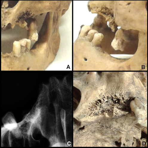
A considerable proportion of the individuals from the historical population of Gródek on the Bug River (n = 50; 32%) exhibited periapical lesions, some of which were potentially life-threatening complications of oral disease (Supplementary material S1, available on request from the corresponding author). Most of these lesions (46 out of 50; 92%) were classified as granulomas, measuring no more than 3 mm in diameter. An example of an extensive lesion was observed in the cranium of an adult male (specimen No. 10; Figure 1). The specimen was generally well preserved. However, most of its dentition had been lost ante mortem, particularly in the maxilla. The teeth extending from the left maxillary lateral incisor to the left maxillary third molar were missing, along with the left part of the maxillary body, and signs of extensive bone remodeling were evident (Figure 1C and Figure 1D). On the right side, further evidence of AMTL was observed, mainly affecting the premolars and molars. The missing teeth in the maxillary alveolar processes corresponded to those missing from the mandible (Figure 1A and Figure 1B).
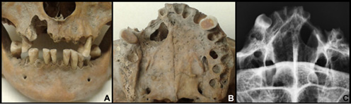
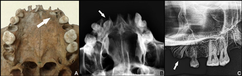
In 4 out of the 50 cases (8%), the lesions exhibited features consistent with periapical cyst formation, involving the uncontrolled proliferation of epithelial cells. In specimen No. 244 (Figure 2), the process appears to have originated from an infection of the pulp of the maxillary canine. As the lesion enlarged, it extended into the body of the maxilla and eventually penetrated the maxillary sinus.
Discussion
In the population from Gródek on the Bug River, individuals in the older age group with AMTL classified as stages III–V predominated (n = 45; 56%), whereas among younger adults, the majority were classified within stages 0–II (n = 56; 73%) (Table 2). As in other historical populations, the severity of AMTL increased with age, as evidenced by the more extensive tooth loss observed in older individuals. Tooth loss is an age-related process, making this finding unsurprising, as repeatedly reported in previous studies.25, 26, 27, 28
Conversely, sex was not found to be a significant factor influencing the severity of AMTL, a finding also reported in some previous studies.29 However, the results of other studies are inconclusive and warrant further discussion. Trombley et al. found that males from a medieval site in Italy had significantly greater tooth loss than females.26 However, this difference was not observed when only young adults (18–29 years) were analyzed, as relatively little AMTL was recorded in this age group.26 Similarly, when analyzing the percentage of AMTL in populations from medieval and modern-period Spanish sites, Lopez et al. found no sex-related differences among medieval individuals.25 However, in the modern-period population, males had fewer remaining teeth than females, indicating a higher percentage of AMTL.25
The present study did not identify sex-related differences in AMTL among individuals from Gródek on the Bug River, in contrast to the findings of some researchers investigating other historical populations. This discrepancy may partly reflect differences in the methodological approaches employed, as the present study evaluated the stage of tooth loss rather than its percentage, as reported in some other studies. The stages of AMTL do not necessarily increase proportionally with the number of teeth lost; rather, they were designed to reflect the increasing functional impact on the SS when the adjacent teeth are lost. Thus, the assessment in the present study focused not simply on the quantity of AMTL, but primarily on the severity of its potential consequences.
The substantial tooth loss observed in the population from Gródek on the Bug River was associated with an increased risk of serious functional consequences. Even the loss of a single tooth can have consequences in the form of the Popov–Godon phenomenon, which occurs when the remaining teeth shift toward an edentulous space following tooth loss, potentially accompanied by the overeruption of the opposing teeth. Such changes in tooth alignment can result in various dental problems, including impaired mastication, increased tooth wear, and potential periodontal complications.30 With further tooth loss, these problems may accumulate and, in the absence of effective prosthetic rehabilitation, lead to permanent changes in the oral cavity.
Various types of prostheses have been used throughout history to replace natural teeth; however, early solutions were generally limited in their functional effectiveness. Dentures with effective retention were first developed in the early 17th century and became more widely used in the 19th and 20th centuries, considerably later than the period represented by the Gródek skeletal series.31 Consequently, this population was exposed to the direct consequences of tooth loss, including changes in facial morphology, TMJ disorders, and digestive problems resulting from impaired initial food processing in the oral cavity.
Tooth loss can also lead to changes in oral hygiene practices, dietary habits, and the overall condition of the remaining dentition, potentially increasing the risk of further dental problems. Individuals with missing teeth may shift toward softer, more cariogenic diets, which could further promote dental caries.32 Tooth loss is also frequently associated with advanced periodontal disease, including stage IV periodontitis, which substantially increases the risk of further tooth loss due to persistent inflammation and the loss of periodontal attachment.33 Other adverse consequences of tooth loss may include nutritional deficiencies, which can impair immune function and tissue healing, potentially exacerbating periodontal disease.34 Thus, the loss of even a single tooth may initiate a cycle of progressive deterioration in oral and general health, ultimately reducing quality of life (QoL).
The classification proposed in the present study takes into account the functional progression of AMTL. In stages I and II, tooth loss is scattered, and the SS retains some adaptive capacity to compensate for the loss of individual teeth (e.g., through the previously mentioned Popov–Godon phenomenon). Stages III and IV, characterized by the clustered tooth loss affecting functional occlusal units, result in substantial functional impairment, whereas stage V represents an almost complete loss of basic stomatognathic functions. Thus, this novel functional approach to AMTL classification can be applied to different populations and chronological periods, extending beyond traditional tooth-by-tooth assessments. It is also worth noting that the assessment of the prevalence of dental diseases, such as dental caries, is often complicated by post-mortem tooth loss (PMTL), whereas the evaluation of the alveolar processes in well-preserved skulls does not present the same difficulties.35
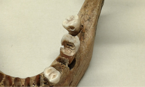
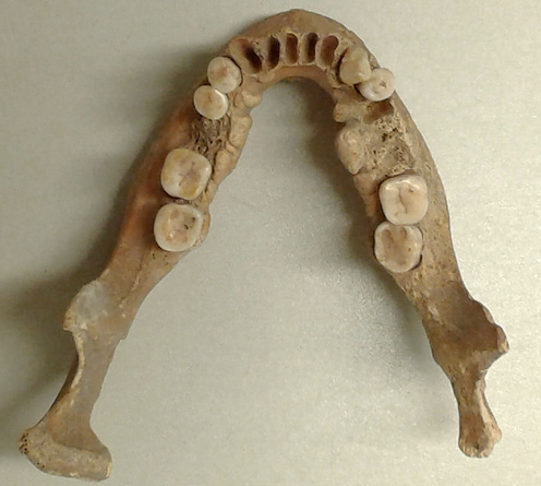
Diseases of the oral cavity, particularly dental caries, were likely the leading cause of tooth loss in the historical population of Gródek on the Bug River. This is supported by cases in which carious lesions are observed alongside the obliterated alveolar sockets (e.g., Figure 3). In some individuals, the periapical inflammatory changes associated with carious lesions are also evident. Most of the observed periapical lesions were classified as granulomas, whereas only a small number were identified as inflammatory radicular cysts.
In the case of individual No. 10 (Figure 1), the traumatic origin of the tooth loss affecting the left part of the maxillary alveolar process should be considered, given the extensive bone loss and pronounced signs of remodeling. It may be hypothesized that the injury occurred during combat with a right-handed opponent, resulting in the loss of the premolars following a blow. Various anatomical abnormalities of the SS may also have contributed to premature tooth loss.
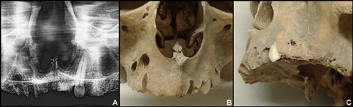
In individual No. 344 (Figure 4), an impacted tooth was observed, accompanied by extensive inflammation in the region of the left alveolar process, at the level of the premolar roots. In individual No. 252 (Figure 5), an impacted lateral incisor was identified in the left maxilla. Individual No. 455 exhibited a mandibular torus (Figure 6), a benign bony protuberance that may have made oral hygiene more difficult, potentially contributing to the faster development of carious lesions and, consequently, tooth loss.
Tooth loss also has significant long-term health consequences that extend beyond the oral cavity, affecting overall health and QoL. Tooth loss, particularly when it occurs early in adulthood, can lead to long-term complications, and is associated with an increased risk of systemic diseases and mortality. In contemporary populations, it has been linked to an increased risk of cardiovascular diseases and stroke.36 Tooth loss also affects the diversity and quality of dietary intake, often leading individuals to favor softer foods that require less chewing. Consequently, they may consume more processed foods and fewer raw vegetables.37, 38 In the Middle Ages, when prosthetic rehabilitation options were limited, and modern processed foods and dietary supplements were unavailable, tooth loss, particularly complete edentulism, could have substantially affected dietary habits, potentially resulting in nutritional deficiencies and further deterioration of general health.37 Thus, AMTL can be regarded as an indicator not only of oral health, but also of broader health conditions and general well-being.39
Oral care has been shown to be inversely associated with mortality among individuals experiencing tooth loss.40 For instance, Adolph et al. demonstrated a strong association between poor oral health, including the loss of at least 10 teeth or a masticatory unit count of fewer than 5, and increased all-cause mortality.41 Particular attention has been directed toward periodontal diseases as a major cause of AMTL. Untreated periodontal diseases tend to become chronic and may lead to persistent low-grade systemic inflammation, potentially contributing to the exacerbation or progression of various systemic disorders, including cardiovascular diseases.39, 42, 43 Chronic inflammation originating in the oral cavity has also been associated with an increased risk of diabetes and stroke.44 Tooth loss is considered an indicator of poor oral health, particularly when an individual has fewer than 20 natural teeth.39
The dental health of the inhabitants of historical Gródek on the Bug River was poor, as evidenced by the extent of tooth loss: 62% of all individuals had experienced substantial AMTL, corresponding to stages II–V of the classification system. This was particularly evident among individuals belonging to the older age categories. Although the population of Gródek on the Bug River appears to have been socially stratified, this differentiation was not significantly reflected in the oral hygiene and dental health of the examined individuals. The widespread occurrence of oral pathologies may have contributed to a substantial chronic inflammatory burden and potentially increased the risk of systemic diseases.
As demonstrated by contemporary research, oral health is increasingly recognized as an important indicator of general health, with numerous studies highlighting the relationships between oral conditions and systemic diseases. Assessments of oral health can provide valuable insights into an individual’s overall health status, as oral pathologies may also reflect the underlying systemic conditions. Assiri et al. demonstrated that individuals with poor oral health, as indicated by factors such as periapical lesions and tooth loss, frequently had a history of systemic diseases, including diabetes, hypertension and anemia.45 Wasterlain et al. demonstrated how dental pathologies reflected living conditions in a skeletal sample from the late 19th and early 20th centuries.46 Similarly, DeWitte and Bekvalac, examining the prevalence of caries and periodontitis in the skeletal remains excavated from the medieval English cemetery of St. Mary Graces, demonstrated associations between oral health status, reduced immune function, and an increased mortality risk in this historical population.47
Thus, the assessment of oral pathologies and their potential impact on QoL can provide valuable insights into an individual’s overall health status and help identify health-related changes over time. In the case of the historical population from Gródek on the Bug River, the extensive AMTL observed may be regarded as a potential indicator of compromised general health and an increased overall health burden. However, further comparative studies involving other historical skeletal populations and incorporating a broader range of paleopathological indicators are necessary to establish more general patterns of health and disease in past populations.
Conclusions
Ante-mortem tooth loss is a common consequence of poor oral hygiene, inadequate diet and advancing age in historical populations. However, AMTL is a more complex phenomenon, as its consequences can have a substantial and long-term negative impact on general health. The present study revealed a high proportion of individuals from Gródek on the Bug River (11th–15th centuries) with moderate to extensive (62%; stages II–IV) and complete (2%; stage V) tooth loss. The oral health status of historical populations can also provide insights into disease patterns that cannot be directly identified through the examination of skeletal remains. The observed patterns of AMTL may reflect difficulties in maintaining oral health, and may be associated with broader health and dietary conditions, although the direct assessment of systemic health status was not possible in the present study.
Ethics approval and consent to participate
Not applicable.
Data availability
The datasets supporting the findings of the current study are available from the corresponding author on reasonable request.
Consent for publication
Not applicable.
Use of AI and AI-assisted technologies
Not applicable.