Abstract
Background. The complete removal of the filling material from the root canal system is essential for effective cleaning and disinfection during retreatment procedures. However, due to the anatomical complexity of the root canal system and the limitations of conventional instruments, achieving complete removal remains a significant challenge.
Objectives. The aim of the study was to assess the efficacy of supplementary techniques, with and without the use of a solvent, for removing the filling material from artificial, standardized grooves in the apical third of root canals.
Material and methods. A total of 120 incisors with single root canals were prepared to size 40/0.04. After longitudinal splitting, standardized grooves were created in the apical third of one half of each root. The grooves were filled with a sealer and gutta-percha, and the roots were then reassembled. The specimens were randomly allocated to 2 control groups (n = 6) and 6 experimental groups (n = 18), according to the removal technique used: passive ultrasonic irrigation (PUI); XP-endo Finisher (XP); or XP-endo Finisher R (XPR), each tested with or without a solvent. Filling material remnants were assessed using a 4-grade scoring system.
Results. Passive ultrasonic irrigation consistently failed to remove the filling material from the grooves, irrespective of solvent use, and yielded significantly poorer scores than all XP and XPR groups (p < 0.001). Without a solvent, neither XP nor XPR achieved the complete removal of the filling material, and no significant difference was observed between the 2 instruments (p = 0.195). However, both instruments demonstrated significantly improved cleaning efficacy when used with a solvent (p < 0.001), with no significant difference between the 2 methods (p = 0.708).
Conclusions. The use of a solvent significantly improves the efficacy of the XP and XPR instruments in removing the filling material from grooves in the apical third of root canals. Both systems exhibited comparable effectiveness, whereas PUI proved ineffective in cleaning the grooves. These findings highlight the critical role of solvents in enhancing cleaning efficacy during retreatment procedures.
Keywords: solvent, passive ultrasonic irrigation, gutta-percha removal, XP-endo Finisher, XP-endo Finisher R
Introduction
Post-treatment endodontic diseases often stem from persistent or secondary intraradicular infections.1 The cornerstone of successful retreatment is the complete removal of the contaminated filling material and thorough disinfection of the root canal system.2, 3, 4 However, the complex anatomy of root canals and the limitations of conventional instruments often make the complete removal of the filling material challenging, if not impossible.5 Although most specially designed instruments and techniques can remove the majority of the filling material from the main canal, none can ensure a completely filling-free root canal system.5, 6, 7, 8 Consequently, the remnants of the filling material may cover the areas harboring residual infection.7, 9 Several supplementary finishing techniques have been proposed to address this problem.10 Passive ultrasonic irrigation (PUI), a widely researched supplementary technique, has been reported to significantly reduce the amount of residual filling material, although complete removal remains elusive.3, 11, 12, 13
XP-endo Finisher (XP) and XP-endo Finisher R (XPR) (FKG Dentaire, La Chaux-de-Fonds, Switzerland) are supplementary instruments made of a nickel-titanium (NiTi) shape-memory alloy and have no taper. They have core sizes of 25 and 30, respectively, and are designed to clean complex canal morphologies and hard-to-reach areas. XP-endo Finisher R was specifically designed for retreatment procedures, in which it is used as a final step in the removal of the filling material. According to the manufacturer (FKG Dentaire), XPR features enhanced expansion capability, allowing its semi-active tip to effectively scrape the canal walls and remove the filling material from challenging locations. Due to its innovative design, the instrument is able to reach and clean areas that are typically difficult to access during root canal retreatment. Several studies have supported the manufacturer’s claims, reporting a significant reduction in residual gutta-percha, following the use of XPR.2, 3, 4, 5, 10 However, recent studies comparing XP and XPR have demonstrated comparable efficacy, although neither instrument achieved complete canal clearance.2, 3 The researchers noted that they had deviated from the manufacturer’s instructions by omitting solvent use and concluded that further investigations should assess the efficacy of XP and XPR with and without solvents.2, 3 Solvents have been recommended when gutta-percha is difficult to remove because of its close adherence to the canal walls.14
The present study aimed to assess the efficacy of XP, XPR and PUI, with and without solvent use, in removing the filling material from artificial, standardized grooves in the apical third of root canals. The researchers hypothesized that there would be no significant differences in efficacy among XP, XPR and PUI, regardless of solvent use.
Material and methods
The study was approved by the Institutional Ethics Committee (IEC approval No. 170.19) at Tel Aviv University, Israel, and adhered to the ethical guidelines outlined in the Declaration of Helsinki.
The study utilized 120 intact human mandibular incisors, each measuring at least 19 mm in length, with fully developed apices and no evidence of apical resorption. The teeth were randomly selected from a pool of recently extracted specimens. Each specimen was secured in a ProTrain endodontic platform (Simit Dental, Mantova, Italy) and radiographically examined in buccal and proximal views to assess the degree of canal curvature, and confirm the presence of a single oval canal configuration, as defined by De-Deus et al.15, 16 Only teeth exhibiting minimal canal curvature (angle <5°) were selected for inclusion in the study.
The researchers prepared access cavities and verified canal patency using a No. 10 K-file (Mani Inc., Utsunomiya, Japan). Canal lengths were determined under a dental operating microscope (Karl Kaps, Asslar, Germany) by advancing the file until its tip became visible at the apical foramen. To standardize the tooth length, the incisal edges were horizontally trimmed to achieve a uniform length of 18 mm, resulting in a working length (WL) of 17 mm.
The specimens were secured in plexiglass tubes using silicone impression material (Coltène/Whaledent, Langenau, Germany). Root canal preparation was performed using the 2Shape instruments (Micro-Mega, Besançon, France) up to size 40/0.04. Between each instrument, the canals were irrigated with 5 mL of 4% NaOCl. Upon completion of the preparation, a final rinse was performed using 5 mL of 17% ethylenediaminetetraacetic acid (EDTA), followed by 5 mL of 4% NaOCl. The canals were then dried with paper points. The teeth were subsequently removed from the impression material, and 2 longitudinal grooves were carefully made using a diamond disc (HORICO, Berlin, Germany), taking care not to penetrate the root canals. Finally, the specimens were split into two halves using a chisel.
A groove measuring 3 × 0.2 × 0.5 mm (length/width/depth) was created in the apical third of one half of each root, 2 mm from the apex, using a size 20 ultrasonic stainless-steel file (Mani Inc.), following the well-established groove-creation protocol.17, 18, 19 Debris was removed from the grooves using a toothbrush.
AH Plus sealer (Dentsply Sirona, Ballaigues, Switzerland) was mixed to a homogeneous consistency and applied using paper points under magnification (Karl Kaps).20 Excess sealer was removed with paper points until a thin, homogeneous layer was obtained. Thermoplastic gutta-percha was then injected into the grooves under magnification using a root canal filling system (SuperEndo β; B&L Biotech USA Inc., Fairfax, USA) and condensed with hand pluggers (Dentsply Sirona). Excess material was removed using a heat carrier plugger (SuperEndo α; B&L Biotech USA Inc.) until a uniform filling corresponding to the dimensions of the grooves was obtained. This technique was intended to simulate a clinical situation in which the filling material remains within a recess in the apical third of the root canal, following its removal from the main canal system.
The root halves were reassembled using a flowable composite (3M ESPE, Seefeld, Germany), which was applied to their external surfaces. The reconstructed specimens were then reinserted into the plexiglass tube molds. The access cavities were sealed using cotton pellets and Cavit™ (3M ESPE). Finally, the specimens were stored under controlled conditions at 37°C and 100% relative humidity to allow the sealer to set.
One week later, the specimens were randomly divided into 8 groups. Six experimental groups, each consisting of 18 samples, were established according to the supplementary removal technique and the use of a solvent: XP and XPR, each with and without a solvent; and PUI, with and without a solvent. Two additional control groups, each comprising 6 samples, were also included. In the positive control group, gutta-percha was applied, but not removed, whereas in the negative control group, gutta-percha was not applied. This grouping strategy enabled a comprehensive evaluation of the different removal techniques under various conditions.
Gutta-percha removal procedure
All attempts to remove gutta-percha were performed with the plexiglass tubes immersed in a digital water bath maintained at 37°C to simulate body temperature conditions.
A torque-controlled motor (Dentsply Sirona) was used to operate the XP and XPR instruments at 1,000 rpm and a torque of 1 N·cm. In the PUI groups, an ultrasonic unit (Satelec P5 Newtron; Acteon Group, Norwich, UK) was used to operate a size 20 Irrisafe instrument at 30% power.
The canals were filled with 0.1 mL of chloroform in the solvent groups and with 4% NaOCl solution in the groups without a solvent. After 2 min, the XP and XPR instruments were inserted to WL, whereas the IrriSafe instrument was inserted to a depth of 1 mm short of WL. All instruments were operated or activated for 30 s using slow in-and-out movements.
The root canals were then irrigated for 30 s with 4% NaOCl solution using a syringe fitted with a 30-gauge needle (NaviTip; Ultradent, South Jordan, USA), inserted to a depth of 1 mm short of WL, with an estimated flow rate of 5 mL/min. The same instrumentation and activation protocol was repeated twice more – the first cycle was followed by 15 s of irrigation with 17% EDTA solution, and the second by a final rinse with 5 mL of 4% NaOCl solution. Any debris adhering to the XP or XPR instruments was removed between the irrigation cycles using gauze soaked in alcohol.
In the PUI groups, care was taken to maintain the IrriSafe instrument in a central position within the canal to minimize contact with the canal walls, which could dampen its oscillatory motion.21 Each instrument was used in a single canal and then discarded.
Cleaning efficacy assessment
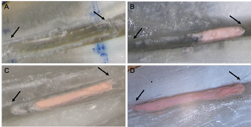
After treatment completion and drying of the canals with paper points, the specimens were disassembled to evaluate the removal of the filling material from the grooves. A microscope (Karl Kaps), set at ×25 magnification and coupled with a digital camera (Sony Alpha A6000; Sony Inc., New York, USA), was used to capture images of the grooves before filling material application and after its removal. To prevent the evaluators from identifying the specimens, the photographs were coded. Two endodontic specialists independently assessed the amount of filling material remaining in the grooves using a 4-point grading scale (Figure 1).22 Score 0 indicated a groove entirely devoid of filling material. Score 1 represented traces of the filling material covering less than half of the groove area. Score 2 denoted traces of the filling material covering more than half of the groove area. Score 3 indicated a groove completely filled with the filling material.
Statistical analysis
Statistical analysis was performed using the Statistical Analysis Program (SAS®) 9.4 (SAS Institute, Cary, USA). Kappa coefficients were calculated to assess intraobserver and interobserver reliability. Differences in filling material scores among the groups were analyzed using the Kruskal–Wallis test, followed by post hoc pairwise comparisons using the Dwass–Steel–Critchlow–Fligner (DSCF) test. Statistical significance was set at p ≤ 0.05.
A sample size of 18 teeth per group provided 85% statistical power at a 5% significance level to detect an 8% difference among the supplementary techniques.
Results
The intraobserver and interobserver differences in scoring did not exceed one score point. The kappa coefficients were 0.928 for the first observer, 0.938 for the second observer, and 0.967 for interobserver agreement.
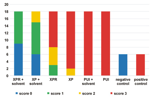
The distribution of scores for filling material removal from the grooves is presented in Figure 2. The results showed that PUI failed to remove the filling material from the grooves, consistently receiving a score of 3, regardless of solvent use. The PUI scores differed significantly from those of all XP and XPR groups (p < 0.001).
In the XP group, 88.9% of the grooves were completely filled with the filling material (score 3), and all specimens (100%) exhibited the filling material covering more than 50% of the groove area (scores 2 and 3). Similarly, more than 50% of the grooves in the XPR group remained completely filled with the filling material (score 3), while 83.3% of the specimens exhibited the filling material covering more than 50% of the groove area (scores 2 and 3). Thus, in the absence of a solvent, neither the XP nor the XPR technique was able to completely remove the filling material from the artificial, standardized grooves. No significant difference was observed between the 2 techniques (p = 0.195).
In contrast, when a solvent was used, the grooves were completely free of the filling material (score 0) in 33.3% and 50% of the specimens treated with the XP and XPR instruments, respectively. Furthermore, 83.3% and 100% of the specimens in the XP and XPR groups, respectively, exhibited the filling material covering less than 50% of the groove area (scores 0 and 1). These results demonstrate that both the XP and XPR instruments removed significantly more filling material from the artificial, standardized grooves in the presence of a solvent (p < 0.001), with no significant difference between the 2 techniques (p = 0.708).
Discussion
The primary objectives of successful endodontic retreatment are to remove the filling material, followed by thorough shaping and cleaning of the root canal space.2 Despite advances in retreatment techniques, achieving root canals entirely free of the filling material remains an elusive goal, as demonstrated by numerous studies.2, 23, 24, 25, 26 The present study aimed to assess the efficacy of supplementary instruments (XP, XPR and PUI), with and without the use of a solvent, in removing the filling material from grooves in the apical third of root canals. The study design, which incorporated an artificial groove model, provided a controlled environment that allowed standardization of the groove size and location, and the filling material volume while simulating complex root canal anatomy.
XPR, a variant of XP featuring a size 30 tip, incorporates an innovative design that, according to the manufacturer (FKG Dentaire), enhances its ability to effectively scrape the canal walls and remove the filling material from difficult-to-access areas. This claim is supported by a study conducted by Machado et al., which demonstrated that the additional use of XPR significantly reduced the volume of residual filling material during retreatment with various instruments.5 Similarly, Silva et al. evaluated the efficacy of XP and XPR in oval canals, and found that both instruments were effective in removing the residual filling material from straight oval-shaped canals.2 Despite differences in the core diameter and the tip angle, which could potentially make XPR more effective than XP in removing the filling material, the authors found no significant differences in the efficacy of the 2 supplementary instruments.2 It should be noted, however, that none of the instruments investigated in these studies was able to completely remove the residual filling material.2, 3 Notably, neither study involved the use of solvents, despite their recommendation by the manufacturer (FKG Dentaire).
The results of the present study confirm that neither XP nor XPR was able to completely remove the filling material from the grooves. However, the efficacy of both XP and XPR in removing the filling material was significantly improved by the use of a solvent, with a similar effect observed for both instruments. Notably, regardless of solvent use, PUI failed to remove the filling material from the grooves, and was significantly less effective than XP and XPR. Therefore, the null hypothesis was rejected.
A recently published study reported that XPR and PUI, when used in combination with a solvent, were similarly effective in removing the residual filling material from root canals.27 The discrepancy between these findings and those of the present study may be attributed to differences in the study objectives. While the previous study focused on cleaning efficacy within the main root canal system, the present study specifically investigated the removal of the filling material from irregular areas. In this context, XP and XPR demonstrated the ability to reach and mechanically dislodge the filling material from the canal walls, particularly in irregular areas. In contrast, PUI relies primarily on fluid dynamics to access such areas.28, 29 Passive ultrasonic irrigation employs a non-cutting stainless-steel tip designed to oscillate freely within the root canal without contacting its walls, thereby maximizing its effectiveness and preventing the restriction of its movement.28, 29 Its cleaning mechanism is based on cavitation and acoustic microstreaming, which enhance the ability of the irrigant to remove debris.27, 28 However, unlike XP and XPR, which directly engage with the filling material, the indirect action of PUI may limit its ability to effectively remove remnants from complex or irregular spaces, such as artificial grooves.
A previous study demonstrated that 3 cycles of PUI combined with chloroform resulted in significantly fewer gutta-percha remnants in simulated uninstrumented areas than the use of NaOCl, chloroform and paper points.22 In the present study, only one cycle of solvent application was performed to replicate the manufacturer’s recommended protocol for XPR. This methodological difference may help explain the discrepancy between the findings of the 2 studies. Multiple solvent-assisted activation cycles may facilitate more effective filling material removal, whereas a single cycle, following the manufacturer’s recommendations, may limit the cleaning efficacy of supplementary techniques. Future studies should investigate the effectiveness of multiple solvent-assisted activation cycles in removing the filling material from irregular root canal areas.
According to a systematic review by Rossi-Fedele and Aly Ahmed, solvents may facilitate the penetration of the file into root canals when used during the initial stages of retreatment, but may hinder cleaning efficacy.30 In contrast, when used after instrumentation, solvents may enhance the removal of the root filling material, thereby reducing the amount of residual material. Therefore, it is not surprising that solvent use in the present study improved the ability of XP and XPR to remove the filling material from the artificial grooves.
Limitations
One potential limitation of the present study is that different activation times were not investigated. Although the study protocol followed the manufacturer’s recommendations, the recommended activation time proved insufficient to completely remove the filling material from all the grooves. Since clinicians cannot control the frequency with which the XP and XPR instruments contact the residual filling material in irregular areas within the recommended working time, longer activation periods may provide more opportunities for mechanical contact, potentially resulting in improved cleaning outcomes. Therefore, future studies should investigate the effects of extended activation times on cleaning efficacy.
Conclusions
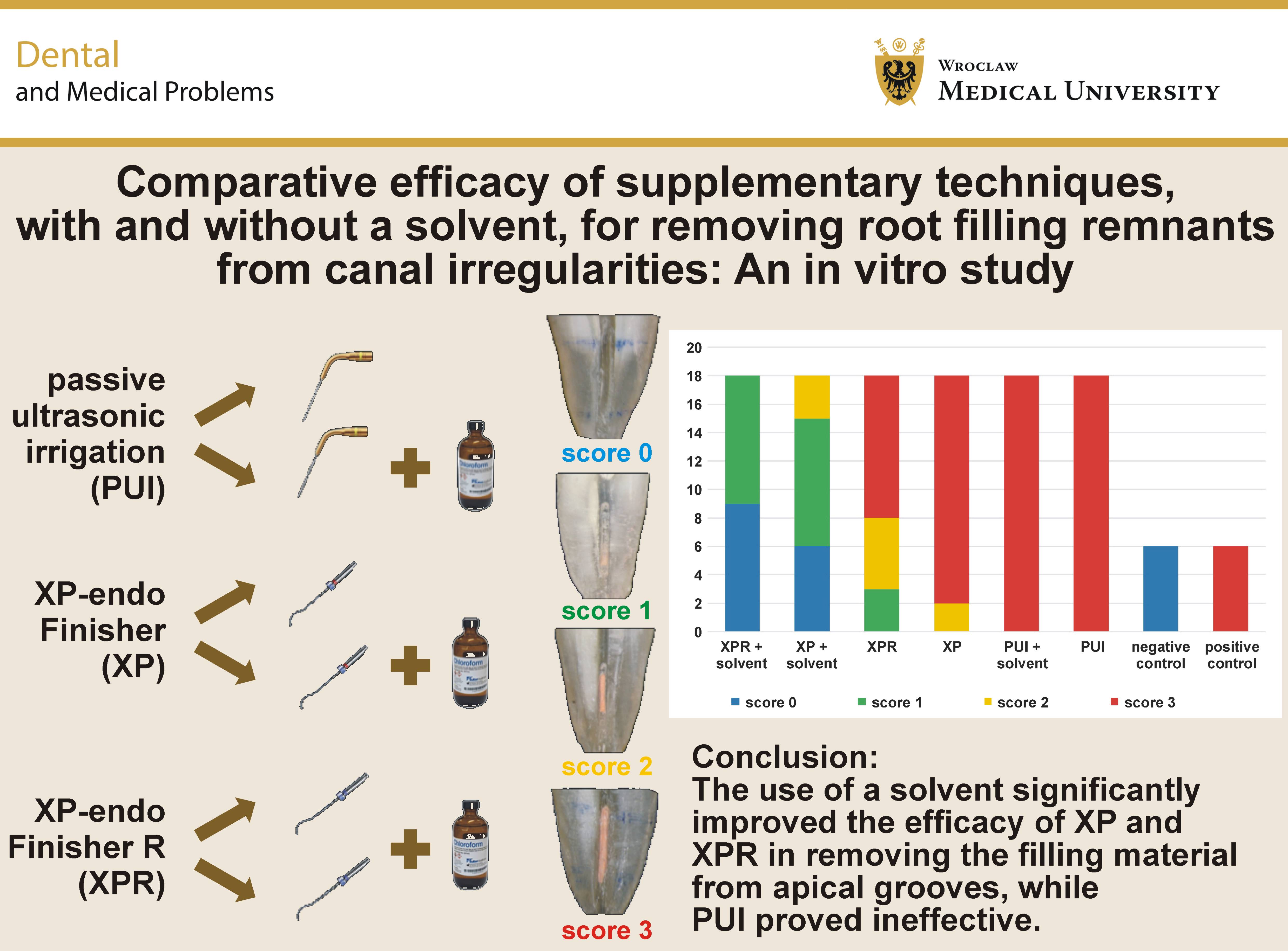
The use of a solvent significantly improved the efficacy of the XP and XPR instruments in removing the filling material from the grooves located in the apical third of root canals. Both instruments demonstrated comparable effectiveness, whereas PUI was ineffective in removing the filling material from the grooves, regardless of solvent use. These findings highlight the importance of solvent use in conjunction with XP and XPR to improve cleaning of irregular areas within the root canal system. However, the potential clinical benefits of this approach remain to be established. Future studies should investigate the effects of extended activation times and multiple solvent-assisted cycles to further optimize the cleaning efficacy of these techniques.
Ethics approval and consent to participate
The study was approved by the Institutional Ethics Committee (IEC approval No. 170.19) at Tel Aviv University, Israel.
Data availability
The datasets supporting the findings of the current study are available from the corresponding author on reasonable request.
Consent for publication
Not applicable.
Use of AI and AI-assisted technologies
Not applicable.