



Abstract
Background. The impact of diabetes mellitus (DM), as reflected by glycemic control, on dental implant outcomes remains debated. Specifically, there is a critical gap in our understanding of the impact of uncontrolled DM on early implant failure (EIF), despite the potential benefits of implant dentistry for these patients.
Objectives. The aim of the present study was to investigate the impact of glycemic control on EIF in patients with DM.
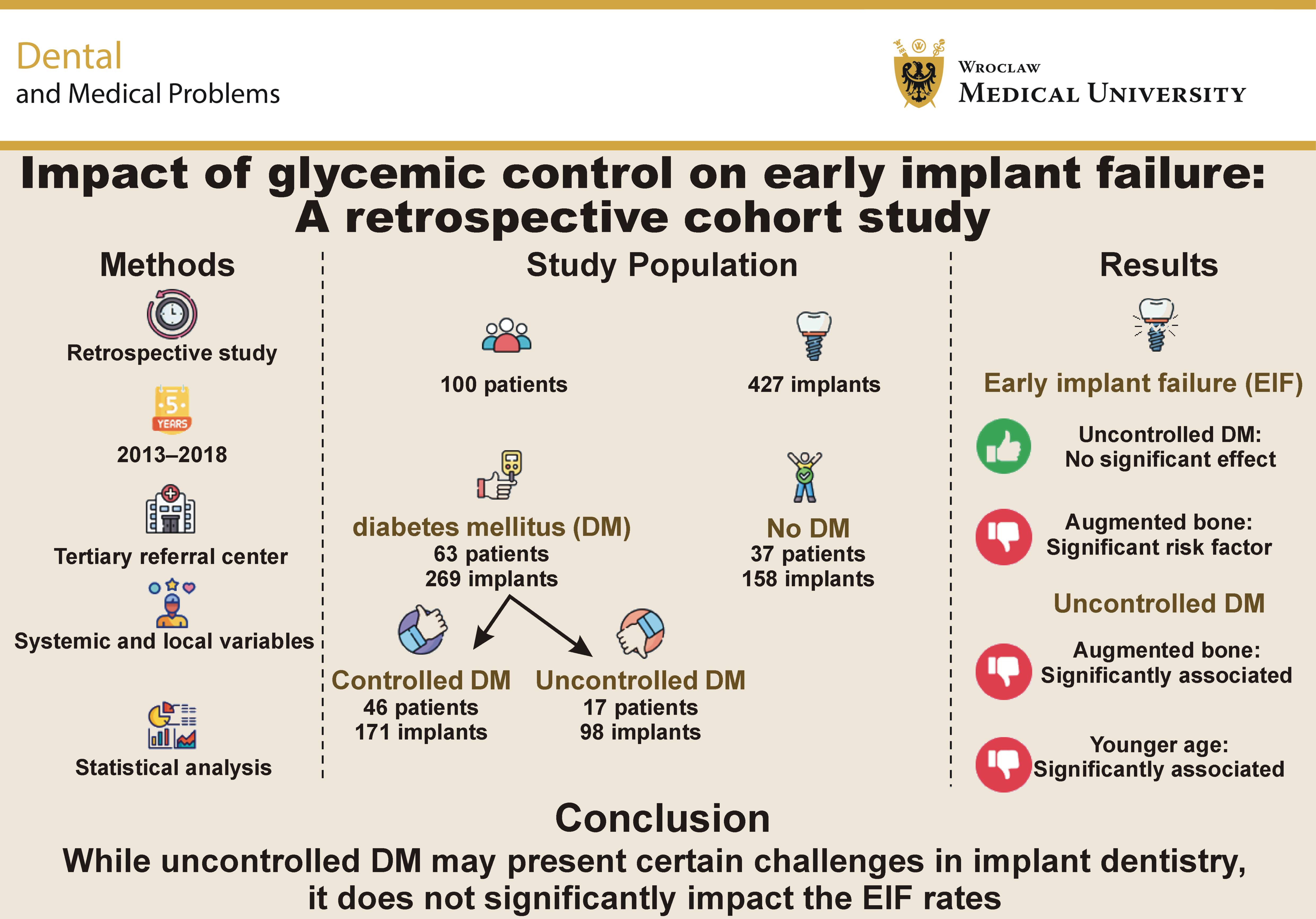
Material and methods. A retrospective analysis of electronic medical records (EMRs) was conducted over a 5-year period at a tertiary referral center. Data from 100 patients with 427 implants were collected, including demographic characteristics, systemic conditions, the glycated hemoglobin (HbA1c) values, procedural details, and implant-related variables. Statistical analyses were performed to assess the associations between glycemic control, the study variables and EIF, with statistical significance set at p < 0.05.
Results. Uncontrolled DM (HbA1c > 8%) was observed in 17 patients and was not a significant predictor of EIF. However, it was associated with a greater need for bone augmentation (90.8% vs. 45.0%), the placement of longer implants (11.8 mm vs. 11.4 mm), a higher proportion of implants placed in the anterior maxilla (22.4% vs. 11.1%), and younger patient age (62.8 years vs. 67.8 years). The EIF rates were higher for implants placed in augmented bone and lower for implants placed in the anterior and premolar regions of the maxilla.
Conclusions. This study indicates that, although uncontrolled DM may present certain challenges in implant dentistry, including a younger age at implant placement, an increased need for bone augmentation, and a preference for specific implant sites, it does not significantly affect the EIF rates.
Keywords: risk factor, implant, glycemic control, diabetes, early implant failure
Introduction
Diabetes mellitus (DM) is a common metabolic disorder that may lead to adverse oral health outcomes, including microvascular disease, delayed wound healing, functional alterations in the salivary glands, periodontitis, changes in the oral microbiota, and an impaired response to infection.1, 2, 3
Dental implants have become a popular treatment option for missing teeth, offering a stable and esthetic solution.4, 5, 6, 7, 8, 9, 10 The influence of DM on implant dentistry has been debated for many years. While some studies have considered dental implant placement in patients with DM a relative contraindication,11 others have reported that implant survival and osseointegration remain predictable and comparable to those in healthy individuals.1, 4, 12, 13 These studies have also highlighted the potential negative effects of hyperglycemia and uncontrolled hemoglobin A1c (HbA1c) levels on implant therapy outcomes.4, 13, 14
Glycemic control is a crucial factor in the management of patients with DM. Adequate control of blood glucose and HbA1c levels may help prevent microvascular and macrovascular complications, wound infections, and impaired bone formation.14, 15, 16 Poor glycemic control has been suggested to compromise osseointegration. For example, Oates et al. conducted a prospective clinical study demonstrating that, in patients with type 2 diabetes mellitus (T2DM), poor glycemic control (HbA1c ≥ 8.1%) was associated with reduced initial implant stability and prolonged healing following implant placement.17 However, after 16 weeks, implant stability was comparable to that in patients without DM, suggesting that successful osseointegration is achievable, although healing may take longer.17 Other studies have reported that glycemic control does not significantly affect implant success.1, 18 Escow and Oates conducted a 2-year observational study involving patients with poorly controlled T2DM (HbA1c levels ranging from 6.3% to 13.1%) and found that elevated glycemic levels did not significantly affect the implant survival rates, challenging the prevailing notion that strict glycemic control is essential for early implant success.18
Although the influence of DM on dental implant outcomes has been investigated in previous studies, an important limitation remains in the current literature. Many studies have excluded patients with uncontrolled DM, despite the ongoing debate on the impact of glycemic control on early implant failure (EIF).19, 20, 21 This exclusion creates a significant knowledge gap regarding the effect of uncontrolled DM on implant outcomes, particularly during the early stages of healing.
Traditionally, some oral surgeons have considered uncontrolled DM a relative contraindication to implant placement.11 This is particularly relevant for patients who struggle to achieve adequate glycemic control, as they may be denied access to implant therapy based on limited evidence. Implant-supported prostheses offer potential benefits for patients with DM, particularly those who are partially or completely edentulous. By restoring masticatory function, dental implants may facilitate healthier dietary choices and potentially improve glycemic control. These benefits may, in turn, contribute to an improved quality of life (QoL), enhanced self-image, and better overall health outcomes in these patients.22, 23
The evolving understanding of DM management, the limited evidence supporting the contraindication of implant therapy in patients with uncontrolled DM, and the growing emphasis on holistic, patient-centered care necessitate a re-evaluation of this approach.
Therefore, the aim of the present study was to assess the impact of glycemic control on EIF in patients with DM. The null hypothesis was that glycemic control is not a significant risk factor for EIF in patients with DM.
Material and methods
A retrospective cohort study, conducted in accordance with the STROBE (Strengthening the Reporting of Observational Studies in Epidemiology) guidelines for observational research, was performed at the Department of Oral and Maxillofacial Surgery, Rabin Medical Center, Petah Tiqwa, Israel, between January 2013 and December 2018. The study complied with the Declaration of Helsinki and its subsequent amendments, and received ethical approval (No. 0674-19-RMC ). Written informed consent was obtained from all participants included in the study.
Patients were eligible for inclusion if they were older than 18 years, had undergone implant dentistry treatment, were treated consecutively during the study period, and had available HbA1c measurements obtained within the 3 months preceding implant placement. Patients with incomplete documentation were excluded from the study.
Surgical protocol
Preoperative radiological assessment was performed using cone-beam computed tomography (CBCT). All bone augmentation and implant placement procedures were performed by well-trained oral and maxillofacial surgeons with a minimum of 10 years of clinical experience. Implant placement began with a mid-crestal incision, followed by the elevation of a full-thickness flap. When the available bone volume was insufficient to accommodate an implant with a diameter of ≥3.3 mm and a length of ≥8 mm, or when augmentation was indicated for esthetic reasons, bone augmentation was performed using one of the following bone substitutes or a collagen membrane prior to implant placement:
– xenogeneic bone substitute (Bio-Oss; Geistlisch Pharma, Wolhusen, Switzerland);
– allogeneic bone substitute (OraGraft®; LifeNet Health, Virginia Beach, USA);
– collagen membrane (Ossix® Plus; Datum Dental Ltd., Lod, Israel).
When adequate primary implant stability could be achieved, implants were placed, with simultaneous bone augmentation when indicated, according to the implant manufacturer’s protocol and using the dedicated surgical bur set. Upon discharge, patients were prescribed antibiotics (amoxicillin 500 mg 3 times daily for 5 days, or clindamycin 300 mg 3 times daily for 7 days in patients with a penicillin allergy), a chlorhexidine mouth rinse and an analgesic regimen.
Data collection
Clinical data were collected using the Chameleon Electronic Medical Records (EMR) system, while medical histories were retrieved from the Ofek EMR system. The Ofek EMR system provides comprehensive access to patients’ medical histories across all Clalit Health Services hospitals and community clinics in Israel. The available information includes diagnoses, medications, complete blood counts (CBCs), histopathology results, and other clinically relevant data. By centralizing this information, the Ofek EMR system helps minimize the risk of disclosure and reporting biases. Data collection was performed independently by D.M. and E.J. Any discrepancies were discussed until a consensus was reached.
Demographic characteristics included patients’ gender and age at the time of the procedure. In addition, systemic conditions, HbA1c levels, and the smoking status were recorded.
Patients were stratified into 3 groups according to HbA1c levels17, 18: non-diabetic (HbA1c < 6.5%); controlled DM (HbA1c between 6.5% and 8%); and uncontrolled DM (HbA1c > 8%).
The following variables were taken into consideration:
– span length: short-span partial edentulism (≤2 missing teeth); long-span partial edentulism (>2 missing teeth; and total edentulism;
– implant:
• brand;
• length;
• diameter;
• location in the maxilla or the mandible, and in particular regions: anterior (central incisor to canine); premolar (first and second premolars); and posterior (first and second molars);
– number of implants per patient;
– need for bone augmentation (yes/no); and
– EIF defined as implant removal ≤1 year after permanent rehabilitation – primary outcome variable.
Statistical analysis
The data were analyzed using IBM SPSS Statistics, v. 25.0 (IBM Corp., Armonk, USA). Descriptive statistics were used to summarize continuous variables as means and standard deviations (M ±SD) and categorical variables as frequencies. Comparisons among the 3 groups (patients without DM, patients with controlled DM, and patients with uncontrolled DM) were performed using the Kruskal–Wallis test for continuous variables and the χ2 test for categorical variables.
To account for multiple implants within the same patient and the resulting within-patient correlation, a generalized estimating equations (GEE) model was used to estimate the risk of EIF. The GEE model accounted for subject-level clustering, binomial distribution and exchangeable correlation. Multicollinearity was assessed using the variance inflation factor (VIF). Variables with univariate p-values <0.1 and no evidence of multicollinearity were included in the multivariable models – multivariable GEE at the implant level and multivariable logistic regression at the patient level. Statistical significance was set at p ≤ 0.05.
Results
Descriptive statistics
Cohort patient level
At the patient level (Table 1), the cohort (N = 100) comprised 40% males and 60% females. The mean age was 67.1 ±11.5 years, and 6% of the patients were smokers. Sixty-one percent of the patients received Zimmer implants (Zimmer Biomet TMT, Parsippany, USA; a microtextured (MTX) surface, treated by grit-blasting with hydroxyapatite, followed by washing with non-etching acid and distilled water), 26% received MIS implants (MIS Implants Technologies Ltd., Karmiel, Israel; a sandblasted and acid-etched surface), and the remainder received Ditron implants (MPI (Molecular Precision Implant); Ditron Dental, Ashkelon, Israel; a blasted and thermally acid-etched endosseous surface). The mean number of implants per patient was 4.3 ±2.9. Most of the cohort (51%) required bone augmentation.
The mean HbA1c level in the entire cohort was 7.10 ±1.08%. Seventeen percent of the patients were classified as having uncontrolled DM (HbA1c > 8%), 46% as having controlled DM (HbA1c 6.5–8%), and 37% as non-diabetic (HbA1c < 6.5%). Twelve patients had at least one EIF.
Cohort implant level
All data were likewise analyzed at the implant level (N = 427) (Table 2). Most implants (58.8%) were Zimmer implants, with 15–20% distributed across each sextant. Twenty-three percent of the implants were placed in patients with uncontrolled DM. The overall incidence of EIF was 2.8% at the implant level.
Statistical analysis
Univariate analyses
At the implant level (Table 3), uncontrolled DM was associated with a substantially higher need for bone augmentation (90.8%) as compared to the controlled DM group (45.0%) and non-diabetic individuals (39.2%). The younger mean age (62.8 ±12.2 years) and female predominance (63.3%) in the poorly controlled DM group were statistically significant. Differences were also observed in implant location, with a higher proportion of implants placed in the anterior maxilla among patients with uncontrolled DM. In addition, implants were significantly longer in this group. Notably, no significant differences were found between the non-diabetic, controlled DM and uncontrolled DM groups.
At the patient level (Table 4), the prevalence of bone augmentation was significantly associated with glycemic control, with a higher need among patients with uncontrolled DM (82.4%) than among those with controlled DM (45.7%) or non-diabetic individuals (43.2%). The number of implants per patient also differed between the groups (p < 0.1), reflecting the greater extent of tooth loss in patients with uncontrolled DM.
Analyses were performed at both the implant and patient levels to identify risk factors for EIF. These analyses revealed that implant placement in the augmented bone was a significant risk factor for EIF. Notably, no EIF events occurred among implants placed in the anterior or premolar regions of the maxilla, suggesting a lower risk of failure at these sites. Conversely, implants placed in the anterior mandible had significantly higher odds of EIF (OR = 3.46). Implants placed in the posterior maxilla showed a borderline statistically significant increase in the odds of failure (OR = 2.85; p = 0.085) (Table 5 and Table 6).
Multivariable analysis for EIF
Similar to the univariate analysis, the implant-level multivariable GEE model showed that implantation in augmented bone was a significant risk factor for EIF (OR = 17.48). Implants placed in the anterior and premolar regions of the maxilla had a significantly lower risk of failure, with no occurrences of EIF observed in these regions (Table 7).
The patient-level model showed that, after controlling for potential confounders, there was no statistically significant difference between the controlled and uncontrolled DM groups (Table 8).
Table 9 illustrates the characteristics of EIF. Notably, 11 of the 12 patients who experienced EIF in this study had augmented bone.
Discussion
Achieving successful osseointegration and long-term implant stability is a primary goal in implant dentistry. Various factors can influence implant outcomes, including systemic conditions such as diabetes.24 Currently, the evidence regarding the impact of uncontrolled DM on EIF remains limited, primarily because individuals with poorly controlled DM are often excluded from clinical studies.19, 20, 21 This study sought to address this gap by analyzing a substantial cohort of patients with varying levels of glycemic control, providing valuable insights into the impact of DM on dental implant outcomes, particularly among patients with uncontrolled DM. The results revealed several significant differences among non-diabetic patients and those with controlled and uncontrolled DM.
Osseointegration is the biological process through which a direct interface forms between bone and an implant.25 Dental implants rely on this process for their initial success; therefore, any disruption may increase the risk of EIF.4 Prolonged hyperglycemia has been associated with several mechanisms that may impair osseointegration and contribute to EIF. These include the tissue damage resulting from insulin-independent glucose uptake by endothelial cells, endothelial dysfunction and vascular impairment. Additionally, hyperglycemia may impair polymorphonuclear leukocyte migration and phagocytosis while enhancing the inflammatory response to microbial stimuli.4 These alterations may compromise angiogenesis and disrupt the inflammatory response, both of which are essential for successful osseointegration. Furthermore, in the context of hyperglycemia, the formation of advanced glycation end products (AGEs) is of particular concern. These compounds result from non-enzymatic reactions between sugars and proteins or lipids, a process accelerated by elevated glucose levels. The accumulation of AGEs has been associated with impaired osteoblast differentiation and function, alterations in bone matrix formation and quality, and compromised angiogenesis, all of which are critical for bone healing around implants.26, 27, 28, 29, 30, 31, 32, 33 By directly affecting bone cells and the structural integrity of bone tissue, AGEs may further compromise the achievement of successful osseointegration in individuals with hyperglycemia.34
The statistical analysis revealed that neither the DM status nor glycemic control was a statistically significant risk factor for EIF at either the patient or implant level. This finding is consistent with several previous studies. Ghiraldini et al. found no significant differences in implant stability 3 months after implantation between patients with well-controlled (HbA1c < 8%) and poorly controlled (HbA1c > 8.1%) DM.35 Oates et al. similarly demonstrated that patients with poorly controlled DM did not exhibit significantly reduced implant stability at 16 weeks after implantation.17 Shi et al. conducted a meta-analysis to assess the association between poorly controlled DM and EIF. Their findings did not demonstrate a significant difference in the implant failure rates between patients with controlled and uncontrolled DM.36
Patients with uncontrolled DM were notably younger, highlighting the potential oral health implications of poor glycemic control, which is associated with an increased risk of periodontitis and tooth loss. These patients may experience tooth loss at an earlier age, leading to the need for implant therapy at a younger age.37 This finding is consistent with the existing literature, as several studies have reported an association between better glycemic control and an older age.38, 39 This observation underscores the importance of effective diabetes management from an early age to help delay or potentially prevent tooth loss and other oral health complications.
Consistent with previous studies, we found that the dental status of patients with uncontrolled DM prior to implant placement was significantly compromised.14 A higher proportion of patients with poorly controlled DM required bone augmentation procedures. Type 2 DM may exacerbate the host inflammatory response to oral plaque, thereby aggravating bone resorption associated with periodontitis and peri-implantitis.39, 40, 41, 42, 43, 44, 45 Patients with DM exhibit increased levels of pro-inflammatory cytokines in bone tissues, including interleukin (IL)-1β, IL-6, and tumor necrosis factor alpha (TNF-α).46, 47, 48, 49 Consequently, increased osteoclast activity may lead to greater bone resorption. Previous studies have also reported that good glycemic control improves osteoclast function, and helps prevent periodontal bone loss and tooth loss.50, 51 These findings are consistent with previous research, as well as with the results of the present study, which indicate a higher prevalence of EIF in patients with augmented bone.22, 52, 53 This evidence underscores the importance of preserving natural bone and preventing bone loss associated with periodontal disease. Additionally, a significant difference was observed in the mean implant length between the 3 groups, with patients with well-controlled DM receiving shorter implants, on average, than those with poorly controlled DM. This difference may be attributable to the additional bone volume gained through the augmentation procedures, which may have allowed the placement of longer implants.
The results also revealed significant differences in implant location between patients with controlled and uncontrolled DM. Uncontrolled DM was associated with a higher likelihood of implants being placed in the anterior maxilla, whereas controlled DM was more frequently associated with implant placement in the mandibular molar region. These findings highlight the importance of considering anatomical factors when evaluating implant placement patterns in patients with DM. They may also reflect more pronounced bone resorption and periodontal damage in the anterior maxilla among patients with uncontrolled DM, although this interpretation requires further investigation. To the best of our knowledge, no previous studies have specifically reported differences in implant location according to glycemic control. However, these findings are supported by a previous study conducted in healthy individuals.54
Another finding regarding implant location was the lower incidence of EIF in implants placed in the anterior and premolar regions of the maxilla, compared with the higher incidence observed in the anterior mandible. This difference may be attributed to the superior vascular supply of the maxilla as compared to the mandible, which may promote osseointegration.55 The relatively limited vascular supply in the anterior mandible may, in turn, contribute to the higher EIF rates observed in this region.55 We hypothesize that this anatomical factor may be particularly relevant in patients with DM, who often experience microvascular damage.1 Impaired blood flow could further compromise osseointegration in areas with inherently limited vascularity, such as the anterior mandible, potentially increasing the risk of EIF. Further studies are needed to investigate this proposed mechanism.
The results of this study call for a reevaluation of the current approach to treating patients with uncontrolled DM who require dental implant therapy. Although achieving optimal glycemic control remains the ideal goal, this is not always attainable, particularly in individuals with complex health conditions or socioeconomic barriers.56 Despite numerous studies investigating implant outcomes in patients with DM, definitive evidence regarding its independent negative impact on EIF remains inconclusive.17, 18, 19, 20, 35, 36, 51, 57 In fact, some studies suggest that implant therapy in patients with DM may indirectly contribute to improved glycemic control. The restoration of masticatory function through implant-supported prostheses may facilitate healthier dietary choices and improve overall health, potentially contributing to a reduction in the HbA1c levels.22
Furthermore, the improvement in QoL associated with implant therapy, both functionally and psychologically, is substantial and should not be underestimated.58 The ability to eat comfortably, speak clearly and smile confidently can have a profound impact on a patient’s overall well-being. Denying these benefits solely on the basis of uncontrolled DM may represent an overly restrictive approach. Our findings suggest that, although uncontrolled DM may present certain challenges in implant dentistry, including a potentially higher risk of peri-implantitis and consequently reduced implant survival, it should not be considered an absolute contraindication to implant therapy. Instead, treatment decisions should be individualized, taking into account the patient’s overall health status, degree of glycemic control, and risk factors for implant complications.
Limitations
The limitations of this study stem primarily from its retrospective design and the involvement of multiple operators, which may have introduced variability in data recording and surgical techniques, thereby limiting the generalizability of the findings. Although this study represents one of the larger investigations of diabetic patients undergoing implant therapy, a larger sample size may help identify additional statistically significant predictors and subtle differences in implant failure risk, as suggested by the wide confidence intervals observed in our analyses. Additionally, data on medication use were unavailable. It is possible that antidiabetic medications may influence implant outcomes and potentially reduce the EIF rates in these patients.
Another potential limitation is the possibility of selection bias. Previous studies have raised concerns regarding the oral complications associated with chronic hyperglycemia, including periodontitis, hyposalivation, impaired inflammatory responses, and potentially EIF. These concerns may have prompted clinicians to adopt a more cautious approach when treating patients with uncontrolled DM. Consequently, such patients may have been advised to improve their glycemic control before undergoing the implant placement or bone augmentation procedures. This selection process could have reduced the observed risk of implant failure, even among patients with elevated HbA1c levels.
Lastly, although the HbA1c levels were measured within 3 months before implant placement, fluctuations in glycemic control over time were not accounted for. This may have resulted in the misclassification of the patients’ glycemic status and potentially influenced the observed associations between glycemic control and EIF.
Conclusions
This study indicates that, although uncontrolled DM may present certain challenges in implant dentistry, including a younger age at implant placement, an increased need for bone augmentation, and a preference for specific implant sites, it does not significantly affect the EIF rates.
Ethics approval and consent to participate
The research adhered to the ethical standards outlined in the Declaration of Helsinki, and received approval from the institutional review board at Rabin Medical Center, Petah Tiqwa, Israel (No. 0674-19-RMC). All study participants provided written informed consent prior to their inclusion.
Data availability
The datasets supporting the findings of the current study are available from the corresponding author on reasonable request.
Consent for publication
Not applicable.
Use of AI and AI-assisted technologies
Not applicable.