Abstract
Background. Zirconia restorations have attracted considerable attention because of their favorable mechanical and esthetic properties, which meet the requirements for both anterior and posterior restorations.
Objectives. This study aimed to evaluate the microshear bond strength (µSBS) between resin cement and zirconia surfaces dip-coated with nanosilica and nanozirconia suspensions of different compositions.
Material and methods. An extra translucent (XT) zirconia disc composed of 5 mol% yttrium-stabilized zirconia was sectioned into 66 samples and divided into 6 groups: untreated zirconia as the control group; zirconia blasted with silica-coated alumina; and zirconia dip-coated with nanosilica–nanozirconia suspensions at volume ratios of 100:0, 75:25, 50:50, and 25:75. The coated surfaces were analyzed using energy-dispersive X-ray spectroscopy (EDS), field emission scanning electron microscopy (FESEM), atomic force microscopy (AFM), and X-ray diffraction (XRD). Resin cement cylinders were bonded to the treated zirconia surfaces, and µSBS testing was performed. The differences between the groups were analyzed using one-way analysis of variance (ANOVA) followed by Tukey’s post hoc test, with the significance level set at p ≤ 0.05.
Results. The highest µSBS was observed for the 50:50 nanosilica–nanozirconia group (51.63 ±5.56 MPa), whereas the lowest values were recorded for untreated zirconia (17.38 ±3.74 MPa). Zirconia blasted with commercially available silica-coated alumina showed µSBS of 31.00 ±6.54 MPa.
Conclusions. Dip coating with nanosilica–nanozirconia suspensions appears to be a feasible method for improving resin–zirconia bonding while preserving the zirconia structure. Among the tested compositions, the 50:50 nanosilica–nanozirconia suspension produced the highest µSBS.
Keywords: microshear bond strength, nanosilica coating, nanozirconia, resin-ceramic bonding
Introduction
Zirconia has become one of the most widely used ceramic materials in dentistry owing to its favorable mechanical properties, high fracture strength, esthetics, and biocompatibility.1, 2 Yttria-stabilized tetragonal zirconia polycrystal (Y-TZP) is widely used as an alternative to metal–ceramic materials for anterior and posterior restorations. Zirconia is available in various brands and translucency levels, which exhibit different mechanical properties.3, 4 Highly translucent zirconia restorations should generally be limited to abutments with a color similar to that of the planned definitive restoration, whereas less translucent zirconia may be used in areas where high translucency is not required; both have demonstrated clinical success.5, 6, 7
However, achieving durable resin bonding to zirconia remains a major challenge because of its chemically inert surface and its resistance to roughening with hydrofluoric acid (HF), owing to the absence of silica, unlike other types of ceramic.3, 4 Conventional methods for zirconia surface treatment have their own limitations and drawbacks, providing a rationale for investigating other approaches.5, 6
Zirconia surface treatments should provide adequate shear bond without adversely affecting the microstructure or integrity of the treated material.7 A widely used protocol for achieving durable bonding to zirconia consists of sandblasting followed by primer application. However, sandblasting may be considered a double-edged approach. Although it can enhance bond strength, it may adversely affect the mechanical properties and reliability of zirconia by causing surface damage, inducing tetragonal-to-monoclinic phase transformation, introducing surface flaws, altering surface topography, and changing surface chemistry through alumina contamination.7, 8
To achieve durable bonding between a ceramic material and resin cement, 2 fundamental requirements should be met: micromechanical interlocking and chemical bonding.9 The former can be achieved through appropriate surface treatment, including sandblasting and HF etching, while chemical bonding is promoted by primers that facilitate adhesion between hydrophobic resin cement and different ceramic computer-aided design/computer-aided manufacturing (CAD/CAM) materials. In silica-based ceramics, bonding protocols generally involve HF etching, which removes the glassy phase of the ceramic matrix and creates surface roughness (Sa) to enhance mechanical bonding, followed by the application of silane coupling agent to promote chemical bonding to resin-based materials.10, 11, 12 For oxide ceramics such as zirconia, various surface-roughening methods can be used to enhance mechanical bonding, whereas primers containing 10-methacryloyloxydecyl dihydrogen phosphate (10-MDP) promote chemical bonding to the zirconia surface.
Resin bonding to zirconia can be improved by applying silica coatings to the zirconia surface,13, 14 although currently available techniques have significant limitations. Tribochemical silica coating introduces silica onto the zirconia surface while simultaneously increasing Sa, thereby enhancing bonding.15 Particle bombardment may also induce phase transformation at the zirconia surface and adversely affect its mechanical properties.16 Another approach is sol–gel deposition, which uses silicon alkoxide precursors to produce smooth silica coatings. However, these uniform films may crack during high-temperature sintering, potentially compromising their performance.17, 18 Other techniques, such as reactive sputtering and chemical vapor deposition, can also be used to deposit silica coatings but require expensive, specialized equipment that is not readily available in dental laboratories.19, 20 More recently, research has focused on nanoscale silica coatings and composite coatings as potential approaches to improving bonding. Fusion sputtering uses an air–water jet to spray zirconia nanoparticles onto unsintered zirconia, creating a porous layer that provides micromechanical retention after sintering.15 However, controlling the thickness and homogeneity of this nanoparticulate layer is difficult. Similarly, applying nanosilica–zirconia composites with a microbrush followed by sintering can improve adhesion, but achieving consistent coating remains challenging.21
Silica coatings have demonstrated the potential to enhance resin bonding to zirconia; however, existing approaches have not yet achieved an optimal balance between strong and durable adhesion and preservation of the favorable surface and mechanical properties of zirconia. Further investigation of novel nanoscale surface modifications is therefore warranted to develop coatings capable of effectively conditioning the chemically inert zirconia surface while minimizing surface damage. Optimized nanoscale coatings may broaden the potential applications of adhesive bonding to zirconia restorations. Dip coating of zirconia with silica has recently emerged as a promising approach because it may improve resin bonding without damaging the zirconia surface while creating a glassy layer that can be chemically modified.22, 23 Microshear bond strength (µSBS) testing is more reliable for clinical applications. Specimens are thought to follow an all-or-none principle, as if any defect introduced during sample preparation may result in immediate failure during testing, which is preferable to obtaining false-positive results. Therefore, the smaller specimen diameter reduces the likelihood of complex stress generation, resulting in a greater proportion of adhesive rather than mixed failures.24
The purpose of this study was to evaluate the effects of coating zirconia with different concentrations of nanosilica on its surface characteristics and to compare these effects with those obtained using commercially available products recommended for zirconia surface treatment. The null hypothesis was that there would be no differences among zirconia specimens coated with different concentrations of nanosilica and those treated with silica-coated alumina blasting in terms of surface characteristics or µSBS.
Material and methods
Extra translucent zirconia (VITA YZ® XT; VITA ZahnFabrik H. Rauter GmbH & Co. KG, Bad Säckingen, Germany), silica and zirconia nanopowders (US Research Nanomaterials, Inc., Houston, USA), silica-coated alumina (CoSil; Veloplex International, London, UK), Z-Prime Plus (BISCO, Inc., Schaumburg, USA), and self-adhesive resin cement (G-CEM ONE™; GC Corporation, Tokyo, Japan) were used in the study.
Zirconia sample preparation
A zirconia disc (VITA YZ® XT), 100 mm in diameter and 18 mm in height, was cut under water cooling into 66 samples measuring 8 mm × 8 mm × 2.5 mm using a cutting machine (MTI Corporation, Richmond, USA). All specimens were polished using dual-speed grinder-polisher (MDP200; Anhui Future International Trading Co. Ltd., Bengbo, China) up to 1,200 grit and then cleaned in an ultrasonic bath (GT SONIC, Shenzhen, China) for 10 min. The specimens were subsequently dried in an oven at 100°C for 1 h.
The samples were grouped according to the composition of the dipping suspension and the proportions of its nano-oxide particles (SiO2 and ZrO2). Eleven specimens from each group were dipped simultaneously in the same suspension to ensure standardization (Table 1).
Preparation of zirconia dipping suspensions
Twenty milliliters of 99% methanol was mixed with the required amounts of nanosilica and nanozirconia for each group. The mixture was stirred using a magnetic stirrer for 30 min, after which the suspensions were sonicated in an ultrasonic bath for 2 h. Subsequently, 1 mL of polyvinyl alcohol (PVA) was added as a binder, and the suspension was mixed again using the magnetic stirrer for 30 min. Finally, a high-power ultrasonic probe (Hielscher Ultrasonics GmbH, Teltow, Germany) was used for 10 min to obtain a homogenous suspension that remained stable for a longer period without settling. The dipping time was set at 6 min based on preliminary trials, in which a dipping time of 6 min provided the highest µSBS.
Zirconia sample sintering
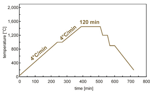
All zirconia specimens were sintered (VITA ZYRCOMAT® 6000 MS; VITA ZahnFabrik H. Rauter GmbH & Co. KG) for 120 min at 1,450°C according to the manufacturer’s instructions (Figure 1).
Characterization
Three specimens were randomly selected from each group for characterization using energy dispersive X-ray spectroscopy (EDS) (Axia ChemiSEM System; Thermo Fisher Scientific, Eindhoven, Netherlands), field emission scanning electron microscopy (FESEM) (Inspect™ F50; FEI Company, Hillsboro, USA), atomic force microscopy (AFM) (NaioAFM; Nanosurf, Liestal, Switzerland), and X-ray diffraction (XRD) (Shimadzu XRD-6000; Shimadzu Corporation, Kyoto, Japan).
Microshear bond strength testing
Microshear bond strength testing was used to quantify the interfacial adhesion between the resin cement and zirconia substrates after different surface treatments. This method applies an increasing shear load to a small cross-sectional bonding area until failure occurs, allowing a localized assessment of bond strength.19 Resin cement (G-CEM ONE™) was used to create cylindrical specimens that were polymerized and bonded to the primed zirconia surfaces. A universal testing machine fitted with a knife-edge shearing tool25 was used to measure the µSBS at a crosshead speed of 0.5 mm/min. The load at failure was recorded in newtons [N] and divided by the cylindrical bond area to calculate the µSBS in megapascals [MPa]. This standardized microshear testing protocol allowed precise, localized measurement of bond strength at the adhesive interface between the resin cement and nanosilica-coated zirconia surface. The test provided a measure of interfacial bond strength and assessed the durability and fatigue resistance of the bonded restorations.
Statistical analysis
Data was statistically analyzed using the IBM SPSS Statistics for Windows software, v. 26.0 (IBM Corp., Armonk, USA). The normality of the data was assessed using the Shapiro–Wilk test. One-way analysis of variance (ANOVA) was used to compare the groups, followed by Tukey’s post hoc test. The significance level was set at p ≤ 0.05.
Results
Microstructure analysis
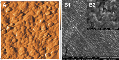
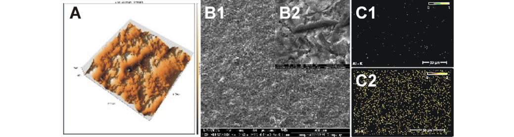
Scanning electron microscopy provided high-resolution images of the zirconia surface morphology after different coating treatments. On untreated polished zirconia, FESEM revealed a smooth, flat surface with only minor scratches and grooves from the polishing process (Figure 2). At high magnification, the zirconia grains were tightly packed and clearly visible, with grain boundaries in close contact and no apparent gaps or defects. In contrast, in group E, in which zirconia was sandblasted with CoSil using the tribochemical silica-coating method, Sa was markedly increased, obscuring the underlying zirconia grain structure (Figure 3). The particle bombardment created an irregular, porous surface with peaks, valleys and protruding features. No smooth or flat areas remained after abrasive blasting with the silica-coated alumina particles.
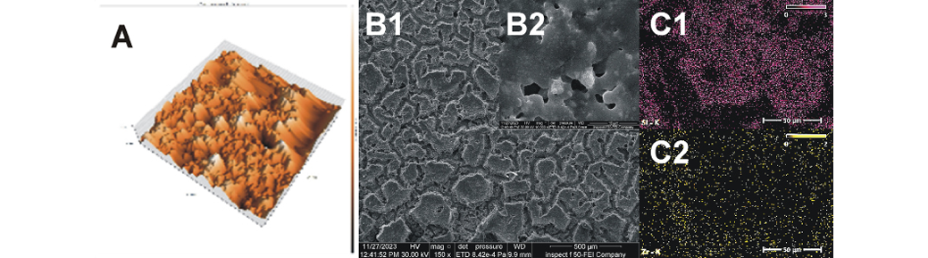
Group A, in which zirconia was dip-coated with a 100% nanosilica suspension, showed an island-like coating that partially covered the zirconia surface (Figure 4). Within this coating, the islands contained pores and cavities, and these nanostructures were perpendicularly oriented. The coating showed incomplete coverage and high porosity, facilitating micromechanical interlocking with resin cement, with some areas of the zirconia surface remaining exposed for chemical bonding interactions.
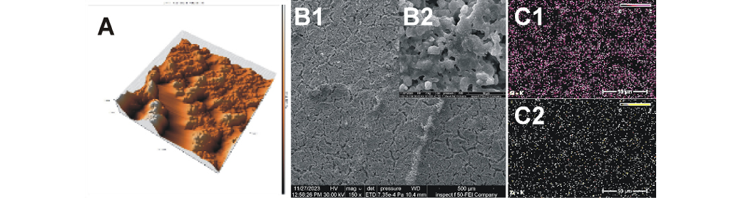
After nanozirconia was added to the dipping suspension (75% SiO2 + 25% ZrO2) in group B, the surface morphology changed from an island-like appearance to a more compact structure, with a decrease in the amount of exposed zirconia substrate (Figure 5). The surface topography of the zirconia coating changed, as indicated by the absence of the scattered pattern observed in specimens coated with pure silica. In addition, the coating islands appeared to be more closely attached to the zirconia substrate, although some areas of the substrate remained uncovered. At high magnification (×10,000), nanozirconia particles were observed dispersed within the melted silica (fused silica).
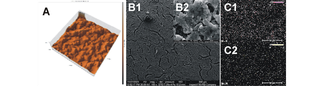
As the proportion of zirconia nanoparticles in the dipping suspension increased to 50 Vf% in group C, the surface topography showed reduced spacing and closer approximation among islands, resulting in a decrease in the amount of exposed zirconia substrate (Figure 6). At high magnification (×10,000), a greater number of zirconia particles were observed distributed within the melted silica, with porosities formed within the coating.
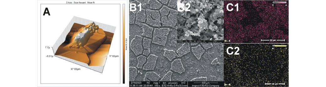
With a further increase in the zirconia content of the suspension to 75% Vf% in group D, no significant changes were observed at low magnification (×150), although a slight reduction in surface coating cracks was noted (Figure 7). At high magnification (×10,000), the melted silica appeared to be densely loaded with nanozirconia particles, with porosities present within the coating.
Elemental analysis
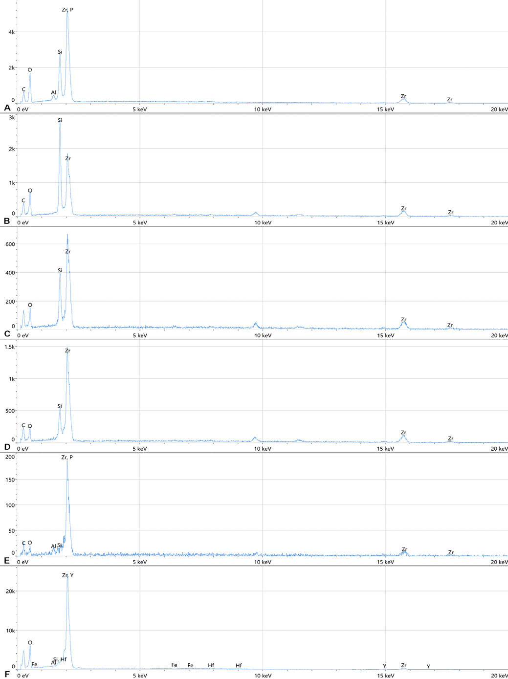
The EDS spectra (Table 2) showed the weight percentages of oxygen, yttrium, hafnium, and zirconium, with an insignificant percentage of detectable silicon and aluminum on the surface of untreated zirconia. For group E, in which the zirconia surface was blasted with silica-coated alumina, EDS revealed the presence of silicon and aluminum in addition to the elements detected on the untreated zirconia surface. In group A, the amount of silicon was higher than that in group E. Groups B, C and D showed different proportions of silicon and zirconium. EDS mapping demonstrated the distribution of the nanoparticles within the coating of each group (Figure 8).
XRD characterization
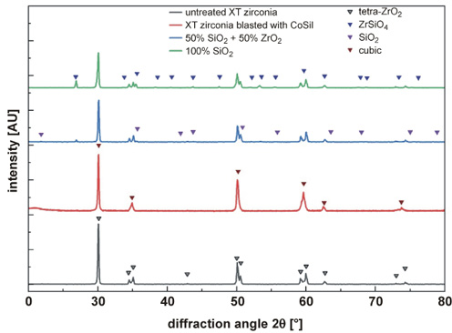
X-ray diffraction analysis provided critical insights on the crystallographic phases present in zirconia after different nanocoating treatments (Figure 9). By measuring diffraction peak angles and intensities, the atomic lattice structure was characterized. For untreated polished zirconia controls, distinct XRD peaks were observed at 2θ values of 30.3°, 35.2°, 50.5°, 60.0°, and 62.8°, corresponding to the (101), (110), (112), (211), and (202) crystal planes of the metastable tetragonal zirconia phase.18 The nanosilica–zirconia-coated samples showed identical peaks, indicating that the coatings did not disrupt the tetragonal lattice structure of zirconia. Preserving the tetragonal phase is crucial for maintaining optimal mechanical properties in zirconia restorations.26 In contrast, tribochemical silica coating induced a phase transformation, with an additional peak at 28.2° representing the monoclinic phase.18 Particle bombardment cause sufficient surface damage to disrupt the tetragonal structure. Such a phase transformation may introduce surface flaws and residual stresses, potentially compromising the mechanical properties of the restoration.27, 28
The 100% nanosilica coatings also exhibited new peaks at 49.0° and 50.5°, which were assigned to the (012) and (110) planes within SiO2.18 For the nanosilica–zirconia coatings, distinct peaks were observed at 30.1264°, 50.1339° and 60.0273°. The presence of characteristic peaks associated with the nanomaterials confirmed the composition of the composite coating on the zirconia surface. The XRD findings indicated that the inert zirconia phase and crystallinity were unaffected by dip coating. By preserving the optimal tetragonal structure, the nanocoatings help maintain the favorable mechanical properties of zirconia for dental applications.
Topographic analysis
The topography of the coated surfaces was evaluated using AFM. The surface roughness of the specimens, assessed over an average scanned area of 60 nm × 60 nm, ranged from 30 nm to 1,000 nm. A comparison of Sa among the groups is presented in Table 3. The average Sa values for groups A–E and the untreated group were 1,552 nm, 707 nm, 792.8 nm, 433 nm, 372 nm, and 193 nm, respectively. The Sa of group A was significantly greater than that of all other groups (p < 0.001).
The reconstructed three-dimensional (3D) surface roughness images representative of each group are shown in Figure 2, Figure 3, Figure 4, Figure 5, Figure 6, Figure 7. Different surface topographies were observed among the groups.
Microshear bond strength
The µSBS values and standard deviations for the different surface treatments are shown in Table 4.
One-way ANOVA showed that the surface treatments significantly affected µSBS (p ≤ 0.05). The nanosilica and nanosilica–zirconia groups with different proportions of nanoparticles in the dipping suspensions exhibited significantly higher µSBS values than the CoSil-treated and untreated zirconia groups (p ≤ 0.05).
Modes of failure
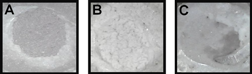
After the μSBS test, the failure modes were examined using a digital microscope at ×250 magnification. Failure modes were categorized into 3 types, as follows (Figure 10):
– adhesive I: failure between the zirconia substrate and the coating (ZrO2–coating interface);
– adhesive II: failure between the coating and the resin cement (coating–resin cement interface);
– mixed failure: a combination of adhesive II failure and cohesive failure.28
The failure mode was evaluated for each group. In group A (100% SiO2), failure occurred between the zirconia substrate and the coating, and was therefore classified as adhesive I failure. In group B (75% SiO2 + 25% ZrO2), 62.5% of failures occurred at the zirconia–coating interface, whereas 37% occurred at the coating–resin cement interface. In group C (50% SiO2 + 50% ZrO2), 62.5% of failures occurred at the coating–resin cement interface, whereas 37% of the specimens exhibited mixed failure. In group D (25% SiO2 + 75% ZrO2), 100% of failures were reported at the coating–resin cement interface. In group E (CoSil), 75% of failures were reported at the coating–resin cement interface and 25% were mixed failures, similarly to the control group (Table 5).
Discussion
The null hypothesis was rejected, since different surface topographies and morphologies were observed on the zirconia surface after dipping in different nanosuspensions and after blasting with silica-coated alumina. In addition, the blasting technique induced a tetragonal-to-monoclinic phase transformation, which may contribute to zirconia surface damage.
The measurement of bond strength at the zirconia–resin cement interface is complex. Microshear bond strength testing was utilized in this study to quantify the interfacial adhesion between resin cement and zirconia after dip-coating surface treatments. Compared with conventional macroscale shear testing, the microshear method provides more reliable and representative bond-strength results.29, 30 Microshear testing is performed by bonding small cylindrical samples and applying an increasing shear force parallel to the interface.19, 24 Failure occurs by debonding at the weakest point, which is often within the microscopic bonding complex. This provides a more representative measure of interfacial adhesive strength rather than the cohesive strength of the cement itself.29 However, the small surface area can introduce challenges related to stress concentrations and non-uniform loading. Despite these limitations, microshear testing remains an accepted and useful technique for comparing bond strengths in localized regions.29, 30
In this study, all nanostructured coatings significantly enhanced zirconia bond strength compared with the polished untreated zirconia surface. The inert zirconia surface lacks retentive features or chemical reactivity with resin cements, resulting in low bond strength. These findings are consistent with those reported in the literature.6, 31, 32, 33 By introducing nanoscale textures and porosities, as demonstrated by FESEM, the coatings increased the surface area and surface energy available for micromechanical retention and adsorption of resin monomers. The 50:50 silica–zirconia ratio provided an optimal balance between chemical and mechanical bonding with the resin cement.
Tribochemical silica coating with CoSil particles increased µSBS compared with polished zirconia. The particle bombardment roughened the inert surface and enhanced micromechanical retention.34 However, a tetragonal-to-monoclinic phase transformation was observed, indicating surface damage. Residual stresses associated with this transformation can compromise the restoration.8, 27 In contrast, dip coating formed homogeneous nanolayers without disrupting the fragile zirconia surface. Porous silica–zirconia composite coatings increased bond strength while preserving the optimal tetragonal phase, as verified by XRD (Figure 9).
Dip coating enables precise control over nanolayer properties by adjusting the coating composition and deposition parameters.35 The approach modifies zirconia surfaces to enhance resin bonding without abrasive blasting or the use of high temperatures. Further optimization of nanocoating composition, thickness, and sintering parameters could produce durable yet microretentive surfaces, expanding the potential applications of high-strength zirconia restorations.
The silica–zirconia-coated surfaces exhibited higher µSBS (p ≤ 0.05) compared with the other groups; this indicates that silica–zirconia coating may be an effective method for improving resin–zirconia adhesion. The observed increase in µSBS could be attributed to increased micromechanical interlocking in addition to chemical bonding.36 The FESEM images (Figure 5, Figure 6, Figure 7) show numerous microgaps between the particles within the silica–zirconia coating. This observation supports the assumption that micromechanical interlocking between the coated zirconia surface and resin cement was enhanced.
The highest µSBS among all groups was observed for the 50% SiO2 + 50% ZrO2 suspension (51.63 ±5.6 MPa) which could be due to the high Sa in this group (792.8 nm), as shown by AFM analysis (Table 3). FESEM demonstrated that the coating formed a network with sharp edges (Figure 5), which could be attributed to the sintering of nanosilica and nanozirconia on the zirconia surface.
Field emission scanning electron microscopy also showed that the coating provided complete coverage of the zirconia substrate. The increased average Sa may have contributed to enhanced micromechanical bonding, while the presence of zirconium at approx. 59.8 wt% on the coating surface could facilitate interaction with 10-MDP in the Z-Prime Plus and thereby promote chemical bonding. The combination of chemical and mechanical bonding may have contributed to the increased µSBS, in agreement with previous findings.37 This high bond strength was also reflected in the failure mode, which showed that the adhesive strength of the coating to the zirconia substrate was high, with no adhesive I failures. Failure occurred at the resin cement–coating interface (adhesive II) in 62.5% of specimens, while 37.5% showed mixed adhesive II/cohesive failure (Table 5, Figure 10).
Dip coating with 100% SiO2 resulted in higher µSBS than tribochemical silica coating and untreated zirconia. The pure silica coating led to the formation of a patch-like coating pattern, leaving some areas of bare zirconia exposed. This surface pattern may have increased the available surface area and enhanced micromechanical bonding.38 In addition, the exposed zirconia substrate could interact with 10-MDP from the primer, thereby enhancing chemical bonding. This finding agrees with previous studies demonstrating that 10-MDP affects the bonding effectiveness of conventional composite resin cement to zirconia ceramics.39 However, this increase in bond strength appeared to be insufficient to ensure strong adhesion between the coating and zirconia substrate, as 100% of failures in this group occurred at the coating–zirconia interface and were classified as adhesive I failures (Table 5).
This simple, laboratory-friendly dip-coating technique shows promise for improving resin bonding to zirconia restorations.33 By combining nanosilica and nanozirconia, bond strength was improved without surface damage. A key challenge was developing stable dispersions of the nanoparticle mixtures to enable homogeneous coatings.40 Nanoparticles tend to agglomerate due to their high surface energy. Adequate particle dispersion and prolonged stabilization of the suspension were achieved by sonication using an ultrasonic probe.41 This allowed consistent dip coating with tunable nanolayer properties. With further optimization of deposition parameters and sintering protocols, this approach could be broadly implemented to tailor zirconia bonding surfaces.
These findings were obtained using only one type of zirconia (XT). Because different types of zirconia have different chemical compositions and physical properties, this technique should also be evaluated using other zirconia types (LT and HT zirconia), to determine whether it similarly improves µSBS between resin cement and these materials.
The study had some limitations, particularly the lack of simulation of clinically relevant intraoral conditions. Aging procedures such as water storage, mechanical loading and thermocycling were not performed. Future studies incorporating thermocycling and mechanical cyclic loading may provide more clinically relevant results by better simulating intraoral conditions.
Conclusions
The new dip-coating method utilizing nanosilica–nanozirconia composite suspensions represents a promising strategy for enhancing resin adhesion to zirconia surfaces. This approach resulted in higher µSBS than silica-coated alumina blasting while preserving the fragile zirconia structure. The ability to modify surface textures and porosities by adjusting the nanoparticle composition of the suspension further demonstrates the versatility and efficacy of this technique. Among the nanosuspensions evaluated, the 50:50 nanosilica–nanozirconia composition produced the most favorable overall results.
Ethics approval and consent to participate
Not applicable.
Data availability
The datasets generated and/or analyzed during the current study are available from the corresponding author on reasonable request.
Consent for publication
Not applicable.
Use of AI and AI-assisted technologies
Not applicable.