Abstract
Background. Non-invasive treatment involves the use of various remineralizing and cariostatic agents, including sodium fluoride, casein phosphopeptide–amorphous calcium phosphate (CPP-ACP), casein phosphopeptide–amorphous calcium phosphate fluoride (CPP-ACPF), silver nitrate, and silver diamine fluoride (SDF).
Objectives. The purpose of the present study was to evaluate the surface morphology and mineral uptake of demineralized dentin following a single application of SDF and CPP-ACPF.
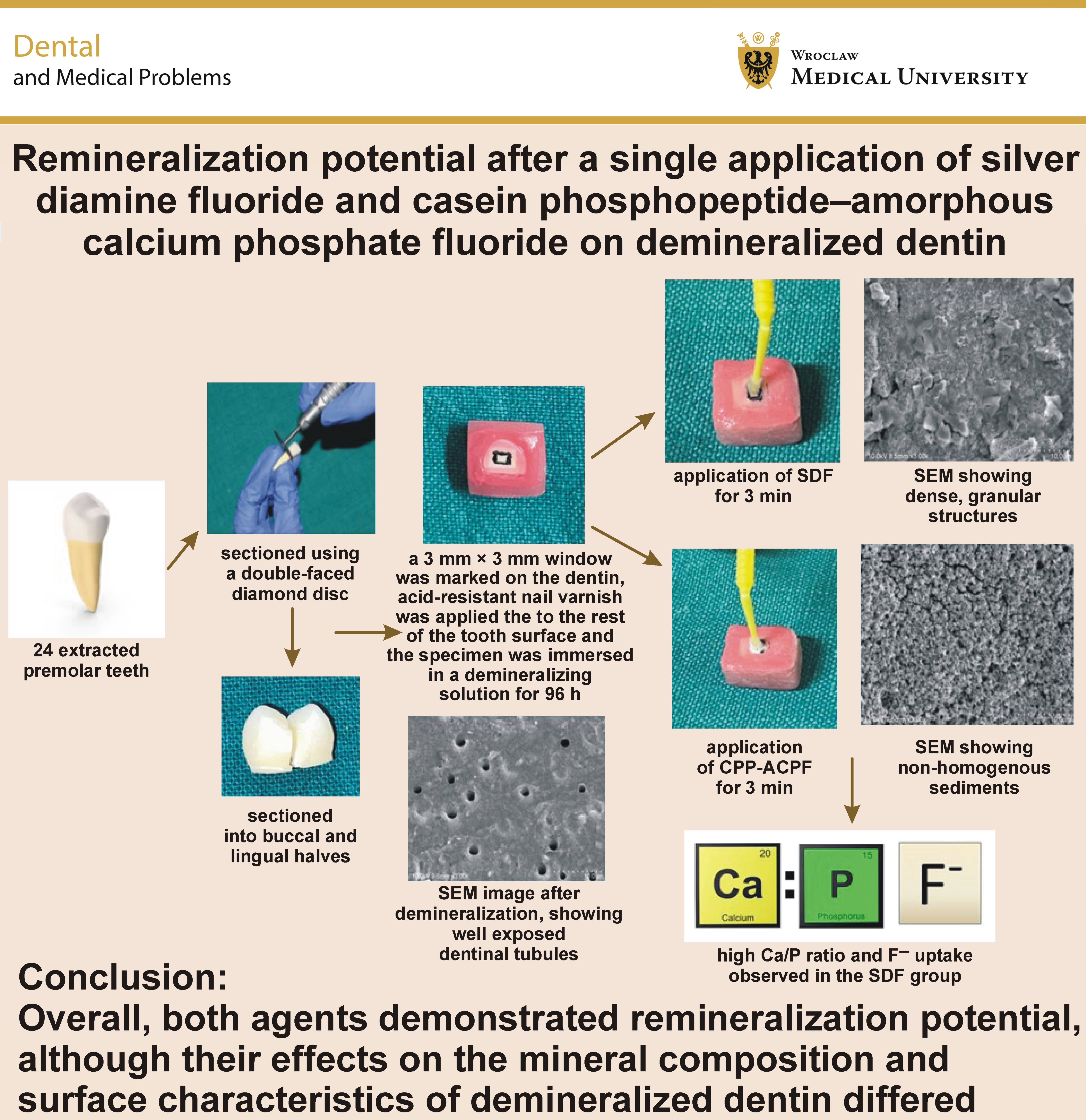
Material and methods. Twenty-four caries-free premolar sections with marked areas of specific dimensions on the dentin surface were exposed to a demineralizing solution for 96 h and randomly allocated to 2 groups: group 1 (SDF); and group 2 (CPP-ACPF). Surface morphology was analyzed using scanning electron microscopy (SEM), while mineral uptake was assessed using energy-dispersive X-ray spectroscopy (EDX) following demineralization and the application of the test agents. Paired and unpaired t tests were used to analyze the continuous data.
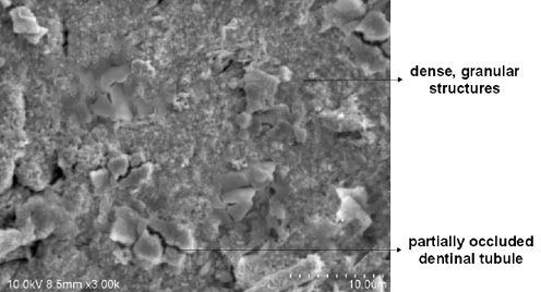
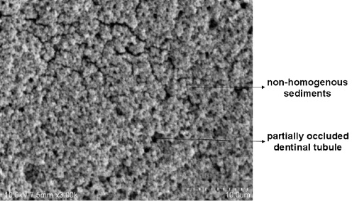
Results. The EDX revealed a difference in the mean increase in the calcium(Ca)/phosphorus (P) ratio between the SDF group (0.34 ±0.92) and the CPP-ACPF group (0.01 ±0.22); however, this difference was not statistically significant (p = 0.400). In contrast, the mean increase in fluoride uptake was significantly higher in the SDF group (2.07 ±0.66 at%) than in the CPP-ACPF group (0.37 ±0.38 at%) (p = 0.001). The SEM images showed dense, granular structures in the intertubular areas, with partial occlusion of the dentinal tubules in the SDF group, whereas the CPP-ACPF group exhibited mineral deposits partially occluding the dentinal tubules, with non-homogeneous sediment formation.
Conclusions. A single application of both SDF and CPP-ACPF demonstrated potential to promote mineral uptake in demineralized dentin, with comparable effects on calcium and phosphate incorporation. However, SDF showed a greater capacity for fluoride uptake and produced a denser, more compact surface morphology, whereas CPP-ACPF resulted in a more porous mineral deposition pattern. Overall, both agents demonstrated remineralization potential, although their effects on the mineral composition and surface characteristics of demineralized dentin differed.
Keywords: CPP-ACP, scanning electron microscopy, silver diamine fluoride, energy-dispersive X-ray spectroscopy
Introduction
Dental caries results from an imbalance between the physiological processes of demineralization and remineralization of dental hard tissues. The current approach to caries management emphasizes the early detection and targeted, non-invasive management of reversible lesions.1 The materials or agents used for this purpose act mainly through 2 mechanisms: preventing further demineralization; or restoring the lost minerals and thereby promoting remineralization.2 In this regard, various remineralizing and cariostatic agents have been introduced, including sodium fluoride, tricalcium phosphate, casein phosphopeptide–amorphous calcium phosphate (CPP-ACP), casein phosphopeptide–amorphous calcium phosphate fluoride (CPP-ACPF), silver nitrate, and silver diamine fluoride (SDF), which have demonstrated varying degrees of effectiveness.
Silver diamine fluoride is a safe and effective caries-arresting agent that was first explored as a treatment option for managing carious lesions in Japan in 1969.3 It contains silver and fluoride ions in an ammoniacal solution, with silver ions stabilized through complexation with ammonia.4 Silver ions exert a strong antimicrobial effect and can react with phosphate and chloride ions to form relatively insoluble silver compounds, contributing to the hardening of soft carious lesions.5 The high fluoride concentration in SDF promotes the remineralization of demineralized dentin, and can contribute to the formation of fluoridated apatite and calcium fluoride. Furthermore, the alkaline nature of the ammoniacal solution may contribute to an acid-buffering effect and promote mineral precipitation.6
Another commonly used non-fluoridated remineralizing agent, casein phosphopeptide–amorphous calcium phosphate (CPP-ACP), is a naturally derived agent obtained from the casein protein of milk.7 CPP-ACP stabilizes and localizes calcium and phosphate ions, maintaining a state of supersaturation with respect to hydroxyapatite, thereby reducing demineralization and promoting remineralization.8 CPP-ACPF is a nanocomplex formed by incorporating fluoride (900 ppm) into CPP-ACP. CPP-ACPF enhances fluoride incorporation into dental biofilm and subsurface enamel, thereby promoting enamel remineralization.9
Kumar and Kumari stated that the silver component of SDF exerts an oligodynamic effect within the dentinal tubules by inhibiting bacterial growth and reducing dentin demineralization.10 According to Cabalén et al., SDF can arrest active dentinal lesions by inhibiting demineralization and promoting remineralization.11
To date, limited evidence is available regarding the effects of a single application of SDF and CPP-ACPF on mineral uptake in demineralized dentin. Therefore, an in vitro study was conducted to evaluate the surface morphology and mineral uptake of demineralized dentin following a single application of SDF and CPP-ACPF, using scanning electron microscopy (SEM) and energy-dispersive X-ray analysis (EDX).
Material and methods
The present in vitro study was conducted in affiliation with the Department of Pedodontics and Preventive Dentistry, Vishnu Dental College, Bhimavaram, India. Surface morphology and mineral content were evaluated using SEM and EDX, respectively, at Osmania University, Hyderabad, India. The study protocol was approved by the institutional ethics committee at Vishnu Dental College (IECVDC/2021/PG01/PPD/IVT/30).
Sample size
The sample size was calculated based on the results of a pilot study using the G*Power 3.1 software (https://www.psychologie.hhu.de/arbeitsgruppen/allgemeine-psychologie-und-arbeitspsychologie/gpower), with the level of significance set at 5% and the statistical power at 80%. A total of 24 teeth were included in the study, with 12 teeth allocated to each group.
Methodology
Extracted non-carious premolars were disinfected in 10% formalin for 1 week12 and cleaned of debris, using an ultrasonic scaler (DTE D3; Guilin Woodpecker Medical Instruments, Guilin, China). The teeth were stored in double-deionized distilled water in a glass beaker at room temperature until the experimental procedure was initiated.
The teeth were decoronated at the cementoenamel junction (CEJ) and the roots were discarded. Each coronal portion was sectioned mesiodistally into buccal and lingual halves, using a double-faced diamond disc (Diatech USA, Mount Pleasant, USA) under continuous water cooling. The enamel was removed using a water-cooled diamond disc to expose the underlying dentin, and the specimens were mounted in self-cure acrylic resin blocks (DPI RR Cold Cure; DPI, Mumbai, India). The specimens were ultrasonicated in deionized water for 5 min, using an ultrasonic cleaner (PHUC-50; Phoenix, USA). A 3 mm × 3 mm window was marked on the dentin surface of each specimen, and the remaining tooth surface was coated with acid-resistant nail varnish (Clarins, Paris, France).
The specimens were randomly allocated into 2 groups: group 1 – SDF (38%, e-SDF; Kids-e-Dental, Mumbai, India); and group 2 – CPP-ACPF (10% w/v, GC Tooth Mousse Plus, GC, Tokyo, Japan). Each specimen was individually immersed in 15 mL of a demineralizing solution containing 2.2 mM calcium chloride, 2.2 mM monosodium dihydrogen phosphate, and 0.05 M acetic acid. The pH was adjusted to 4.5, using 50% sodium hydroxide. The specimens were maintained in the demineralizing solution for 96 h at 37°C.
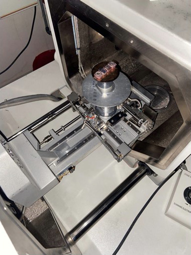
Following demineralization, the specimens were subjected to SEM with EDX (Hitachi S-3700N; Hitachi Ltd., Tokyo, Japan) after dehydration, gold sputtering and conductive adhesive fixation using a sputter coater (Hitachi E-1010 Ion Sputter, Hitachi Ltd.). The SEM was used for the qualitative evaluation of surface morphology, whereas EDX was used for the quantitative assessment of mineral content. The specimens were evaluated before and after the application of the respective test agents for 3 min at a beam voltage of 10 kV and a magnification of ×3,000 (Figure 1).
Statistical analysis
Statistical analysis was performed after assessing the normality of data distribution. The data were tabulated and analyzed using parametric tests. An independent-samples t test was used to compare the 2 groups, while a paired-samples t test was used to compare the measurements obtained after demineralization with those obtained following the application of the respective test agents. A p-value ≤0.05 was considered statistically significant.
Results
Comparison of the mean Ca/P ratio
On intragroup comparison, group 1 showed a higher mean calcium(Ca)/phosphorus (P) ratio after the application of SDF as compared to the baseline value; however, the difference was not statistically significant (p = 0.400). Similarly, group 2 showed a higher mean Ca/P ratio after the application of CPP-ACPF as compared to the baseline value, although the difference was not statistically significant (p = 0.920) (Table 1).
On intergroup comparison, the SDF group demonstrated a greater increase in the mean Ca/P ratio than the CPP-ACPF group; however, the difference between the groups was not statistically significant (p = 0.400) (Table 2).
Comparison of the mean fluoride uptake
On intragroup comparison, the SDF group showed a higher mean fluoride content value than at the baseline, and the difference was statistically significant (p = 0.001). The CPP-ACPF group also showed a higher mean fluoride content value than at the baseline; however, the difference was not statistically significant (p = 0.064) (Table 3).
On intergroup comparison, the SDF group demonstrated a greater increase in the mean fluoride content value (2.07 ±0.66 at%) than the CPP-ACPF group (0.37 ±0.38 at%), and the difference between the groups was statistically significant (p = 0.001) (Table 4).
Scanning electron microscopy evaluation
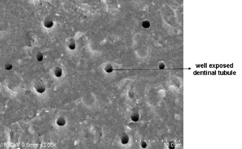
On evaluation at ×3,000 magnification, all demineralized dentin specimens showed clearly exposed dentinal tubules (Figure 2). The SDF-treated specimens exhibited dense, granular structures in the intertubular areas, with partial occlusion of the dentinal tubules (Figure 3). The CPP-ACPF-treated specimens showed mineral deposits partially occluding the dentinal tubules, with non-homogeneous sediment deposits observed on the dentinal surface (Figure 4).
Discussion
Decades of research have shifted the focus of caries management toward the early detection of lesions and the use of non-invasive treatment approaches.13 Ideally, the natural buffering capacity of saliva would help reverse early tooth demineralization. When these natural protective mechanisms are insufficient to compensate for mineral loss, several approaches can be employed to arrest or reverse lesion progression.14 Remineralization is one such approach, involving the replenishment of the lost minerals through the availability of calcium and phosphate ions, often aided by remineralizing agents. This process helps restore the mineral content of dental hard tissues and maintain the optimal Ca/P ratio of hydroxyapatite, approx. 1.67, thereby contributing to the strengthening of the tooth lattice network.15 Fluoride varnishes have been widely used for the professional application of topical fluoride for more than 25 years, and were developed to prolong the contact time between fluoride and the tooth surface.16
Currently, SDF has attracted considerable attention from both researchers and dental practitioners. It has been proposed that SDF can simultaneously inhibit the progression of the existing carious lesions and reduce the development of new carious lesions.17 Silver diamine fluoride has demonstrated antimicrobial activity, including the inhibition of cariogenic microorganisms and the disruption of dental plaque biofilm, and may also promote calcium uptake and mineral deposition.18 SDF-treated carious lesions have been reported to exhibit significantly greater surface microhardness and increased resistance to acid-induced demineralization.19 A 38% concentration of SDF is commonly used for caries management and contains approx. 44,800 ppm fluoride.4 However, lower concentrations, including 30% and 12%, have also been investigated. A review by Gao et al.reported that increasing the SDF concentration from 12% to 38% may enhance its effectiveness in arresting caries.17
Casein phosphopeptide–amorphous calcium phosphate is another widely studied and established non-fluoridated remineralizing agent. CPP-ACP is a nanocomplex that was patented in the United States in 1991; CPP-ACP acts by stabilizing calcium and phosphate ions in the presence of fluoride, maintaining them in a bioavailable form.20 According to Reynolds et al., CPP-ACP has demonstrated greater potential for the remineralization of subsurface enamel lesions as compared to other forms of calcium phosphate.21
Dentin remineralization is more challenging than enamel remineralization because of differences in their composition and structure. The dentin contains a substantially higher proportion of organic matrix, primarily type I collagen, as well as a tubular microstructure.22 Dentin mineralization is regulated by non-collagenous proteins, and involves the deposition of calcium and phosphate ions within and around the collagen fibrils, followed by the formation and growth of hydroxyapatite crystals.23 Fluoride is an important ion in the formation of fluoridated apatite, which is more resistant to acid dissolution and cariogenic challenges than hydroxyapatite.24
Hence, CPP-ACPF was compared with SDF to evaluate their remineralization potential on demineralized dentin. Scanning electron microscopy combined with energy-dispersive X-ray analysis (SEM-EDX) is a useful microanalytical technique for evaluating surface morphology and elemental composition. The SEM provides detailed, high-resolution images by scanning a focused electron beam across the specimen surface and detecting secondary and backscattered electron signals. When combined with EDX, SEM can provide the qualitative identification and quantitative assessment of the elemental composition of specific areas of the specimen. Therefore, SEM-EDX was used in the present study to evaluate changes in surface morphology and mineral composition following the application of the test agents.
In the present study, a single application of SDF resulted in improvement in the mean Ca/P ratio and significantly higher fluoride uptake as compared to CPP-ACPF. Richards reported significantly greater remineralization following the application of SDF under net demineralizing conditions.25 Similarly, Yadav et al. demonstrated the greatest changes in the Ca/P ratio following remineralization in the SDF group as compared to the other treatment groups.26
Sinha et al. reported that the treatment of deep carious lesions with SDF increased the concentrations of calcium, phosphate and fluoride ions in caries-affected dentin.27 Yilmaz et al. suggested that the superior remineralizing effect of SDF may be attributed to its high fluoride concentration and the alkaline nature of the solution.28 Chibinski et al. reported that, after 12 months, the caries-arresting effect of SDF was 66% greater than that of other active materials and 154% greater than that of placebo; overall, SDF demonstrated an 89% greater caries-arresting effect as compared to the other treatment groups.29
The CPP-ACPF samples showed improvement in the mean Ca/P ratio and increased fluoride uptake in comparison with the demineralized dentin samples. Cross et al. reported similar findings, suggesting that the localization of CPP-ACPF at the surface layer facilitates the sustained release of calcium, phosphate and fluoride ions while preventing their rapid precipitation as fluorapatite, thereby promoting mineralization within the demineralized lesion.30 Hedge et al. reported that CPP-ACPF demonstrated a marginally greater remineralization potential as compared to the other agents evaluated.31
The addition of fluoride (0.2% NaF) to CPP-ACP has been reported to produce a marginally greater degree of remineralization than CPP-ACP alone, possibly through the formation of fluorapatite in the presence of calcium and phosphate ions.32 Yadav et al. reported that CPP-ACPF demonstrated greater remineralization than fluoride-free toothpaste, fluoride-containing toothpaste, and CPP-ACP alone.26 This effect may be attributed to the incorporation of fluoride into the CPP-ACP complex, resulting in a synergistic effect that enhances remineralization in comparison with CPP-ACP alone, which is consistent with the findings of the present study. Kamath et al. also observed an increase in the mean fluoride content from the post-demineralization stage to the post-remineralization stage following treatment with CPP-ACPF in the enamel.1
The variation in the Ca/P ratio and fluoride content values may be attributed to differences in the inorganic composition of the specimens, as well as the absence of the complex oral environment and the controlled experimental conditions. In vivo conditions present more dynamic and multifactorial challenges to the tested materials. Since the existing literature provides evidence supporting the beneficial effects of SDF and CPP-ACPF, further studies should evaluate these agents under conditions that more closely mimic the oral environment. Such investigations may provide a better understanding of their remineralization potential and clinical effectiveness in the management of demineralized dental hard tissues.
Conclusions
A single application of both SDF and CPP-ACPF demonstrated potential to promote mineral uptake in demineralized dentin, with comparable effects on calcium and phosphate incorporation. However, SDF showed a greater capacity for fluoride uptake and produced a denser, more compact surface morphology, whereas CPP-ACPF resulted in a more porous mineral deposition pattern. Overall, both agents demonstrated remineralization potential, although their effects on the mineral composition and surface characteristics of demineralized dentin differed.
Ethics approval and consent to participate
The study protocol was approved by the institutional ethics committee at Vishnu Dental College, Bhimavaram, India (IECVDC/2021/PG01/PPD/IVT/30).
Data availability
The datasets supporting the findings of the current study are available from the corresponding author on reasonable request.
Consent for publication
Not applicable.
Use of AI and AI-assisted technologies
Not applicable.