Abstract
Background. There is growing interest in the use and processing of natural products for potential applications in dentistry, including endodontic treatment. Anchovy (Stolephorus insularis) is one such natural product.
Objectives. The aim of the present study was to evaluate odontoblast-like cells (OLCs) and alkaline phosphatase (ALP) expression in the pulp of Wistar rats after the administration of a combination of calcium hydroxide (CH) and the anchovy nano-extract as a potential material for pulp regeneration.
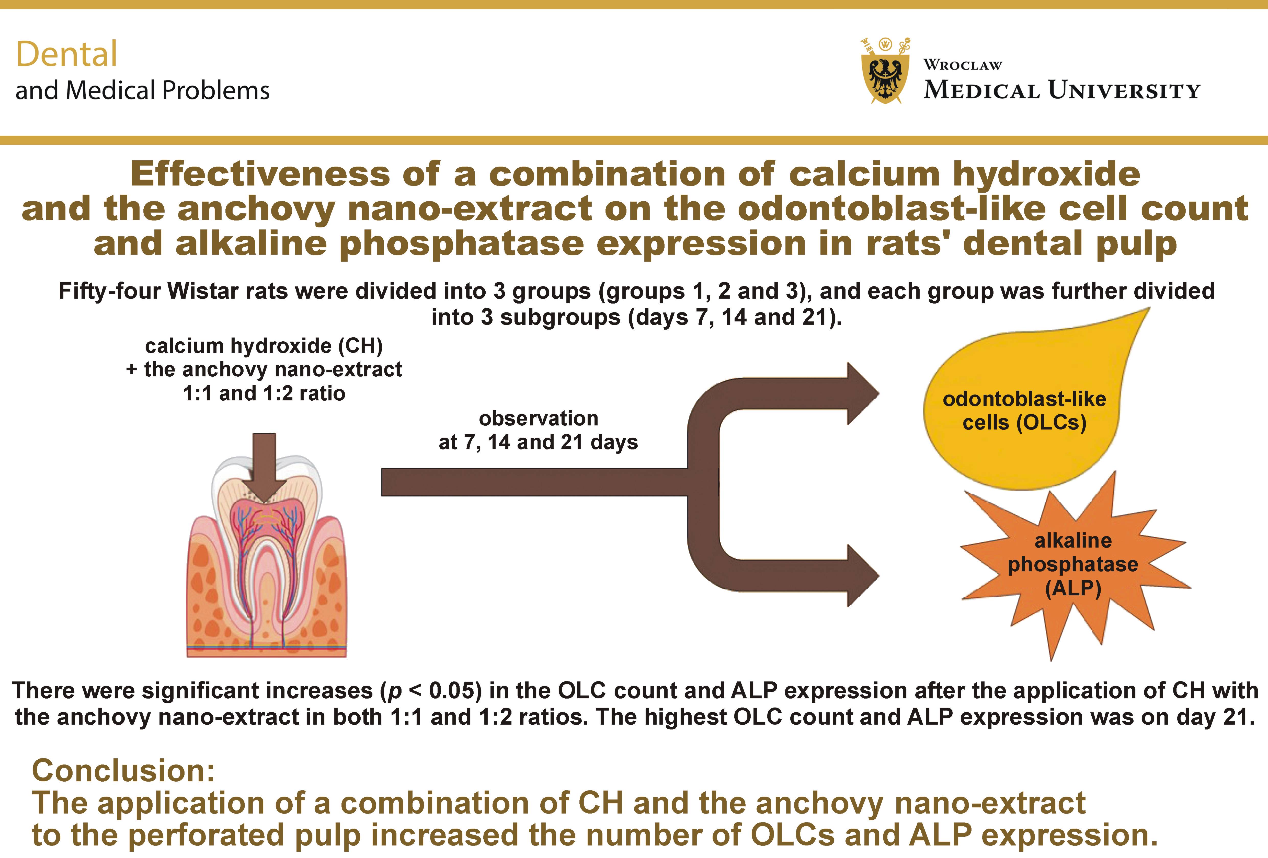
Material and methods. Fifty-four Wistar rats were divided into 3 groups: the control group (group 1) received CH and distilled water; the 1st treatment group (group 2) received a 1:1 ratio combination of CH and the anchovy nano-extract; and the 2nd treatment group (group 3) received a 1:2 ratio combination of CH and the anchovy nano-extract. Each group was further divided into 3 subgroups according to the observation periods of 7, 14 and 21 days. Pulp perforation was performed on the rats’ maxillary right first molars, followed by the placement of the tested materials into the perforated pulp. The rats were sacrificed according to the respective observation periods. The specimens were processed, and evaluated histopathologically (staining with hematoxylin and eosin (H&E)) and through immunohistochemistry (IHC). The data were analyzed using the one-way ANOVA followed by the Tukey HSD post hoc test. Statistical significance was set at p < 0.05.
Results. There were significant increases in OLCs and ALP expression following the application of the anchovy nano-extract at both 1:1 and 1:2 ratios, with significant differences as compared to the control group (p < 0.05). The highest OLC count and ALP expression were observed on day 21. The higher 1:2 ratio resulted in faster OLC formation, which occurred on day 14; in the case of the 1:1 ratio, increased OLC formation occurred on day 21. Both ratios increased ALP expression on day 21.
Conclusions. The application of a combination of CH and the anchovy nano-extract to the perforated pulp increased the number of OLCs and ALP expression.
Keywords: regeneration, medicine, alkaline phosphatase, calcium hydroxide, odontoblast
Introduction
Oral health has a significant impact on a person’s quality of life (QoL), affecting their ability to speak, eat, and maintain overall health. Therefore, comprehensive treatment is necessary to address oral health holistically.1 Vital pulp exposure can occur due to trauma, caries removal, or during mechanical cavity preparation.2 Consequently, prompt pulp capping is imperative when pulp exposure occurs.3 Preserving pulp vitality is crucial in all of these scenarios to ensure that the tooth remains vital, receives adequate nutrients, maintains innervation, and has effective immunological defense. Pulp capping treatment aims to promote the regeneration of the damaged pulp by employing bioactive substances.2
The regeneration process in the pulp, involving stem cells, odontoblasts, inflammatory cells, and fibroblasts, takes place following pulp capping treatment. An increase in stem cell activity occurs during the pulp healing phase, characterized by the proliferation and differentiation of these cells into odontoblast-like cells (OLCs). To initiate mineralization, alkaline phosphatase (ALP) is synthesized by these newly formed OLCs.3 Alkaline phosphatase plays an important role in the tissue response to injury.4, 5 It is a marker of OLC differentiation, and its presence indicates that the cells are undergoing differentiation.6
Bioactive substances have been reported to support tissue regeneration.7 These substances may be in the form of stem cells or bioceramic materials, such as mineral trioxide aggregate (MTA) and Biodentine™, which are currently considered among the most reliable materials for pulp capping. They offer several advantages over calcium hydroxide (CH), but are relatively expensive.8 A recent study reported that both bioceramic materials and CH are effective for the direct pulp capping of permanent teeth.9 Calcium hydroxide has been widely used for direct pulp capping.10, 11 One of the advantageous characteristics of CH is its high pH, which contributes to stem cell activation.12, 13 It increases the pH of an acidic environment, inhibits the growth of microorganisms, and supports the healing process and defense mechanisms of the pulp tissue.14 However, several studies have reported various drawbacks of CH, including its high alkalinity, which may result in pulp necrosis and inflammation, as well as its high solubility, inadequate adhesion and poor sealing ability.2, 3
In recent decades, there has been growing interest in the use of pharmaceuticals and natural products in dentistry.15, 16, 17 Several natural products have been explored for use in endodontic treatment.18, 19, 20, 21 Anchovy (Stolephorus insularis) is one such natural material that has been investigated as a potential alternative treatment option.22, 23 The mineral composition of 100 g of anchovy includes 500 mg of calcium, 500 mg of phosphorus and 1 mg of iron.24 The anchovy extract is currently being investigated for its potential to stimulate bone and tooth formation. The bioactivity of the anchovy extract is attributed to its mineral composition, particularly its calcium and phosphorus content.22 Previous studies investigating the anchovy extract against Streptococcus mutans have demonstrated its antibacterial efficacy.24, 25
The present study aimed to analyze the effect of 1:1 and 1:2 combinations of CH and the anchovy nano-extract on the OLC count and ALP expression following application to the perforated vital pulp in rats on days 7, 14 and 21. The null hypothesis was that the combination of CH and the anchovy nano-extract would have no effect on the number of OLCs or ALP expression following application to the perforated vital pulp in rats.
Material and methods
Animals
This post-test-only control group study followed the ARRIVE (Animal Research: Reporting of In Vivo Experiments) 2.0 guidelines. Ethical approval for the study was obtained from the institutional ethics committee at the Faculty of Dental Medicine, Airlangga University, Surabaya, Indonesia (approval No. 835/HRECC.FODM/XI/2022). This in vivo experimental study used 54 healthy adult male rats (Rattus norvegicus) aged 12–16 weeks, with body weight ranging from 250 to 300 g. The sample size was calculated using the Lemeshow formula based on the estimated proportion from a previous study.26 The animals were randomly assigned using a lottery system to 9 groups, with 6 rats in each group. The animals were divided into 3 main groups: group 1 as the control group; group 2 receiving a 1:1 ratio combination; and group 3 receiving a 1:2 ratio combination of CH and the anchovy nano-extract. Each main group was further divided into 3 subgroups according to the observation periods of 7, 14 and 21 days. The rats had free access to food and water, and were housed under a 12-hour light/dark cycle at a constant temperature of 25°C and relative humidity of 45–55%.26 The rats were continuously cared for and monitored by 3 laboratory veterinarians.
Preparation of the anchovy nano-extract
The anchovy nano-extract was prepared using 1 kg of anchovy, which was boiled for 1 h and then oven-dried for 24 h. A total of 100 g of dried anchovy was macerated in 1 M hydrochloric acid for 2 h, followed by neutralization through washing according to the standard maceration procedure.27 The anchovy was then desiccated in an oven at 105°C for 2 h. Subsequently, the dried anchovy was pulverized using a mortar, passed through a 60-mesh sieve, and further processed using high-efficiency milling for 1 h at 3,000 rpm with a milling ball diameter of 0.5 mm. The milling process was continued until nanosized particles were achieved, with periodic particle-size assessments performed using dynamic light scattering.28
In this study, the anchovy nano-extract was used in 2 ratios – a 1:1 ratio and a 1:2 ratio of CH to the anchovy nano-extract, both dissolved in distilled water. The control group (group 1) received CH dissolved in distilled water. Each rat received 0.1 mg of the respective mixture.
Pulp capping
The rats were anesthetized with ketamine 40 mg/kg (Ketalar; Pfizer Healthcare, Dubin, Ireland) and xylazine 5 mg/kg (Xyla; Interchemie, Venray, The Netherlands) by intraperitoneal injection. Povidone–iodine 1% (Betadine; Watsons Indonesia, South Jakarta, Indonesia) was applied to the gingiva and oral mucosa using cotton pellets. The involved maxillary right first molar was then swabbed with Consepsis™ (Ultradent Products Inc., South Jordan, USA) for aseptic preparation. The tooth was prepared using a dental handpiece (NSK, Kanuma, Japan) with a No. ¼ round diamond bur (Edenta, Au, Switzerland) to perforate the pulp.29 The perforated pulp was identified by the appearance of a pink spot. The respective material was then placed directly over the perforated area. Group 1 (control) received CH only, group 2 received a 1:1 mixture of CH and the anchovy nano-extract, and group 3 received a 1:2 mixture of CH and the anchovy nano-extract. The material was placed over the perforated pulp, and the cavity was restored with glass ionomer cement (GIC) (GC, Tokyo, Japan) to cover the capping material and prevent contamination. Following the pulp capping procedure, the rats were euthanized on days 7, 14 and 21 for the evaluation of OLCs and ALP expression. Euthanasia was performed by an overdose of intraperitoneal anesthetic injection consisting of ketamine 450 mg/kg (Ketalar) and xylazine 45 mg/kg (Xyla), in accordance with the American Veterinary Medical Association (AVMA) guidelines, followed by a standard decalcification process as described in a previous study.26
Histopathological and immunohistochemical analysis
Histological sections were prepared at an approximate thickness of 4 μm and oriented parallel to the long axis of the tooth. Odontoblast-like cells were evaluated histopathologically, while ALP expression was evaluated using immunohistochemistry (IHC). Primary monoclonal antibodies (Santa Cruz Biotechnology, Dallas, USA) were used to specifically detect the target protein through antigen–antibody binding. The enzyme was then reacted with a chromogenic substrate, and the resulting staining was observed using an Olympus BX51 light microscope (Olympus, Tokyo, Japan) at ×1,000 magnification.
Statistical analysis
The data obtained in this study were analyzed using SPSS Statistics for Windows, v. 24 (IBM Corp., Armonk, USA), and presented as mean ± standard deviation (M ±SD). The Shapiro–Wilk test was used to assess data normality, followed by Levene’s test to assess the homogeneity of variance. The one-way analysis of variance (ANOVA) was performed to determine significant differences among the groups. Subsequently, the Tukey honestly significant difference (HSD) post hoc test was conducted to identify specific differences between the groups. Statistical significance was set at p < 0.05.
Results
The OLC and ALP data were normally distributed and homogeneous. The one-way ANOVA was performed to assess the effects of the treatment on OLCs and ALP expression. The results showed a significant difference among the groups (p < 0.05). Therefore, the null hypothesis was rejected. The mean values for the OLC count and ALP expression are presented in Table 1, while the comparisons of OLCs and ALP expression among the different groups and observation periods are presented in Table 2 and Table 3, respectively.
Significant differences in OLCs and ALP expression were observed between both groups receiving CH with the added anchovy nano-extract at 1:1 and 1:2 ratios and the CH-only group (p < 0.05). No significant difference was observed within the CH-only group with regard to different observation periods (p > 0.05). Significant increases in the OLC count and ALP expression were observed on days 14 and 21 as compared to day 7 (p < 0.05).
Odontoblast-like cells
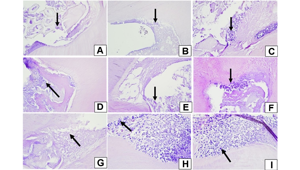
The histopathological identification of OLCs on days 7, 14 and 21 is shown in Figure 1. The arrows indicate the OLCs observed in each group.
Significant differences in the OLC count were observed between both groups receiving CH with the added anchovy nano-extract at 1:1 and 1:2 ratios and the CH-only group (p < 0.05). The addition of the anchovy nano-extract to CH at a 1:1 ratio significantly increased the OLC count as compared to the CH-only group on days 7, 14 and 21 (p = 0.000 for all). Similarly, the 1:2 ratio significantly increased the OLC count as compared to the CH-only group on day 7 (p = 0.001), day 14 (p = 0.000) and day 21 (p = 0.000). A significant difference in the OLC count between the 1:1 and 1:2 ratios was observed on day 14 (p = 0.034) (Table 2).
No significant change in the OLC count throughout the whole observation period was observed within the control group, which received CH only (p > 0.05). In group 2, a significant increase in the OLC count was observed on day 21 as compared to day 7 (p = 0.014) and day 14 (p = 0.023). In group 3, a significant increase in the OLC count was observed on days 14 (p = 0.016) and 21 (p = 0.008) as compared to day 7, while no significant difference was observed between days 14 and 21 (p = 0.934) (Table 3).
Alkaline phosphatase expression
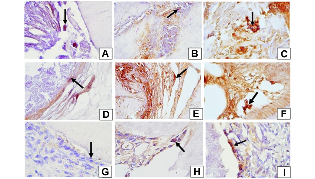
The immunohistochemical detection of ALP on days 7, 14 and 21 is shown in Figure 2. The arrows indicate ALP expression in macrophage cells in each group.
Significant differences in ALP expression were observed between both groups receiving CH with the added anchovy nano-extract at 1:1 and 1:2 ratios and the CH-only group (p < 0.05). The addition of the anchovy nano-extract to CH at a 1:1 ratio significantly increased ALP expression as compared to the CH-only group on day 7 (p = 0.008), day 14 (p = 0.000) and day 21 (p = 0.000). Similarly, the 1:2 ratio significantly increased ALP expression as compared to the CH-only group on day 7 (p = 0.001), day 14 (p = 0.001) and day 21 (p = 0.000). No significant difference in ALP expression was observed between the 1:1 and 1:2 ratios on any of the observation days (Table 2).
No significant difference in ALP expression throughout the whole observation period was observed within the control group, which received CH only (p > 0.05). In group 2, a significant increase in ALP expression was observed on day 21 as compared to day 7 (p = 0.031) and day 14 (p = 0.023). Also in group 3, a significant increase in ALP expression was observed on day 21 as compared to day 7 (p = 0.008) and day 14 (p = 0.009) (Table 3).
Discussion
In this study, the rats were subjected to mechanical pulp injury in the form of perforation to stimulate the formation of reparative dentin.29 Rats were selected, as the structure of their teeth resembles that of human teeth. Several previous studies have used rat maxillary and mandibular molars as experimental models.26, 29 The formation of reparative dentin is the primary goal of vital pulp treatment following injury.30 Stem cells migrate from richly vascularized tissues to the injured area to facilitate tissue repair. In general, the repair process following pulp capping involves moderate inflammation, the recruitment and migration of stem or progenitor cells, cell proliferation, and subsequent differentiation. Stem cells play a crucial role in tissue regeneration.31 Various materials and methods have been investigated to optimize the proliferation and differentiation of mesenchymal stem cells (MSCs).32, 33, 34, 35 Exploring new materials and approaches to enhance the proliferation and differentiation of MSCs, including dental pulp stem cells, is important for supporting pulp regeneration.36, 37, 38, 39 Previous studies have demonstrated the positive effects of MSCs on tissue regeneration, including in medically compromised subjects.40, 41, 42, 43, 44 Odontoblast-like cells originate from dental pulp stem cells, which are activated by the cytokines released during the inflammatory process.30 However, excessive or prolonged inflammation, particularly in the presence of bacteria and their by-products such as lipopolysaccharides, may interfere with the proliferation and differentiation of these cells and consequently impair pulp regeneration.45, 46 Therefore, pulp capping materials should not only protect the injured pulp from bacterial contamination, but also support the cellular processes involved in pulp repair and regeneration.
This study evaluated ALP expression and the number of OLCs as markers involved in the formation of reparative dentin. The ability of the anchovy nano-extract to stimulate reparative dentin formation in the perforated pulp of Wistar rat molars may be attributed to its calcium and phosphorus content, which can contribute to the formation of calcium phosphate and hydroxyapatite. Hydroxyapatite plays an important role in supporting reparative dentin formation and maintaining the vitality of the injured pulp. Calcium hydroxide mixed with distilled water showed lower OLC count and ALP expression than the anchovy nano-extract treatment groups. Calcium hydroxide has a strongly alkaline pH.47 Its high alkalinity causes the formation of a necrotic region, known as the cauterization zone, shortly after contact with the pulp tissue, subsequently leading to inflammation in the adjacent tissue.48 Other limitations of CH as a pulp capping material include its high solubility and inadequate sealing ability, which may compromise the long-term integrity of reparative dentin.49
In this study, significant differences in OLC formation were observed between the CH–anchovy nano-extract 1:1 combination group and the CH–anchovy nano-extract 1:2 combination group between days 14 and 21. This finding may be attributed to the phosphorus content of anchovies, which may contribute to odontoblast cell proliferation. Anchovies contain phosphorus, which, when mixed with water, can form phosphate. Phosphate can react with calcium to form calcium phosphate compounds, which play an important role in the differentiation of stem cells into odontoblasts. The anchovy extract contains 76% calcium phosphate compounds in the form of hydroxyapatite.25 Calcium and phosphate ions are essential for the mineralization of the reparative dentin matrix. The results of a previous study indicated that the number of odontoblast cells increased with increasing concentrations of the anchovy extract.24 Calcium and phosphate support mineralization by promoting protein adsorption.50 This process is beneficial for dentinogenesis and provides a three-dimensional (3D) framework for pre-odontoblasts and odontoblasts, thereby supporting new dentin formation. During the early stage of MSC differentiation, Runt-related transcription factor 2 (Runx2) and bone morphogenetic protein-2 (BMP-2) expression is significantly increased, followed by the expression of ALP, osteopontin (OPN) and osteocalcin (OC) as later markers of mineralization.50
In this study, ALP expression increased among the treatment groups. Alkaline phosphatase is an enzyme that promotes mineralization, reported to be expressed after 14 and 21 days, with greater expression observed on day 21.51 These findings are consistent with our study. The anchovy nano-extract contains calcium and phosphate as bioactive components. Calcium and phosphate may influence the Wnt/β-catenin signaling pathway or activate the p38/MAPK signaling pathway, thereby promoting the differentiation of MSCs in the pulp and potentially increasing the expression of mineralization-related genes.52 The expression of ALP by pre-odontoblasts and odontoblasts indicates that these cells are undergoing the mineralization phase.
The nanoparticle size of the anchovy provides finely dispersed calcium and phosphate bioactive particles, which may result in a larger surface area and greater interaction with the pulp, and may also contribute to a dose-dependent effect. Microenvironmental factors, such as the degradation or dissolution of calcium and phosphate from the anchovy nano-extract, may also influence the results. In the in vivo environment, a series of biological events, including immune regulation and immune responses, may affect the observed outcomes. Further studies are needed to evaluate the interaction between the anchovy nano-extract and the inflammatory responses of the pulp.
Conclusions
Our study demonstrated that the application of CH and the anchovy nano-extract at both 1:1 and 1:2 ratios increased the number of OLCs and ALP expression in the dental pulp of rats. The higher 1:2 ratio resulted in faster OLC formation as compared to the 1:1 ratio. Both ratios increased ALP expression on day 21.
Ethics approval and consent to participate
Ethical approval for the study was obtained from the institutional ethics committee at the Faculty of Dental Medicine, Airlangga University, Surabaya, Indonesia (approval No. 835/HRECC.FODM/XI/2022).
Data availability
The datasets supporting the findings of the current study are all contained within the article.
Consent for publication
Not applicable.
Use of AI and AI-assisted technologies
Not applicable.