Abstract
Background. The problem of avulsed teeth often being brought to the clinic in a dry state remains unresolved in the literature. Before an avulsed tooth is replanted, the periodontal ligament (PDL) cells require an appropriate storage medium to preserve their viability and biological properties. Hank’s balanced salt solution (HBSS) is the reference medium recommended for this purpose in the literature.
Objectives. The aim of the present study was to compare the effects of the reference solution – HBSS – and injectable platelet-rich fibrin (I-PRF) on the differentiation of PDL fibroblasts by examining the differentiation markers associated with osteoblastic, osteoclastic, fibroblastic, and cementoblastic lineages.
Material and methods. Twenty freshly extracted third molar teeth were used for cell isolation. After being left to dry for 30 min, the teeth were immersed for 30 min at room temperature in HBSS (n = 7) or I-PRF (n = 7). For the positive control group (n = 3), cells were isolated immediately after extraction, whereas for the negative control group (n = 3), cells were isolated after 30 min of dry time. Viable cells were counted, and immunofluorescence staining was performed to assess cell differentiation, using antibodies against Runt-related transcription factor 2 (RUNX2), receptor activator of nuclear factor kappa-B ligand (RANKL), collagen type I (COL1), and cementum attachment protein (CAP). Integrated density (IntDen) was measured using the ImageJ software.
Results. There were no statistically significant differences between the HBSS and I-PRF groups in the expression of RUNX2, RANKL or CAP. The I-PRF group showed significantly higher expression of COL1 (p < 0.01).
Conclusions. Injectable PRF may contribute to the regeneration of PDL cells when used as a rejuvenating medium before replantation following an extraoral dry period.
Keywords: replantation, cell differentiation, tooth avulsion, collagen type I, platelet-rich fibrin
Introduction
Replantation is often the primary treatment for avulsed teeth, although its success is significantly influenced by the timing of intervention and the immediate actions taken.1, 2 A key factor for a favorable outcome is the viability and regenerative potential of the periodontal ligament (PDL), as the survival of a replanted tooth largely depends on the integrity of its PDL. However, the viability of PDL cannot be directly assessed by dental professionals through a clinical examination alone.3
The duration that a tooth remains outside the oral cavity, either in a dry state or in a storage medium, critically affects the viability of PDL cells. Studies have shown that PDL cells generally become non-viable after being maintained in a dry state for more than 30 min.4 However, evidence suggests that permanent teeth with closed apices may still be replanted when the extraoral dry time is less than 60 min, although they may eventually develop ankylosis or replacement resorption.5
The International Association for Dental Traumatology (IADT) guidelines classify PDL cells as “may be viable but compromised” in avulsed permanent teeth with closed apices that have been stored in media such as milk, Hank’s balanced salt solution (HBSS), saliva, or saline for less than 60 min of extraoral dry time.5 When an avulsed tooth is brought to the clinic by a patient, parent, guardian, or other caregiver in a dry condition, there is a concern that the heterogeneous PDL cell populations remaining on the root surface may differentiate into osteoblasts, osteoclasts or cementoblasts following replantation.6 Root resorption is closely related to the nature of these heterogeneous PDL cell populations.7, 8
Studies have shown that immersing an avulsed tooth in a nutrient-rich medium before replantation can rejuvenate the surviving cells, reduce the inflammatory response and decrease the risk of root resorption.9 Hank’s balanced salt solution has been commonly used by dental professionals because of its ability to support the recovery of compromised PDL cells after extended dry periods, unlike milk or saline.10
Blood derivatives, such as platelet-rich plasma (PRP) and platelet-rich fibrin (PRF), are rich sources of growth factors that play important roles in tissue repair, making them valuable in dental treatment. Researchers have investigated the antimicrobial and anti-inflammatory effects of pure platelet-rich plasma (P-PRP) and leukocyte platelet-rich plasma (L-PRP) in periodontal treatment.11 Leukocyte platelet-rich fibrin promotes angiogenesis, chemotaxis, mitosis, and cell proliferation.12, 13 Titanium-prepared platelet-rich fibrin (T-PRF), which incorporates titanium particles, has also gained attention in regenerative medicine.14, 15 These findings highlight the potential of platelet concentrates to both combat infection and promote tissue regeneration, suggesting their potential for more effective treatment.11 Similarly, recent studies on tooth replantation have indicated that platelet concentrates may support tissue regeneration.3, 16, 17, 18 An important advantage of these materials is that they can be obtained from the patients themselves. Recent modifications to the centrifugation protocols used for PRF preparation have led to the development of injectable platelet-rich fibrin (I-PRF). It is a flowable platelet concentrate that can release growth factors for up to 14 days and contains approx. 1.5 times more platelets than whole blood.19 These characteristics suggest that I-PRF may be a practical storage medium for avulsed teeth, as it can be obtained from the patient’s own blood and may serve as a rejuvenating medium to help maintain the viability of PDL cells.
There are many studies concerning the effects of storage solutions on the prognosis of replantation. However, no pretreatment of the root surface that is effective in restoring the depleted cellular components of PDL before replantation has yet been suggested.5 Although studies have reported on the growth factors released from I-PRF and their effects on fibroblast and osteoblast behavior,20, 21 no study has examined the differentiation mechanisms of PDL cells from avulsed teeth in response to I-PRF.
The present study aimed to improve the prognosis of replanted avulsed teeth under the care of dental professionals. The avulsion treatment method proposed in this study seeks to provide an effective pretreatment approach for avulsed teeth that arrive at a dental clinic in a dry state. Therefore, we tested a novel procedure for treating avulsed teeth by comparing I-PRF and HBSS as pretreatment media. Since PDL cell differentiation can be assessed by the expression of specific markers associated with particular cell lineages, Runt-related transcription factor 2 (RUNX2), receptor activator of nuclear factor kappa-B ligand (RANKL), collagen type I (COL1), and cementum attachment protein (CAP) were used to assess differentiation toward osteoblastic, osteoclastic, fibroblastic, and cementoblastic lineages, respectively.
Material and methods
The study was conducted in accordance with the principles outlined in the 2013 revision of the Declaration of Helsinki. Participant recruitment and selection procedures were approved by the Ethics Committee for Clinical Research at the Faculty of Medicine, Kütahya Dumlupınar University, Turkey (approval date: August 16, 2017; approval No. 2017-10/3). All participants were informed about the study and voluntarily provided written informed consent before participation.
Sample preparation
Twenty third molars were prophylactically extracted from systemically healthy patients aged 18–35 years, and the freshly extracted teeth were immediately used for PDL fibroblast isolation. The teeth had no caries, periapical infection, restorations, periodontal disease, or hypoplasia. Patients were excluded from the study if they were taking anticoagulants, antiplatelet medications or antiresorptive drugs (such as bisphosphonates), had blood or bone metabolism disorders, or were pregnant or lactating.
Four 9-milliliter samples of peripheral venous blood were collected from each participant into non-coated tubes without anticoagulants (Vacusera; Disera, Izmir, Turkey) to obtain I-PRF. The white-capped polyethylene terephthalate tubes were centrifuged using a centrifuge device (PRF Duo Quattro; Process for PRF, Nice, France) at 1,300 rpm for 3 min, with modifications to previously described protocols.22 The rotor angulation of the device was 40°, with a relative centrifugal force (RCF) of 119 g at the minimum (RCFmin), 208 at the maximum (RCFmax), and 145 at the clot (RCFclot). Injectable PRF was then collected from the tubes using a sterile syringe, taking care to avoid contamination with red blood cells (RBCs).
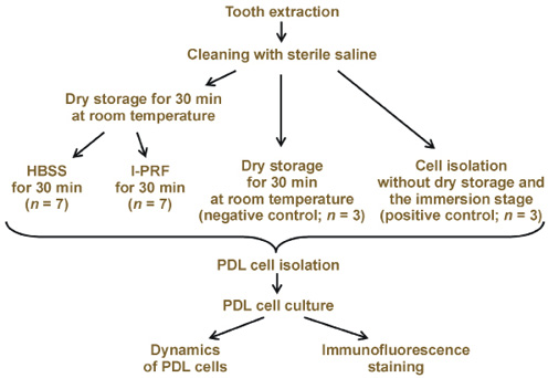
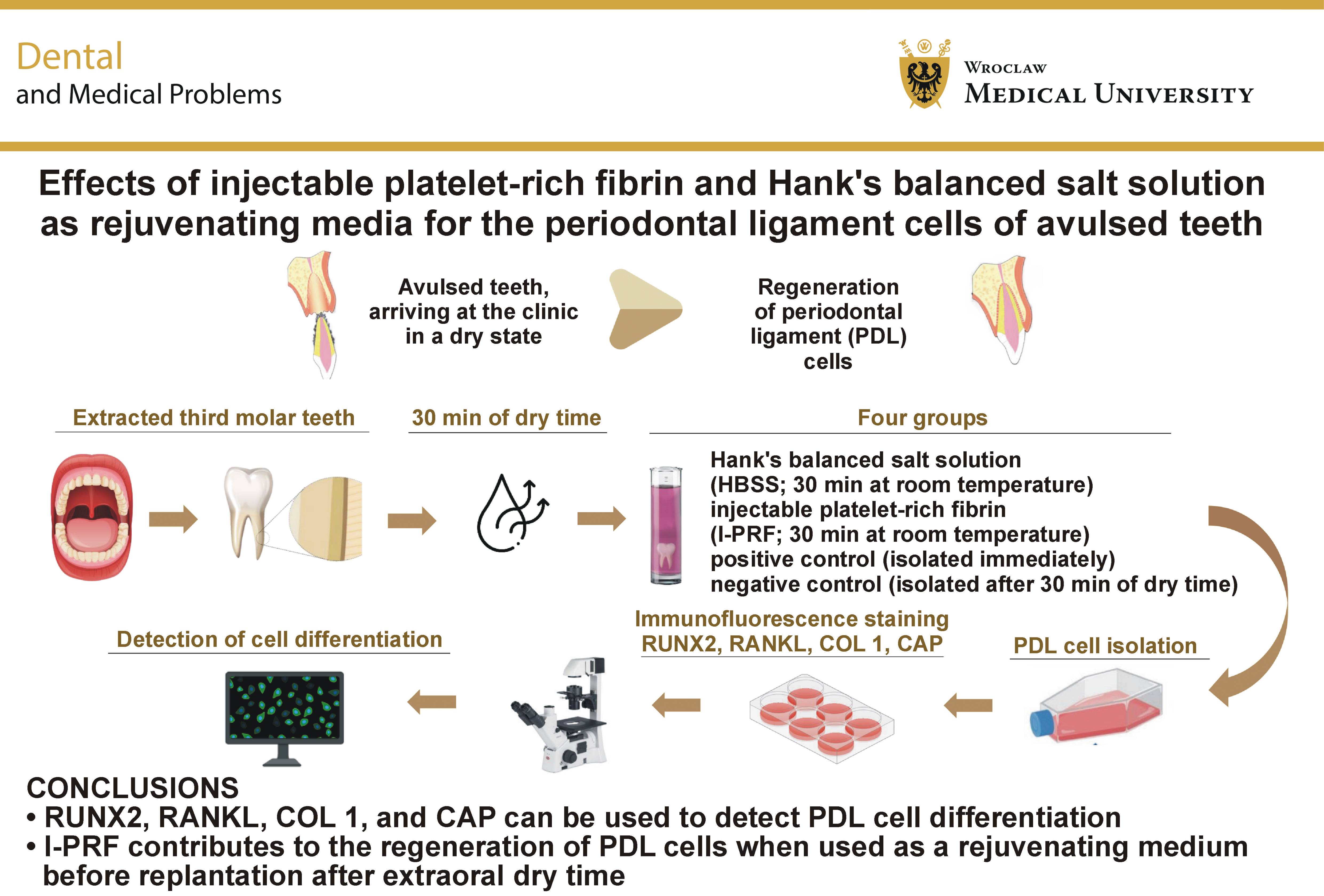
The extracted third molar teeth were cleaned with sterile saline, and all teeth except those in the positive control group were stored dry for 30 min at room temperature. For the negative control group, 3 teeth (n = 3) were used for PDL cell isolation after a 30-minute dry period. For the experimental groups (n = 7 teeth per group), the teeth were immersed in either HBSS (Capricorn Scientific, Ebsdorfergrund, Germany) or the I-PRF obtained from the blood of the respective patients for 30 min at room temperature following the 30-minute dry period. For the positive control group, 3 teeth (n = 3) were used immediately after extraction for PDL cell isolation, without a dry period or the immersion stage. Figure 1 outlines all stages of the study.
Cell isolation
Following the storage period, the teeth were thoroughly rinsed with phosphate-buffered saline (PBS) (Capricorn Scientific). The PDL tissue was carefully removed from the middle third of the root surface, using a scalpel blade. The collected tissue was placed in Eppendorf tubes containing 1 mL of Dulbecco’s Modified Eagle’s Medium/Nutrient Mixture F-12 Ham (DMEM/F-12; Wisent Bioproducts, Saint-Jean-Baptiste, Canada), supplemented with 10% fetal bovine serum (FBS) (Capricorn Scientific) and 1% penicillin-streptomycin-amphotericin B solution (Capricorn Scientific). The tissue samples were then dissected into small fragments using sterile scissors and centrifuged at 500 rpm for 1 min. The resulting pellet was resuspended in 1 mL of a solution containing 2 mg/mL collagenase type I and 2 mg/mL dispase (Roche, Mannheim, Germany) and incubated in a 37°C water bath for 30–45 min. The released cells were collected by centrifugation at 111 × g for 2 min and the resulting cell pellet was transferred to a T25 tissue culture flask. The culture flasks were maintained at 37°C in a humidified atmosphere containing 5% CO2 and 95% air. The culture medium was replaced every 3 days until the cells reached full confluence. The cells were detached from the flask surface using 0.05% trypsin-ethylenediaminetetraacetic acid (EDTA) in Dulbecco’s phosphate-buffered saline (DPBS) (Capricorn Scientific). Following detachment, the cells were stained with trypan blue and counted using a hemocytometer under an inverted microscope. The cells were seeded at a density of 5 × 10³ cells per T25 flask, and evaluated at the 3rd or 4th passage (P3 or P4).23
A total of 5,000 cells were seeded onto coverslips placed in a six-well plate. Once the cells reached confluence, the culture medium was aspirated and the cells were washed 3 times with PBS. The cells were then fixed with 3.5% paraformaldehyde (Merck, Darmstadt, Germany) for 30–35 min at room temperature, followed by 3 additional washes with PBS.24
Immunofluorescence staining
Three positive-control coverslips and 1 negative-control coverslip were selected for each tooth for each of the antibodies RUNX2, RANKL, COL1, and CAP. Following fixation, the coverslips were incubated with a blocking solution containing 0.1% Triton™ X-100 (BioShop Canada, Burlington, Canada) at 37°C for 30 min.
The primary antibodies against RUNX2 (polyclonal antibody; 5 µg/mL, ab114133; Abcam, Cambridge, UK), RANKL (polyclonal antibody; 1 µg/mL, ab9957; Abcam) and COL1 (polyclonal antibody; 1:500, ab34710; Abcam) were added to the respective groups and incubated overnight at 4°C. The CAP primary antibody (monoclonal antibody; 1:50, sc-53947; Santa Cruz Biotechnology Inc., Dallas, USA) was added and incubated at room temperature for 1 h. All samples were then rinsed 3 times with PBS. For the RUNX2, RANKL and COL1 treatment, a fluorescein isothiocyanate (FITC)-conjugated secondary antibody (1:50, SA00003-2; Proteintech Group Inc., Rosemont, USA) was applied at room temperature for 1 h. For the CAP treatment, an FITC-conjugated secondary antibody (1:50, SA00003-1; Proteintech Group Inc.) was applied at room temperature for 1 h. Following rinsing with PBS, the cell nuclei were stained by incubating the cells with 4’,6-diamidino-2-phenylindole (DAPI) (FluorLast™ with DAPI; BioVision Inc., Milpitas, USA) at room temperature for 5 min. Control samples were processed without the addition of primary antibodies.
The stained samples were examined using a fluorescence microscope (Nikon ECLIPSE Ci-E, Nikon Instruments Inc., New York, USA). Images were acquired using a 5.0-megapixel charge-coupled device (CCD) camera (DS-Fi1c; Nikon Instruments Inc., Melville, USA). Image acquisition was performed using the NIS-Elements software, v. 4.10 (Nikon Instruments Inc., Melville, USA). The images were acquired at a resolution of 1,280 × 960 pixels with a bit depth of 24 bits. For fluorescence imaging, FITC (excitation filter: 480/30; barrier filter: 535/45) and DAPI (excitation filter: 375/28; barrier filter: 460/60) filter sets were used, with a filter diameter of 25 mm for both.
Image analysis
Image analysis was performed on the photographs acquired at a standard magnification of ×40. Fluorescence intensity was assessed using the ImageJ software, v. 1.51v (https://imagej.net/ij). Integrated density (IntDen) was used to quantify the total fluorescence intensity within the selected area, and was subsequently normalized to the cell count. For each experimental condition, a minimum of 3 slides were prepared, and fluorescence intensity was quantitatively assessed in the acquired microscopic images. The presented images are representative of at least 3 independent experiments.
Statistical analysis
Statistical analysis was performed using IBM SPSS Statistics for Windows, v. 20.0 (IBM Corp., Armonk, USA). Descriptive statistics were calculated as minimum–maximum values (min–max), and the Kolmogorov–Smirnov test was used to assess the normality of data distribution. The one-way analysis of variance (ANOVA), followed by Tukey’s post hoc test, was performed for comparisons among the groups. A p-value <0.05 was considered statistically significant.
Results
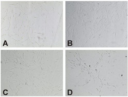
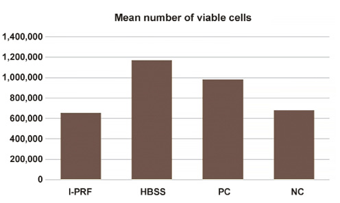
The cells derived from the digested tissue exhibited consistent adhesion to plastic surfaces across all experimental groups, and displayed similar growth patterns. No discernible differences were observed among the groups in terms of the ability of cells to adhere to plastic surfaces (Figure 2). Consequently, the cells were successfully maintained through successive passages, with comparable mean cell viability observed across all groups (p > 0.05) (Figure 3).
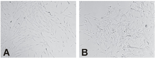
The microscopic images of PDL fibroblasts were captured following exposure to different rejuvenating media, including HBSS and I-PRF. In all experimental groups, PDL fibroblasts demonstrated the ability to form cell colonies from a single cell, indicating their clonogenic potential. The morphological evaluation of the P3 cells revealed that the cells from the I-PRF-treated group retained their typical fibroblastic, spindle-shaped morphology. In contrast, the cells from the HBSS-treated group exhibited early signs of senescence, and the typical fibroblastic morphology was no longer evident (Figure 4).
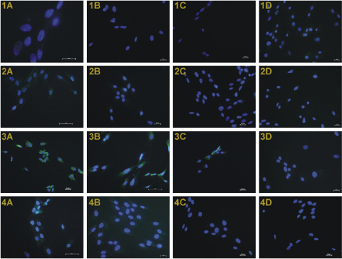
The RUNX2 antibody was used to assess PDL cell differentiation toward the osteoblastic lineage, RANKL to assess differentiation toward the osteoclastic lineage, COL1 to assess differentiation toward the fibroblastic lineage, and CAP to assess differentiation toward the cementoblastic lineage. All markers were evaluated in all experimental groups (Figure 5).
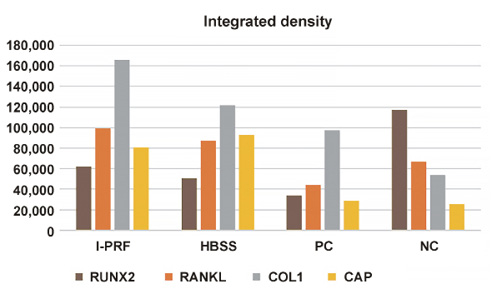
RUNX2 expression was observed as bright spots within the nucleus. The IntDen values in the I-PRF, HBSS and positive control groups differed significantly from that of the negative control group. The negative control group exhibited a higher IntDen value than the other groups (p < 0.01) (Figure 5).
RANKL expression was significantly higher in the I-PRF and HBSS groups than in both control groups (p < 0.01). Although RANKL expression was slightly lower in the I-PRF group than in the HBSS group, no statistically significant difference was observed between these 2 groups (Figure 5).
COL1 expression was significantly higher in the I-PRF group than in both the positive and negative control groups (p < 0.01). The IntDen value for COL1 expression in the HBSS group was intermediate between those of the I-PRF and positive control groups (Figure 5).
CAP expression was deficient in both control groups, whereas mild expression was observed in both the I-PRF and HBSS groups (p < 0.01) (Figure 5).
Fluorescence microscope images for all groups with antibody expression are presented in Figure 6.
Discussion
Since the awareness of the need to immediately replant avulsed teeth or store them in an appropriate liquid medium is still limited in the general population, avulsed teeth are frequently brought to the clinic wrapped in napkins. Although dry teeth can be replanted by the clinician, alternative treatment approaches should be investigated to improve the prognosis. Optimal recovery following dental trauma or regenerative periodontal treatment depends on the complete regeneration of PDL and the establishment of a balanced connection between the cementum and the alveolar bone. In cellular regeneration following avulsion, it is essential to restore the depleted cellular metabolites and maintain cell viability.25 Therefore, we investigated a novel approach for the rejuvenation of avulsed teeth by comparing I-PRF with HBSS. In the present study, the early-passage primary PDL cultures obtained from extracted human permanent teeth were used to mimic the cellular conditions of an avulsed tooth.
Although the highest cell number was observed in the HBSS group, no statistically significant difference was found among the groups. These findings demonstrate the successful cultivation of a substantial proportion of viable precursor cells derived from the PDL of representative avulsed teeth. Moreover, the data indicate that the PDL cells obtained from teeth that had not been stored in a rejuvenating medium and had an extraoral dry time of 30 min remained viable. As these cells were classified as “may be viable but compromised” with regard to their regenerative potential, the present study investigated whether I-PRF could provide a greater rejuvenating effect than HBSS. The literature suggested that I-PRF may be superior to HBSS as a rejuvenating medium.5
The periodontal tissue response to tooth avulsion involves mechanisms related to cell viability, regeneration and resorption, which are influenced by cellular identity and differentiation. The rationale for using HBSS as a rejuvenating agent in the present study was its proposed ability to maintain and replenish the depleted cellular components of PDL cells.10 Hank’s balanced salt solution has been shown to maintain cell viability and reduce root resorption when used as a storage medium prior to replantation.3 In a previous study, HBSS enhanced osteogenic differentiation in PDL fibroblasts by upregulating RUNX2 expression, whereas milk preserved cell viability and cellular identity.23 However, milk is not known to restore the depleted cellular metabolites.26 Given the distinction between storage and rejuvenating media, milk was not used as a rejuvenating medium in the present study. Further research into its potential to restore the depleted cellular functions in avulsed teeth would be of interest.
Ideal healing would involve the regeneration of collagen fibers capable of attaching to the newly formed cementum and bone.27 Collagen type I is the main component of PDL fibers, and its expression can be used as an indicator of PDL tissue regeneration. A reimplantation study demonstrated that PRF promoted the proliferation of human periodontal ligament stem cells (PDLSCs) and inhibited their osteoblastic differentiation by upregulating COL1 mRNA expression.16 Consistent with the effects of PRF, the present study demonstrated that I-PRF helped PDL cells maintain their fibroblastic identity, as evidenced by significantly higher COL1 expression compared with the positive control.
The present study suggests that I-PRF helps maintain the fibroblastic phenotype of PDL cells, as evidenced by the strong expression of COL1. The concomitant high levels of COL1 and RANKL expression in the I-PRF group may indicate a balance between bone resorption and regeneration. However, the precise mechanisms underlying PDL cell-induced osteoclastogenesis remain unclear. The immersion of avulsed teeth in HBSS has been reported to induce osteoclastogenesis in PDL cells, resulting in the formation of osteoclast-like cells independently of RANKL.28 Previous research also demonstrated stable RANKL expression in HBSS-treated teeth despite prolonged storage periods.23 When PDL cells are co-cultured with CD14+ monocytes, osteoclast formation can occur, at least in part, through RANKL-mediated mechanisms.23, 29 In the present study, RANKL expression was higher in the PDL cells treated with I-PRF than in the control groups. Given that osteoclast activity is associated with inflammatory root resorption, further investigation of osteoclastogenesis, inflammatory cytokines, and the effects of I-PRF on the RANKL/RANK (receptor activator of nuclear factor kappa-B)/OPG (osteoprotegerin) system may provide important insights into the mechanisms underlying periodontal healing following avulsion.
Injured periodontal tissues can regenerate through the action of proteins such as cementum-derived growth factor (CDGF), CAP and cementum protein-1 (CEMP1), which promote cell adhesion and differentiation, and may contribute to the formation of new cementum.30 Platelet-rich fibrin has been shown to enhance human PDLSC proliferation while inhibiting osteoblastic differentiation by increasing the mRNA expression levels of COL1 and CEMP1.16 Furthermore, studies investigating PRP have demonstrated that regeneration is enhanced when stem cells, fibrin glue and PRP are used in combination.31 To our knowledge, the present study is the first to evaluate the effects of I-PRF on PDL cells in relation to cementogenesis. The significant difference in CAP expression between the experimental and control groups is consistent with previous findings, and suggests that healing following tooth avulsion may involve a regenerative response that helps minimize the resorption of the damaged root surface.32, 33
A key advantage of I-PRF is that it can be prepared at a low centrifugation speed (such as 700 rpm, 60 × g in original protocols or 1,300 rpm in modified protocols) within a short period (3 min). In contrast, the preparation of other platelet concentrates, such as PRF, requires more time, potentially increasing the extraoral dry time of avulsed teeth. The lower centrifugation speed also increases the proportion of platelets and leukocytes recovered, which may enhance tissue regeneration and promote the activity of PDL fibroblasts in the repair process.22, 34
Limitations
Although the small sample size is a major limitation of the present study, another limitation is that the effects of centrifugation force and duration, which may result in different PRF-based matrices, were not evaluated in relation to osteoclastogenesis. Although numerous studies have investigated the use of I-PRF in cells of heterogeneous origin, the inability to assess individual cellular defense mechanisms, such as macrophage (M) differentiation, may also be considered a limitation of the present study. Studies evaluating macrophage polarization toward the M1 or M2 phenotypes are needed to comprehensively assess the effects of I-PRF on the RANKL pathway and osteoclastogenesis. Further in vitro and in vivo studies are warranted to elucidate the molecular mechanisms underlying the effects of I-PRF on osteoclastogenesis and to determine whether different PRF isolation protocols influence this process. In addition, before I-PRF can be used clinically as a rejuvenating medium, its platelet content should be characterized through biochemical and cellular analyses, and the platelet concentrates obtained from individual patients should be quantitatively evaluated to determine the biological activity and composition of I-PRF.
Conclusions
The present study investigated the use of I-PRF as a rejuvenating medium following tooth avulsion. One of the most common challenges encountered in avulsion cases is the lack of public awareness regarding the need to store avulsed tooth in an appropriate liquid medium; consequently, families frequently present to the clinic with the tooth in a dry condition. In conclusion, I-PRF may be considered as a regenerative medium for avulsed teeth presenting to the clinic after an extraoral dry period. Further studies involving different extraoral dry times and larger sample sizes are warranted to confirm these findings.
Ethics approval and consent to participate
The study was conducted in accordance with the principles outlined in the 2013 revision of the Declaration of Helsinki. Participant recruitment and selection procedures were approved by the Ethics Committee for Clinical Research at the Faculty of Medicine, Kütahya Dumlupınar University, Turkey (approval date: August 16, 2017; approval No. 2017-10/3). All participants were informed about the study and voluntarily provided written informed consent before participation.
Data availability
The datasets supporting the findings of the current study are available from the corresponding author on reasonable request.
Consent for publication
Not applicable.
Use of AI and AI-assisted technologies
Generative AI technology was used solely to assist in language editing and proofreading of the manuscript. ChatGPT 3.5 was used.