



Abstract
Background. Clear aligner treatment (CAT) may induce masticatory muscle tenderness and trigger temporomandibular disorders (TMD). Considering that CAT may interfere with the masticatory system, it is important to understand the relationship between CAT and occlusal changes.
Objectives. This study aimed to analyze occlusal changes during CAT and assess the influence of post-orthodontic occlusion on masticatory performance. A secondary objective was to determine whether the case complexity, the facial biotype and malocclusion were associated with occlusal alterations, masticatory performance, or the onset of TMD signs and symptoms during CAT.
Material and methods. Forty-two individuals who underwent and completed CAT were evaluated at 3 time points: before treatment (T0); at the end of treatment (T1); and 3 months after treatment, during which time clear aligners (CAs) were used only at night (T2). Temporomandibular disorders were assessed using Axis I of the Diagnostic Criteria for Temporomandibular Disorders (DC/TMD) at T0, T1 and T2. Masticatory performance was evaluated at T1 and T2 using a two-colored chewing gum test and colorimetric analysis. Occlusal area and anterior and posterior occlusal contacts were evaluated digitally. Non-parametric statistical tests were used, with the significance level set at p < 0.05.
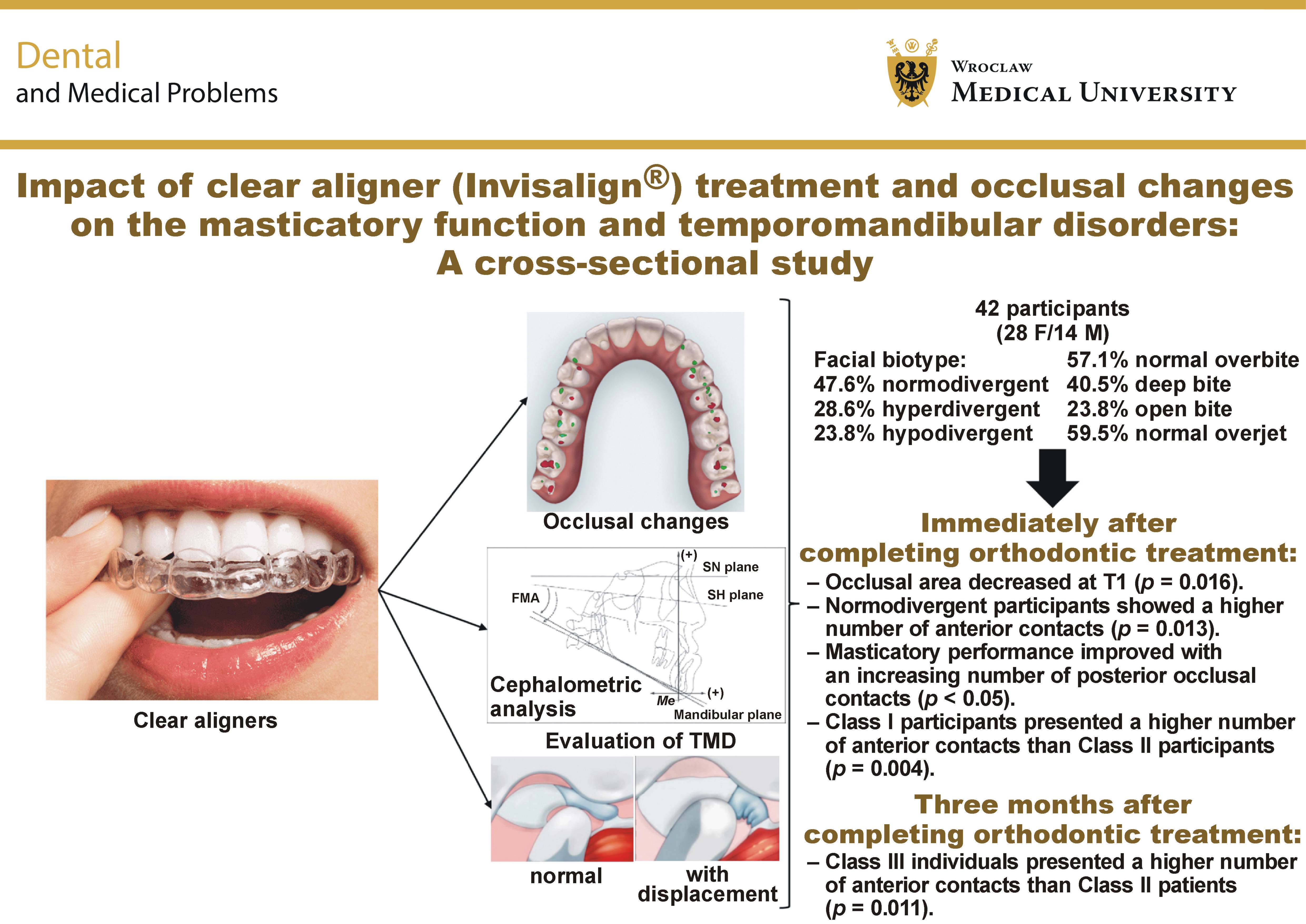
Results. The occlusal area decreased significantly from T0 to T1 (p = 0.016). At T1, normodivergent individuals had significantly more anterior contacts than hyperdivergent patients (p = 0.013). Masticatory performance increased with the number of posterior occlusal contacts (p < 0.05), and individuals with Class I molar relationship had more anterior contacts than Class II patients (p = 0.004). At T2, Class III individuals showed higher values than Class II patients (p = 0.011).
Conclusions. The number of occlusal contacts and occlusal contact area decreased during treatment. Nighttime use of CAs slightly enhanced occlusal contact recovery. Complex cases presented more posterior contacts at T0; simple cases presented a greater occlusal area at the end of CAT. Class I individuals presented more anterior contacts at T0. These movements increased the anterior contacts in Class III molar cases at the end of treatment and 3 months later. No significant differences in masticatory performance were found according to the molar relationship, the facial biotype or the case complexity; no TMD signs and symptoms were found during or after CAT.
Keywords: TMD, masticatory function, clear aligners, masticatory performance, occlusal contacts
Introduction
Within the twenty-first century’s digital era, the merging of new technology gave rise to modern orthodontic treatments (OTs). Aside from the conventional OT with fixed appliances, options such as clear aligners (CAs) have emerged.1 Orthodontic treatment ensures the proper alignment of teeth and improves the occlusal and jaw relationships.2 For the orthodontist, the esthetic characteristics of malocclusions are an important factor in determining the demand for OT3, 4 and achieving a stable and functional occlusion. Appropriate masticatory function at the end of OT is another critical goal that cannot be overlooked.
Equally distributed contacts that are softer anteriorly and stronger posteriorly are among the occlusal goals. In order to achieve proper occlusion at the end of OT, the clinician needs to create heavy contact on the splint during treatment.5 Respecting the abovementioned objectives is a key factor in ensuring the long-term stability of OT. Orthodontic pain and occlusal changes during treatment are the main factors influencing how the masticatory muscles adapt to OT.6 During treatment, the CA covers the entire occlusal surfaces of the teeth, promoting a bite-block effect for approx. 22 h per day.7, 8 This duration does not allow the occlusion to settle as intended, creating artificial interference within the occlusal space.9
The failure to obtain stable and solid occlusal contacts during treatment has been identified as the main limitation of CA treatment (CAT).8 This lack of posterior contacts can be resolved during the settling of the occlusion. Previous studies have documented that complete settling requires time and that the number of occlusal contacts increases during the post-treatment period.10 However, occlusal disharmonies are present during CAT, and its relationship with potential issues such as temporomandibular disorders (TMD), periodontal disease, cervical abfraction, or less effective mastication remains controversial.11, 12
Temporomandibular disorders are an umbrella term that involves dysfunction and pain in the masticatory muscles and/or temporomandibular joint (TMJ), causing damage to the masticatory function.13, 14 The potential impact of OT on the onset and evolution of TMD has been controversially discussed over the years.15 Several studies suggest that OT increases the likelihood of the onset of symptoms and signs of TMD.16, 17 In contrast, others indicate positive effects on the resolution of TMD or, most crucially, on reducing the patient’s risk of their development.18
Correction of malocclusions, often accompanied by mechanical transformation as well as skeletal changes, may alter the muscular and articular stability of the craniomandibular structures. Clear aligner interference has been reported to increase the frequency of wake-time tooth clenching episodes, trigger masticatory muscle tenderness, produce wear facets on aligner trays, and even induce the development of TMD.19, 20 Individuals undergoing CAT may have temporary TMD due to repetitive clenching on their trays to relieve orthodontic pain.21 One potential drawback is that wake-time tooth clenching could contribute to masticatory muscle pain and TMD.21
It has been well established that masticatory performance is related to occlusion and occlusal contact areas.6 Individuals with malocclusion present lower masticatory performance than those with normal occlusion, as the occlusal areas of contact and near-contact are positively correlated with masticatory function. A critical issue of functional connectivity in the masticatory system is the influence of occlusal conditions on masticatory function.6
Considering that CAT may interfere with the masticatory structures, it is essential to understand the correlation between OT-related occlusal changes and their influence on mandibular function. Thus, this study aimed to analyze occlusal changes during CAT and assess the influence of post-orthodontic occlusion on masticatory performance (primary outcome); moreover, it assessed whether the complexity of CAT, the facial biotype, and the malocclusion may be related to possible alterations and the onset of TMD signs and symptoms during the use of CAs (secondary outcomes). The positive hypothesis is that CAT achieves the desired result without causing any TMD; the null hypothesis is that even if the result was reached, TMD were detected. The clinical significance of the study lies in understanding the functional outcomes of CA (Invisalign®) treatment by evaluating its effects on (1) chewing efficiency; (2) occlusal changes, to determine whether it achieved a stable occlusion, promoting optimal function; (3) TMD, which may be influenced by changes in occlusion and muscle activity; and (4) long-term stability and patient comfort. These findings may guide clinical practice and treatment protocols, as well as help dentists and, specifically, orthodontists tailor treatments to prevent masticatory dysfunction and TMD-related complications. Its relevance is supported by the limited number of studies in the literature that have investigated CAs while evaluating 4 variables: occlusal changes; the facial biotype; chewing performance; and their possible correlation with TMD.
Material and methods
This quantitative, comparative, and observational longitudinal cohort study was conducted in accordance with the recommendations of the Declaration of Helsinki (2024) and the World Health Organization (WHO)22 regarding research involving human participants and followed the STROBE (Strengthening the Reporting of Observational Studies in Epidemiology) guidelines.23 Ethics approval was obtained from the Ethics Committee of the University Institute of Health Sciences (IUCS), Gandra, Portugal (approval No. 26/CE-IUCS/2020). All participants provided informed consent before the commencement of the study.
Study sample
A non-probability convenience sample was recruited from individuals aged between 16 and 45 years with complete permanent dentition (excluding third molars) undergoing CAT. The individuals were recruited from 2 independent private orthodontic clinics, Clínica Médico Dentária de São João da Madeira (São João da Madeira, Portugal) and Clínica de Medicina Dentária Dr. Manuel Neves (Porto, Portugal), from October 2019 to December 2022. All individuals were treated under the supervision of a specialist in both Orthodontics and Pediatric Dentistry who was also an Invisalign® Diamond Provider. The study comprised a sample of 42 individuals (28 female and 14 male) undergoing CAT; the sample was limited due to practical constraints. The participants were evaluated at 3 different time points: before starting treatment (T0); at the end of treatment with CAs without misfits (T1); and 3 months after the end of treatment, during which time CAs were only used at night (T2). The patients were invited to participate in the study in order to evaluate occlusal changes during CAT and assess the influence of post-orthodontic occlusion on masticatory performance. All invited individuals agreed to participate in the proposed analysis.
Sample size calculation
Considering the study design, a prospective cohort study comparing pre-treatment and post-treatment outcomes, the following parameters were adopted: a significance level (α) of 0.05, power (1−β) of 80% (0.80), and effect sizes (Cohen’s d for the analysis of continuous variables) of 0.5 (medium effect) for occlusal changes, 0.4 (small-to-medium effect) for temporomandibular dysfunction, and 0.5 (medium effect) for chewing performance. The standard deviation (σ) was assumed to represent 10% variation. The following formula was used for the calculation of the sample size:
N = ((((Zα/2) + Zβ) × σ) / Δ)2
where:
Zα/2 = 1.96 (for 95% confidence);
Zβ = 0.84 (for 80% power);
σ = 10%; and
Δ = 5% (expected mean difference).
Assuming an effect size of d = 0.5, the sample size was calculated as follows: N = ((1.96 + 0.84)2 × 10²) / 52; N = 31.36. Therefore, the minimum sample size required for this study was 32 patients. Because of the elevated risk of dropout, 30% more patients were included, resulting in a total sample of 42 patients.
Eligibility criteria
Two professionals (VM and TP) conducted the evaluation of patients. The inclusion criteria were as follows: individuals who started and completed CAT; individuals whose masticatory performance was assessed at the end of treatment and 3 months after treatment; individuals who used nighttime aligners without maladjustments between the end of treatment and 3 months after treatment; and individuals with available and complete cephalometric analyses.
The exclusion criteria encompassed individuals who did not have complete dentition, patients whose occlusal records were incomplete, individuals presenting TMD signs or symptoms before OT, patients requiring mandibular advancement with the use of special wings (Invisalign®), those with cognitive or neurological changes or identified syndromes, individuals with a history of trauma and/or tumors of the head and neck or metabolic diseases affecting the joints and/or muscles, patients who were taking anti-inflammatory, analgesic or psychiatric medications, and individuals who suffered from dental pain or periodontal problems during the previous year.
Data collection for TMD diagnosis
The diagnosis of TMD was made before starting CAT (T0), after completing OT (T1), and 3 months after treatment (T2), in accordance with international guidelines using Axis I of the Diagnostic Criteria for Temporomandibular Disorders (DC/TMD).24 Before starting the study, a pilot study was conducted to assess intra- and inter-examiner reliability in 30 participants with the same characteristics as the primary study sample. These individuals were not included in the main study sample because the goal of the pilot study was the intra- and inter-examiner calibration of the professionals who would collect the diagnostic data on TMD after OT. Three examiners (VM, MPG, GVOF) independently performed the evaluation procedure on 2 occasions separated by a one-week interval, demonstrating excellent reliability, with an intraclass correlation coefficient (ICC) >0.90.
After appropriate calibration of the examiners who applied the DC/TMD, information on TMD signs and symptoms was collected at 3 time points (T0, T1 and T2), with the aligners removed during the assessment.
Data collection for masticatory performance
Masticatory function was evaluated using a masticatory performance test with two-colored chewing gum. Participants sat upright and chewed 2 pieces of gum (Hue-Check Gum®; Orophys GmbH, Muri bei Bern, Switzerland), one pink and one blue, for 20 chewing cycles.25 The operator counted the chewing cycles silently. The gums were initially moistened in water and bonded together with moderate force. The chewing gum was placed in the patient’s mouth, with the blue side facing the tongue. Participants were instructed to chew normally, and no further instructions were given. After 20 chewing cycles, the operator asked the patient to stop chewing, and the gums were removed from the oral cavity, placed in a new clear plastic bag, and subsequently flattened using a 1-mm thick plate. Both sides of the specimen were scanned immediately after collection to minimize enzymatic degradation, and the images were saved in JPEG format.
The composite images were then evaluated using a specifically developed program that is freely available (Viewgum software; dHAL Software, Kifissia, Greece). The software first transformed the images into the HSI color space and then semi-automatically calculated the hue value for each pixel in the segmented images. The hue variation (VOH) was the measure of mixing calculated by the software. The VOH is also considered a measure of masticatory performance because it has a logarithmic association with the number of chewing cycles.26 A high VOH results from poorly mixed colors due to deficient chewing, whereas adequate chewing leads to well-mixed colors and a low VOH. The hue variation was considered a measure of mixing, with values closer to 0 indicating better color mixing and, consequently, better masticatory performance. The Hue-Check Gum® method was used.27 Information was collected at T1 and T2 and recorded.
Digital measurements
After analyzing the clinical records, the characteristics of each participant were collected and evaluated, including gender and age at the beginning of CAT. Cephalometric tracing, overbite, overjet, and facial biotype were measured by 1 professional (VM). The facial biotype classification was performed using the Frankfort-mandibular plane angle (FMA). Facial biotypes were classified as hypodivergent with values equal to or below 22 degrees, normodivergent with values between 23 and 27 degrees, and hyperdivergent with values equal to or above 28 degrees.28 Intraoral photographs and images of the occlusal contacts planned in the ClinCheck® software (Align Technology, Tempe, USA) for CAT were collected for each participant.
Subsequently, intraoral photographs and images obtained using the intraoral scanner at T0, T1 and T2 were compared using the iTero® software (Align Technology). The files in PLY format obtained using the same software were downloaded and analyzed. The PLY file of each participant had the following characteristics selected in the iTero® software: “open Shell”, “arches combined (arches locked in bite relation)”, and “PLY (color)”. Cephalometric measurements, the number of anterior and posterior occlusal contacts (T0, T1 and T2) and occlusal areas were organized in a Microsoft Excel (Microsoft Corp., Redmond, USA) file. The collected data was later used for statistical analysis.
To obtain the area and the number of occlusal contacts at T0, T1 and T2, the files in PLY format were processed using the MeshLab® software (https://www.meshlab.net). The ClinCheck® software was used to quantify the number of occlusal contacts. Any opposing pair of maxillary and mandibular teeth with the same tooth number was counted as 1 occlusal contact, and the counting was performed on the lower arch. The maximum expected number of occlusal contacts in a 28-tooth dentition was 14. The maximum number of posterior occlusal contacts was 8, and the maximum number of anterior occlusal contacts was 6. Third molars were excluded from the occlusal area and contact measurements.
Orthodontic intervention
The individuals were instructed to use each CA for as many hours as possible (20–22 h/day), as instructed by the provider, and to remove the aligners only when eating and performing oral hygiene. The aligners were changed every 7 days from the beginning of treatment, as recommended by Align® protocols. Compliance with aligner use was verified with an aligner wear monitor based on fading compliance indicators and was confirmed by asking the patient about their use. Follow-up visits were conducted every 2 months. Additional aligners were required at the end of the study to serve as a retention method and were used only at night to improve the occlusal contacts.
For the present investigation, only 1 type of aligner system was used for all individuals to reduce confounding factors linked to the mechanical properties of the material. It has already been demonstrated that differences in thermoplastic material composition and thickness result in different flexibilities, force activation, and consequent stress-relaxation behavior.29 After completing the treatment, the additional aligners were used without mismatch between the upper and lower arches and were worn only at night.
Clinical assessment and complexity of the case
Based on the clinical data (clinical photographs, radiographs and dental casts), the complexity levels of cases receiving CAT were assessed independently by 2 professionals (VM and TP). In cases of discrepancy, a third researcher was consulted (TP). For this purpose, an online assessment tool, Invisalign® Doctor Site, was used.30 The orthodontic malocclusion traits were classified as requiring simple, moderate or complex correction based on Align® recommendations and the Invisalign® evaluation tool.31 According to the Invisalign® criteria, patients need to present only 1 complex problem for their case to be classified as complex. This classification of complexity considers different parameters, namely the type of dentition, the need for surgery, the amount of spacing and crowding, rotations, narrow arches, posterior crossbite, anteroposterior correction, anterior crossbite, anterior open bite, deep bite, and the need for extraction. Each of these parameters has sub-parameters, which were also evaluated.
Statistical analysis
Data was analyzed using the IBM SPSS Statistics for Windows software, v. 29 (IBM Corp., Armonk, USA). Descriptive statistics were calculated to estimate frequencies, percentages, means, medians, standard deviations, and minimum and maximum values. The Shapiro–Wilk test was used to assess the normality of the variables under study.
Due to the non-normal distribution of the data, Friedman’s two-way analysis of variance (ANOVA) was used to compare the occlusal contact areas and the number of anterior and posterior occlusal contacts among T0, T1 and T2. This is a non-parametric statistical test used to determine whether 3 related groups differ from one another. The comparison of masticatory performance between the end of the first series of aligners and 3 months after nighttime use was performed using the Wilcoxon signed-rank test (a non-parametric test used to compare 2 periods within the same sample to assess whether their population mean ranks differ). The contact area and the number of anterior and posterior occlusal contacts at T0, T1 and T2, as well as masticatory performance at T1 and T2, were compared among the different groups according to the facial biotype, the case complexity, the molar class, and the vertical bite pattern using the Kruskal–Wallis test (a non-parametrical test), followed by Dunn’s test with Bonferroni correction to determine which groups differed significantly from one another. The Bonferroni correction adjusts the alpha level to account for the number of pairwise comparisons performed. To evaluate the relationship between occlusal area, anterior and posterior contacts, and masticatory performance at T1, as well as the corresponding relationships at T2, Spearman’s correlation coefficient was used (a non-parametric measure of the association between 2 variables). The significance level was set at 0.05.
The effect size (Cohen’s d, with bias correction using Hedges’ method) was calculated to compare groups and examine the correlation between variables. A Cohen’s d of 0.0–0.2 was considered a small effect size; d of 0.5 was considered a medium effect size; and d ≥ 0.8 was considered a large effect size. Therefore, if the means of the 2 groups did not differ by more than 0.2 standard deviations, the difference was considered trivial, even if it was statistically significant.
Results
Sample characteristics
Participants were aged between 16 and 45 years (mean age of 22.19 ±10.51 years), with 28 females (66.7%) and 14 males (33.3%). None of the patients presented TMD signs or symptoms. Regarding the facial biotype, 20 participants (47.6%) had a normodivergent biotype, 12 (28.6%) were hyperdivergent and 10 (23.8%) were hypodivergent. Most participants had a normal overbite (57.1%) and a normal overjet (59.5%). The clinical assessment of the sample revealed 17 individuals (40.5%) with a deep bite, 15 (35.7%) with a normal bite, and 10 (23.8%) with an open bite. Concerning the complexity of the cases, 27 cases (64.3%) were of moderate complexity, 9 cases (21.4%) were considered complex, and 6 (14.3%) were simple.
Changes in occlusal contacts and areas throughout CAT
The changes in occlusal area and posterior and anterior contacts throughout CAT are presented in Table 1. The comparison of the occlusal area throughout treatment showed that the initial occlusal area (T0) decreased significantly by the end of CAT (T1) (p = 0.002), with a large effect size (d = 0.94).
Comparison of masticatory performance at the end of CAT and 3 months after nighttime use only
When comparing the median values of masticatory performance at the end of CAT (T1) and 3 months later, when CAs were used only at night (T2), a reduction in the median values was observed, indicating better masticatory performance. However, no statistically significant differences were found between the 2 assessment time points, and the effect size was considered small (d = 0.23) (Table 2).
Comparison of occlusal area, occlusal contacts and masticatory performance throughout CAT
Case complexity
At T1, individuals with simple cases presented higher occlusal area values than those with moderate or complex cases (p = 0.027). A statistically significant difference in occlusal area was found between moderate and simple cases (p = 0.023). Significant differences were also found in posterior contacts at T0, with complex cases showing lower median values than moderate and simple cases (p = 0.046) (Table 3).
Facial biotype
Table 4 compares occlusal area, anterior and posterior contacts, and masticatory performance according to the facial biotype of individuals undergoing CAT at the 3 assessment time points. Regarding anterior contacts, statistically significant differences were found at T1, with normodivergent individuals presenting a significantly higher number of contacts than hyperdivergent individuals (p = 0.013), with a large effect size (d = 1.45).
Molar class relationship
At T0, individuals with Class I molar relationship had significantly more anterior contacts than those with Classes II and III. Dunn’s test showed that these differences were significant between the Class I and Class II groups (p = 0.004). At T1, individuals with Class III molar relationship had a significantly higher number of anterior contacts than those with Class II molar relationships, with the effect size of d = 1.51 (p = 0.011). At T2, individuals with Class III molar relationship presented higher numbers of anterior contacts than those with Classes I and II. These differences were statistically significant between the Class I and Class II groups (p = 0.024) and between the Class II and Class III groups (p = 0.011) (Table 5).
Vertical bite trend
According to Table 6 and with regard to the vertical bite trend, no statistically significant differences were found in occlusal area, masticatory performance, or anterior and posterior contacts among the deep-, normal- and open-bite groups. Small effect sizes were observed for the occlusal area at T2 between deep and normal bites (d = 0.27) and normal and open bites (d = 0.14); for anterior contacts at T0 between deep and open bites (d = 0.24), at T1 between deep and normal bites (d = 0.00), and at T2 between deep and normal bites (d = 0.00); for posterior contacts at T0 between deep and open bites (d = 0.00), and at T2 between normal and open bites (d = 0.00); and for masticatory performance at T1 between normal and open bites (d = 0.00), and at T2 between deep and normal bites (d = 0.17).
Relationship between changes in occlusal area, occlusal contacts and masticatory performance
Table 7 shows the relationships between occlusal area and occlusal contacts with masticatory performance. The number of posterior contacts at T1 showed a weak, negative and statistically significant correlation with masticatory performance at T1 (r = −0.378, p < 0.05). Increases in occlusal area and in the number of anterior and posterior contacts were associated with lower masticatory performance values, which represented better masticatory performance.
Discussion
Esthetics is a key consideration for orthodontists. However, it is crucial to keep in mind that masticatory function and orthopedic principles should also be considered. Orthopedic stability in the masticatory structures should focus on reducing the risk factors associated with the development of TMD and promoting good masticatory function.32 Thus, awareness of adequate occlusion is important for OT success since the two are directly related. Also, occlusion is strongly connected with the health of the TMJ and masticatory muscles.33
A statistically significant decrease in occlusal area was recorded between T0 and T1. These results are in line with the study by Horton et al., which reported a similar occurrence in the interocclusal contact area after the use of an occlusal-covering appliance (Essix) compared with the use of a Hawley splint (without occlusal coverage).34 Following CAT, a lack of posterior contacts is common and is expected to be resolved during occlusal settling. As suggested by Sultana et al., this could be explained by the fact that during the retention period, functional accommodation of the occlusion occurs, leading to an increase in the number of contact points.35 According to the results obtained, although the differences were not statistically significant, the use of a CA only at night for 3 months after the completion of treatment appeared to enhance the restitution of occlusal contacts, including both the occlusal area and posterior occlusal contacts. This agrees with other studies that reported similar results despite using other retaining devices.36
In this sense, the idealized static occlusal objectives can be accomplished using CAs with careful planning, knowledge of the limits of this OT system, and, most importantly, knowledge of how to overcome them. Restoring physiologically normal masticatory function after OT is an important goal that should not be overlooked. Reducing foods to smaller particles is a key component of mastication and occurs at the interface between occlusal areas of antagonistic teeth. As described in the literature, mechanical masticatory performance is closely related to several factors, such as the areas of interocclusal contact and near-contact between post-canine teeth during maximum intercuspation.37, 38 The outcome variable of the technique used in this study to assess masticatory function was the degree of color mixing of the chewing gum, expressed by VOH and measured by electronic colorimetric analysis. Poorly mixed colors resulting from insufficient chewing produce a high VOH, whereas well-mixed colors resulting from sufficient chewing produce a low VOH. In the present study, no improvement in masticatory performance was observed between T1 and T2, contrary to what was expected and described in the literature.39 A systematic review highlighted that OT significantly improves oral health-related quality of life, with a noted enhancement in masticatory performance after treatment. This improvement was attributed to the regulation of muscle activity, which promotes the symmetrical function of the masticatory muscles.40 Similarly, other authors have emphasized that orthodontic interventions lead to measurable changes in occlusion and masticatory function, reinforcing the concept that orthodontic appliances positively affect masticatory efficiency.37 Furthermore, Gameiro et al. conducted a longitudinal study that objectively assessed masticatory and swallowing performance, revealing that OT provides functional benefits beyond mere esthetic improvement.3 Their findings suggest that the primary goals of mastication were achieved after treatment, as evidenced by improved performance metrics. This aligns with the observations of Lee et al., who reported that the degree of improvement in masticatory performance was positively correlated with the degree of improvement in occlusal area,38 and with Alshammari et al.,4 who noted that orthodontic interventions can improve problems related to jaw motor function and chewing, particularly in children with malocclusion.
The relationship between occlusal area or occlusal contacts and masticatory performance has garnered considerable attention, with studies consistently indicating that a larger occlusal contact area is positively correlated with enhanced masticatory efficiency. Lee et al. found a significant relationship between occlusal contact area and masticatory performance, particularly highlighting the posterior occlusal area, which demonstrated a stronger correlation with masticatory outcomes than the anterior area.41 This notion is further supported by Bunpu and Changsiripun, who concluded that a greater occlusal contact area consistently facilitates improved chewing efficiency among patients.42 Miura et al. also highlighted that an increased occlusal contact area was pivotal in enhancing masticatory efficacy, particularly in individuals with pathological conditions such as cleft lip and palate, noting that low occlusal force and abnormal jaw movement can negatively impact masticatory efficiency.43 Additionally, Gameiro et al. demonstrated that a reduced occlusal contact area in individuals with malocclusions could lead to diminished masticatory performance due to decreased muscle activity and stabilization during mastication.3 These findings underscore the critical role of occlusal area in determining masticatory performance, suggesting that maintaining or enhancing occlusal contacts should be a focal point in OT and functional restorative dentistry.
Kawai et al. reported that masticatory efficiency and maximal occlusal force can significantly improve with continued masticatory exercises, such as gum chewing, which enhance blood flow and reduce discomfort in the periodontal ligament.44 This is further supported by the findings of Jang et al., who noted that eliminating unfavorable occlusal conditions post-extraction leads to smoother chewing cycles and improved masticatory performance.27 Moreover, studies have shown that while masticatory performance improves, it may not reach the levels observed in individuals with normal occlusion. Magalhães et al. pointed out that although OT generally enhances masticatory function, some impairments may persist compared with those observed in individuals without malocclusion.45 This suggests that while OT is beneficial, further interventions or exercises may be necessary to fully optimize masticatory performance. This may be one of the reasons for the lack of statistically significant results observed in the present study between T1 and T2. The findings may also indicate stabilization of the occlusion, which was achieved after treatment and maintained after 3 months of nighttime CA use.
Complex cases had fewer posterior occlusal contacts at the beginning and end of CAT. This may be explained by the fact that this type of case generally presents a greater number of anatomical and functional parameters that determine this classification, making its correction more difficult. Cassinelli et al. demonstrated a significant correlation between case complexity and treatment difficulty, which is consistent with the present results.39
Strong relationships exist between the degree of skeletal divergence (hypodivergent, normodivergent or hyperdivergent), the number of occlusal contacts and masticatory function. Skeletal divergence has also been shown to be negatively correlated with maximum bite force. This means that a hypodivergent patient tends to present higher bite forces. The opposite is also true for hyperdivergent individuals. In addition to the bite-block effect theory,46 the amount of force and the pattern of masticatory muscle activation, as well as changes in masticatory function, are important variables. Therefore, various effects can be expected in individuals with different degrees of skeletal divergence, since different bite forces are observed in these facial types. It would be expected that individuals with a hypodivergent biotype would tend to have a higher bite force, leading to the development of stronger occlusal contacts and, consequently, better mechanical masticatory performance compared with hyperdivergent individuals, who have lower facial muscle strength.47, 48
Contrary to expectations, the results showed no statistically significant relationship between facial biotypes and masticatory performance.48 However, even in the absence of statistical significance, together with the small improvement observed in masticatory performance, slight increases between occlusal area and posterior contacts were observed between T1 and T2 for normodivergent and hyperdivergent biotypes. These slight increases align with findings reported by other authors, showing that normodivergent individuals normally present a higher number of anterior occlusal contacts than hyperdivergent individuals at the end of treatment. This may be explained by the fact that the hyperdivergent individuals often present a tendency toward an open bite.49 Moreover, the literature indicates only limited improvement in masticatory performance between the immediate post-OT period and 3 months thereafter, highlighting the complexity of neuromuscular adaptation processes. While some studies report initial increases in masticatory efficiency, long-term gains may not be as pronounced due to individual variations in neuromuscular adaptation and the potential need for a more extended adaptation period beyond 3 months.40 The authors noted that improvements in masticatory performance can be inconsistent and dependent on multiple factors, including the patient’s age and pre-existing conditions. Additionally, the findings of Cohen-Lévy et al. suggest that re-establishing normal function after treatment may involve considerable adaptation, with marked increases in bite force occurring initially and subsequently tapering off after several months.50 Furthermore, Magalhães et al. emphasized that although OT often ameliorates certain aspects of mastication, overall performance may still be suboptimal compared with that of individuals without occlusal discrepancies, suggesting that observed changes after treatment may reflect a settling period rather than lasting improvement.45 Collectively, these findings highlight the necessity of comprehensive longitudinal studies to clarify the time course of recovery in masticatory performance following OT.
According to Angle’s malocclusion classification, Bae et al. reported that the masticatory performance may differ among malocclusion classes.49 These authors concluded that masticatory performance was higher in individuals with Angle’s Class I malocclusion,51 followed by those with Angle’s Class II and, finally, Angle’s Class III malocclusion. The results obtained in the present study are consistent with the contrasting findings of Watanabe-Kanno and Abrão, who stated that occlusal contacts cannot be standardized according to malocclusion type.52 However, although Angle’s classification system is the most universally accepted, it has several shortcomings. Malocclusion is a three-dimensional (sagittal, transverse and vertical) condition, whereas Angle’s system considers only anteroposterior deviation. Since the present study evaluated masticatory performance according to Angle’s classification, the applicability of its results is limited because this classification does not consider transverse and vertical problems,49, 51 which may also influence masticatory performance.
The results show that at the beginning of treatment, individuals with Class I molar relationship presented significantly higher numbers of anterior contacts than those with Class II. The fact that individuals with Class II frequently present overjet may help explain these results, since the anterior teeth are not in direct contact with one another, resulting in fewer anterior contacts.53 Consistent with these findings, at the end of CAT, the present study showed that individuals with Class III molar relationship had a significantly higher number of anterior contacts compared to Class II individuals.
The thickness of the CA within the interocclusal space promotes a bite-block effect, leading to anatomical changes and inherent difficulties in achieving posterior contacts.7 Fan et al. showed that intruding posterior teeth with CAs allowed anterior mandibular rotation.54 Reducing facial height and improving pogonial projection with correct torque, inclination and maxillary molar distalization may exacerbate Class III malocclusion.55, 56 However, throughout OT, the goal is to reposition the lower front teeth to a more posterior position relative to the upper teeth. As the teeth are moved into their new positions, it is common for patients to experience more anterior occlusal contacts at the end of OT. This is consistent with the results obtained. It is important to remember that the bite-block effect refers to the intrusion of posterior teeth due to the thickness of the aligners, which can influence occlusal contacts and vertical dimensions. Some patients using CAs experience transient open-bite tendencies after treatment due to insufficient occlusal settling and a reduction in posterior contacts.32 Therefore, brackets and wires do not act as a uniform bite block, although bite turbos or elastics can be used intentionally to manage deep or open bites. Hence, occlusal settling is generally better because the teeth can naturally come into contact throughout treatment, particularly after debonding.57 Conversely, another study concluded that for the treatment of mild malocclusion in adolescents, CAs were as effective as fixed appliances, with significantly better outcomes with CAT in terms of tooth alignment, occlusal relationships and overjet.58
The etiology of TMD is complex and multifactorial,59, 60 a fact recognized by the scientific community. In addition, occlusion is known to play a role in TMD, but this relationship is still controversial.61 Studies have indicated that this role may be more important than generally accepted, especially in internal derangements of the TMJ.21 The literature has already shown that occlusal changes resulting from CAT can alter the orthopedic stability of the craniofacial system and may create conditions conducive to the onset of TMD.5 However, our results showed that the use of CAs did not affect the onset of TMD signs and symptoms. This could be explained by the fact that a biomechanical environment tends to induce an adaptative response of the system, whether through remodeling of its structural components or changes in function, because the sensorimotor system is not static but plastic and has the capacity to adapt to the functional demands.62, 63, 64 It should also be remembered that cognitive, affective and emotional factors often determine whether and how a patient adjusts to dental treatment.65, 66
In this context, Hifny et al. suggested a direct association between TMD and occlusion, indicating that patients with a history of TMD and specific occlusal interferences may be more susceptible to maladaptive disorders following OT, potentially increasing the risk of developing complications if these factors are not adequately addressed during treatment.67 A study by de Kanter et al. supported these results and also advocated for a strong focus on occlusal considerations during the diagnostic and treatment phases of orthodontics.68 Nevertheless, the degree to which occlusion contributes to TMD remains debated, as discussed above. According to Kannan and Padmanabhan, a “stable occlusion” is an essential goal of OT; however, they emphasized that not attaining a “perfect occlusion” does not inherently lead to TMD signs and symptoms.69 This notion is further reinforced by Manfredini et al., who describe orthodontics as generally neutral with respect to the risk of TMD, suggesting that while occlusal factors might play a role in some cases, they are not deterministic predictors of TMD outcomes.70 The systematic review by Fernández-González et al. showed that the multifactorial nature of TMD cannot be overlooked; TMD may arise from various factors, such as trauma, emotional stress and parafunctional activities, alongside occlusal discrepancies.15 In this context, the relationship between occlusion and TMD symptoms may reflect a complex interplay rather than a clear causative chain. Moreover, some authors have suggested that while OT may indeed alter TMJ function, the clinical changes resulting from treatment may not significantly differ from those observed in untreated controls.71 Similarly, in a cohort study, Sim et al. indicated that although Class II malocclusion is associated with higher rates of TMD, the overarching conclusion was that OT does not definitively exacerbate or mitigate TMD symptoms.72 Additionally, it is essential to consider that not all types of malocclusion affect TMD signs equally. Kaku et al. reported that deep bites were significantly correlated with TMD symptoms, suggesting that specific occlusal patterns might warrant more focused investigation of their role as potential risk factors for TMD development.73
With advances in healthcare technology, it is essential to discuss the types and properties of materials used in dentistry.74, 75, 76 A study comparing conventional resin and 3D-printed resin concluded that conventional heat-cured poly(methyl methacrylate) (PMMA) displayed superior flexural properties, while 3D-printed resin showed the highest hardness.77 All tested materials were vulnerable to deterioration in their mechanical properties due to artificial aging, but conventional PMMA exhibited the greatest resistance to changes, making it the optimal choice for successful clinical performance. Another study78 compared the mechanical properties of 3 different 3D-printed resins: IBT Resin (Formlabs Inc., Somerville, USA); BioMed Amber Resin (Formlabs Inc.); and Dental LT Clear Resin (Vertex-Dental, Soesterberg, the Netherlands). Tensile strength testing showed that BioMed Amber Resin yielded the most repeatable results, with similar force values required to fracture all resin specimens. The lowest tensile force was required to damage IBT Resin, which was the least stable and least rigid material, whereas Dental LT Clear Resin was the most resistant of the 3 resins. Complementing the previous study,78 other authors79 evaluated the properties of Dental LT Clear Resin and concluded that polishing had minimal influence on the properties of the resin but increased the resistance of the specimens to artificial aging, while artificial aging significantly affected the compressive modulus and tensile properties of the material. All these factors should be considered when using the aligners during treatment. Furthermore, another study80 concluded that 3D-printed aligners can be successfully printed in-house and used in mildly crowded cases, with tooth movement accuracy comparable to that of other aligners.
The materials used in CA fabrication, specifically 3D-printed and thermoformed aligners, can significantly influence occlusal recovery outcomes during OT. Research indicates that thermoformed aligners, which are often thinner and may exhibit variable rigidity, can lose their fit against the teeth, particularly at the gingival edge. This loss of fit may impair effective tooth movement and occlusal adjustments.81 This loss of fit is linked to a reduction in the efficacy of the applied forces, as observed by Gaddam et al., who noted that variations in aligner thickness can lead to different amounts of torque being generated within the dental arch.82 Conversely, advancements in 3D printing technologies aim to produce aligners with improved fit and mechanical properties, potentially enhancing their ability to facilitate effective occlusal recovery.83 Therefore, while traditional thermoformed aligners may adversely affect occlusal dynamics and recovery due to fit issues and stress distribution, 3D-printed aligners could provide a more precise and adaptable treatment modality, promoting better patient outcomes and post-treatment occlusal function.84
In the context of ongoing technological developments and the importance of articulators in dental treatment planning, including systems such as CADIAX® compact 2 and MODJAW™,79 one study compared mechanical and computerized registration methods using 2 selected kinematic facebows: the Gerber Dynamic Facebow; and the computerized ARCUSdigma II axiograph. The authors concluded that the use of a mechanical facebow was associated with a higher risk of manual measurement errors during the tracing procedure. Moreover, because of the substantial discrepancies between the results, the authors recommended using an articulator compatible with the facebow used for the measurements.
Limitations
One limitation of the present study was its small sample size resulting from the inclusion criteria, which limited the statistical power and reduced the reliability of the findings. A larger sample would provide better representation of the population and more accurate results, together with a longer follow-up period. Also, there was no control group (e.g., fixed appliance group), which should be considered and included in future studies for comparison with CAT. As the aim of the present study was to conduct a cohort analysis of CAT outcomes, specifically assessing occlusal changes and their influence on masticatory performance, a control group was not included at this stage. Such a comparison could improve understanding of the impact of different types of OT on occlusal outcomes. Moreover, no baseline (T0) data were recorded regarding the relationship between occlusal area, occlusal contact and masticatory performance, and these variables should be assessed in future studies. This may represent a potential confounding factor. Other potential confounding factors include variability in patient compliance with aligner use, dietary habits that may influence masticatory performance, and parafunctional habits such as bruxism.
Conclusions
The results demonstrated that during CAT, the number of occlusal contacts and the occlusal contact area decreased. It was also observed that using CAs only at night slightly enhanced the recovery of occlusal contacts. Regarding case complexity and occlusal factors, complex cases presented fewer posterior contacts at the beginning of CAT. In contrast, simple cases showed a greater occlusal area at the end of CAT. A higher number of anterior contacts was observed at the beginning of treatment in individuals with Class I molar relationship. These changes were associated with an increase in anterior contacts in Class III cases at the end of treatment and 3 months later. No significant differences in masticatory performance were found according to the molar relationship, the facial biotype or the case complexity. Furthermore, no TMD signs and symptoms were observed during or after CAT. Future studies should include a control group, better account for potential confounding factors, increase the sample size, and conduct long-term follow-ups beyond 3 months to assess occlusal settling more thoroughly.
Ethics approval and consent to participate
Ethics approval was obtained from the Ethics Committee of the University Institute of Health Sciences (IUCS), Gandra, Portugal (approval No. 26/CE-IUCS/2020). All participants provided informed consent before the commencement of the study.
Data availability
The datasets generated and/or analyzed during the current study are available from the corresponding author on reasonable request.
Consent for publication
Not applicable.
Use of AI and AI-assisted technologies
Not applicable.