Abstract
Background. There are no studies in the Colombian population assessing the prevalence and severity of apical lesions (ALs), or the relationship between the frequency and quality of endodontic treatment (ET) obturation and the presence of ALs in patients with coronary atherosclerosis (CA).
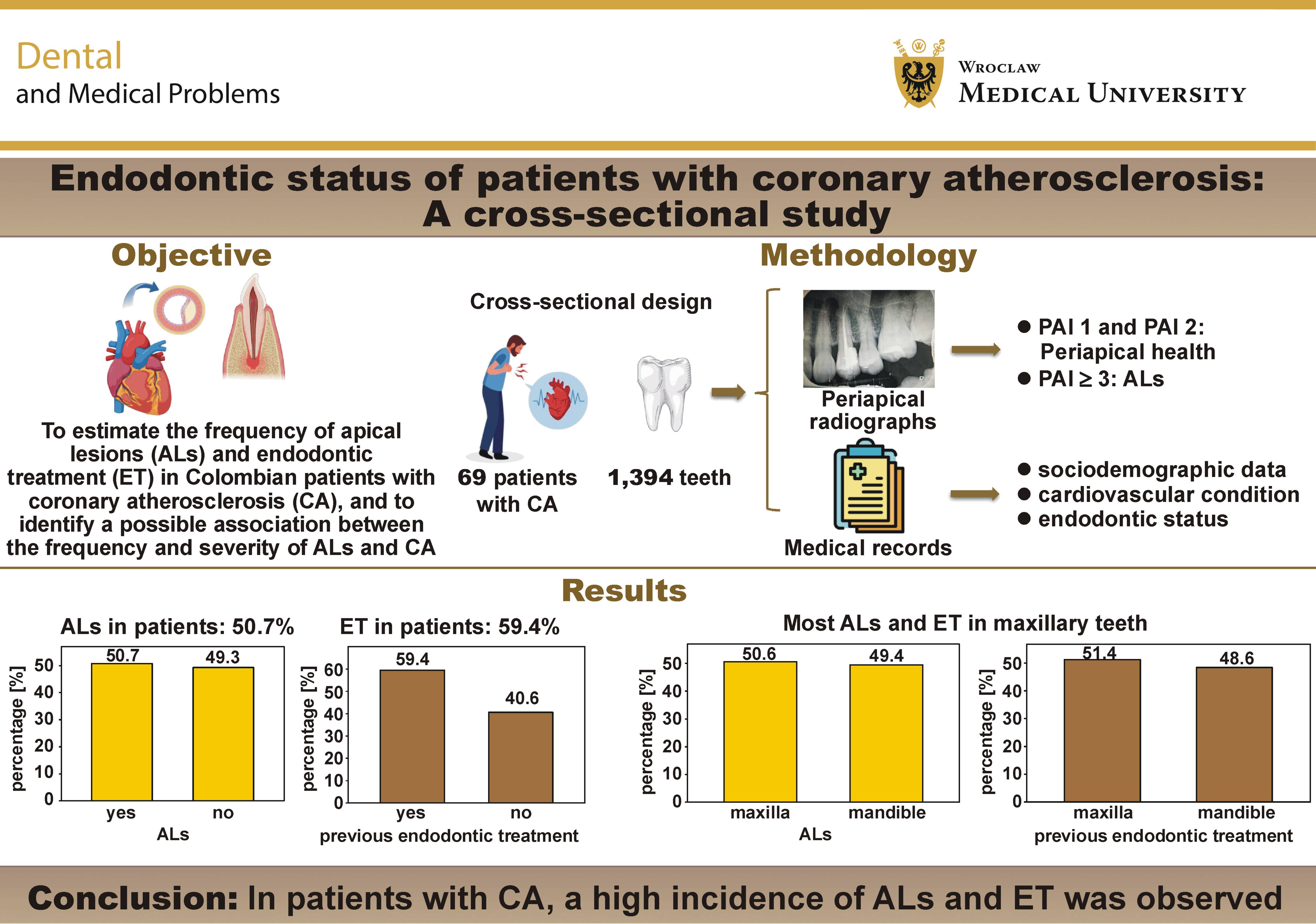
Objectives. The present study aimed to estimate the frequency of ALs and ET in Colombian patients with CA, and to identify a possible association between the frequency and severity of ALs and CA.
Material and methods. The study had a cross-sectional design. We included patients with the evidence of CA diagnosed by coronary angiography. The main outcome variables were the number of compromised coronary arteries and their degree of obstruction, as well as ET, including ALs and previously treated teeth (PTT). The periapical index (PAI) was assessed using periapical radiographs.
Results. A total of 1,394 teeth from 69 patients were evaluated (δ = 20.2 teeth/patient). The frequency of ALs and ET was 5.7% and 10.1%, respectively, considering all teeth evaluated, with a greater presence in the maxilla (50.6% and 51.4%, respectively). Apical lesions were most frequently observed in premolars and molars (35.4%), whereas ET was more frequent in molars (38.6%). Among teeth with ALs, PAI 3 was the most frequent score. Apical lesions were present in 36.6% of endodontically treated teeth; 72.9% of these teeth had inadequate obturation, of which 40 (39.2%) had ALs (p = 0.021).
Conclusions. In patients with CA, a high incidence of ALs and ET was observed, with no evidence of an association between the frequency of ALs, the severity of coronary vessel occlusion, and the number of coronary arteries affected. More ALs were observed in teeth with inadequate ET. However, as we did not have a control group for comparison, these findings should be interpreted with caution.
Keywords: atherosclerosis, coronary artery disease, coronary disease, periapical periodontitis, root canal obturation
Introduction
Coronary atherosclerosis (CA) is the underlying cause of several cardiovascular diseases (CVDs). It is characterized by the narrowing of the arterial lumen due to the accumulation of fatty deposits and the loss of elasticity of the arterial walls, resulting in vessel stenosis and reduced blood flow.1, 2 Coronary atherosclerosis has been defined as a lesion of the intimal layer of blood vessels, comprising a fibrous cap and an atheromatous core. The plaque is composed of smooth muscle cells, the extracellular matrix (ECM), inflammatory cells, calcifications, lipids, and necrotic debris.3 Cardiovascular diseases are among the leading causes of disability and mortality worldwide.4 According to Joseph et al., in 2015, deaths attributable to CVDs accounted for approx. ⅓ of all deaths globally.5 In Colombia, CVDs are a leading cause of death. According to the Pan American Health Organization (PAHO), in 2021, 190.2 deaths from CVDs were reported in Colombia per 100,000 inhabitants.6
Apical periodontitis (AP) is an acute or chronic inflammatory lesion around the apex of a tooth caused by a polymicrobial infection of the root canal system,7, 8 characterized by periapical bone resorption, referred to as an apical lesion (AL).9 This condition is one of the persistent inflammatory disorders of the oral cavity and may contribute to systemic inflammation.10, 11 It has been associated with platelet aggregation and hypercoagulability.8, 12 The etiological factors of AP include various forms of dental pulp damage, such as caries, trauma, iatrogenic injury, and the failure of endodontic treatment (ET).13 Endodontic treatment can prevent AP or restore dental health by eliminating intraradicular infection,14 promoting bone repair15 and maintaining proper tooth function.16 Adequate root canal filling plays an important role in successful ET by preventing bacterial reinfection through the reduction of coronal leakage and, consequently, halting disease progression.17 The impact of ET quality has been evaluated based on radiographic criteria, and root canal fillings can be classified as adequate or inadequate.18
The reported prevalence of AP and ET varies worldwide.19, 20, 21 However, there are no studies in the Colombian population evaluating the frequency and severity of ALs, their relationship with the frequency and quality of ET obturation, or their association with CA. Therefore, this study aimed to estimate the frequency of ALs and ET in Colombian patients with CA, and to identify a possible association between the frequency and severity of ALs and CA.
Material and methods
This was an observational, descriptive, cross-sectional study that included patients with the angiographic evidence of CA, recruited between 2017 and 2020 at the Fundación Valle del Lili, Cali, Colombia.
The study was approved in November 2017 by the Biomedical Research Ethics Committee of the Fundación Valle del Lili (approval No. 730-217). All patients provided written informed consent prior to enrollment in the study. The study was conducted in accordance with ethical considerations and the Declaration of Helsinki.
Coronary angiography was performed using the Innova 3100 IQ system (GE HealthCare, Waukesha, USA). The procedure began with arterial puncture (femoral or radial), followed by the insertion of a specialized catheter into the left chambers of the heart. A contrast medium was then injected through the catheter, allowing the visualization of the coronary artery lumen with the use of specialized imaging techniques, and enabling the presence or absence of atherosclerosis to be determined.
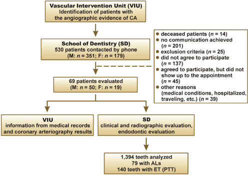
Convenience sampling was used to recruit patients during the study period. We included patients over 18 years of age who had at least one tooth in the mouth, a confirmed diagnosis of CA, and who agreed to participate in the study and signed the informed consent form. Further details of the study population are presented in Figure 1. Patients with contraindications to X-ray exposure, pregnant women, and patients who had received dental treatment within the previous 3 months were excluded.
Sociodemographic data and medical information related to the cardiovascular condition were collected, including the number of compromised coronary arteries and their degree of occlusion, classified as non-significant (≤50% occlusion), significant (51–70%), severe (71–90%), or critical (>90%). Information regarding ET was also collected, including the presence of ALs and previously treated teeth (PTT). The periapical index (PAI) was assessed using periapical radiographs22; this index allows the evaluation of the condition of the periradicular tissues. For this study, PAI scores of 1 and 2 were considered indicative of periapical health, whereas PAI scores ≥3 were considered indicative of AP.23, 24, 25 According to the American Association of Endodontists (AAE), teeth that had undergone ET were defined as PTT.26 The quality of root canal obturation was classified as adequate or inadequate.18
Statistical analysis
For statistical analysis, absolute and relative frequencies were calculated for categorical variables. The measures of central tendency and dispersion were calculated for clinical, radiographic, and clinical history variables. Associations between the categorical variables related to PAI and endodontic diagnoses were assessed using the χ2 test. The Shapiro–Wilk and Levene tests were used to assess the distribution and homogeneity of variance, respectively, when comparing the mean numbers of teeth according to endodontic diagnoses and PAI. The severity of coronary artery occlusion in patients with CA was compared using the Kruskal–Wallis test. The confidence level was set at 95%, and statistical significance was defined as p < 0.05.
Results
A total of 530 patients with CA were identified through coronary angiography. Of these, 114 met the inclusion criteria, 69 agreed to participate and attended the scheduled assessment, and 1,394 teeth were evaluated (δ = 20.2 teeth/patient) (Figure 1).
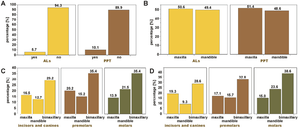
Among the 69 included patients, the mean age was 65.7 ±8.6 years, and 50 (72.5%) were men. At the patient level, ALs and PTTs were present in 35 (50.7%) and 41 (59.4%) patients, respectively (Table 1). At the tooth level, ALs and PTT were observed in 79 (5.7%) and 140 (10.1%) teeth, respectively (Figure 2A), with a greater proportion located in the maxilla, accounting for 40 (50.6%) ALs and 72 (51.4%) PTT (Figure 2B). In both jaws, ALs were most frequently observed in premolars and molars, accounting for 28 (35.4%) cases (Figure 2C), whereas PTT were more frequently observed in molars, accounting for 54 (38.6%) cases (Figure 2D).
Among patients with ALs, 10 (28.6%) had at least one coronary artery with a significant atherosclerotic lesion, and the same proportion had at least one artery with a severe lesion. Additionally, 8 (22.9%) patients had at least one artery with a non-significant lesion, and the same proportion had at least one artery with a critical lesion (Supplementary Table 1, available on request from the corresponding author). Severe and critical atherosclerotic lesions were more frequently observed (in 34 and 25 patients, respectively), with PAI 3 being the most frequent score in both groups (0.68 ±1.32 and 0.80 ±1.04 teeth, respectively) (p > 0.05) (Supplementary Table 2).
Among teeth with ALs, PAI 3 was the most frequent score, with no significant difference between endodontic diagnoses with regard to PAI (Table 2). Of the 140 PTT, 47 (36.6%) presented with ALs. Inadequate obturation was observed in 102 (72.9%) PTT, of which 40 (39.2%) had ALs (p = 0.021). Most patients did not recall the timing of ET or the type of operator who performed ET (Table 3).
Discussion
The prevalence of AP in patients with CA reported in the literature ranges from 50.8% to 85.3%,23, 27 similar to the findings of this study. An AP frequency of 39.2% in males and 44.2% in females has been reported in previous studies.28 In this study, a greater presence of ALs was observed in males, which may be partially related to the higher prevalence of CA among male patients.29 The presence of ALs in teeth was 5.7%, with asymptomatic AP being the most prevalent periapical diagnosis (5.5%). These results are similar to those reported by Tibúrcio-Machado et al., who found a global prevalence of AP of 52% in patients without systemic compromise and 5% in teeth.30 Likewise, de Oliveira et al. reported a prevalence of AP of 42.6% in patients with CA and 40.1% in endodontically compromised teeth,28 which is similar to the findings of this study. The prevalence of ET reported in previous studies ranges from 41.0% to 60.8% in patients, and from 2% to 9% in teeth,31, 32 which is also similar to our findings. Moreover, the prevalence of AP in teeth with ET found in this study is consistent with the previously reported global prevalence of 41%.33
De Oliveira et al. reported a greater presence of AP in the maxilla, upper incisors and lower premolars (40.3%, 52.8% and 46.2%, respectively).28 In the present study, the maxilla, upper premolars and lower molars had a higher frequency of ALs. Previous studies have reported a higher prevalence of ET in the maxilla,34, 35, 36, 37, 38, 39, 40 and a high prevalence of ET in molars.34, 38 These findings are consistent with those of the present study.
Apical periodontitis and periodontal disease differ in terms of their etiology and pathogenesis; however, they share similar characteristics. Both are chronic oral infectious diseases, share a common Gram-negative microbiota and result in increased levels of inflammatory mediators.28, 39 A potential relationship between periodontal disease and CVDs has been reported previously40, 41, 42; therefore, it has been hypothesized that AP could also impact patients with cardiovascular compromise due to its close relationship with periodontal disease.28 Several studies have demonstrated an increased risk associated with AP in patients with CVDs and CA, suggesting that an increase in the number of teeth with AP is associated with a higher risk of CVDs.23, 24, 27, 28, , 43, 44, 45, 46
There are no reports in the literature on the relationship between the frequency and severity of ALs, the number of coronary vessels affected, and the severity of their occlusion. Virtanen et al. conducted a cross-sectional study in which medical information was collected from randomly selected subjects, who were classified as patients with or without AP, and found a greater tendency and severity of ALs in patients with CVDs.47 They showed that AP was significantly associated with CVDs and suggested that the presence of AP in subjects who also regularly had periodontal disease might indicate a higher oral inflammatory load.47 In contrast to the findings of Virtanen et al.,47 but in line with those reported by Frisk et al.48 and Frisk,49 we found no association between the severity of ALs and CA. This may be explained by the fact that both AP and CVDs can occur independently without directly affecting each other.10 It is important to highlight that the quality of the current evidence is moderate to low, and a causal relationship between these two diseases could not be established.50
Regarding the quality of ET, no studies exclusively evaluating populations with CA were found. Kielbassa et al. reported a prevalence of adequate ET of 9.9%, while 50.3% of ET was considered inadequate.37 Virtanen et al. found that, among the 60.8% of patients with ET analyzed, 38.3% were considered to have satisfactory ET, whereas 51.7% were considered to have unsatisfactory ET.47 Moreno et al. reported that 66.7% of the teeth evaluated had inadequate ET; of these, 54.0% presented ALs.35 These results are similar to our findings, in which a statistically significant association was found. Different studies have reported a significant association between the quality of ET and the status of the periapical tissues,35, 36, 37, 51 suggesting that inadequate ET may act as a risk factor for the development of ALs.33, 52 This may indicate that ET has a protective effect on the periapical tissues when performed adequately.
Various studies have evaluated the success rate of ET, which is characterized by the restoration of dental health (i.e., no evidence of AP), the prevention of AP, the continued retention of the tooth, and the restoration of its function. Alley et al. reported a higher success rate for ET performed by endodontists (98.1%) than by general dentists (89.7%) over a 5-year period.53 Lazarski et al. found no differences in the ET success rates according to the type of operator, reporting an overall success rate of 94.4% over a period of 3.5 years.54 Burry et al. analyzed the survival of teeth with ET, and found a decrease in the survival rates at 1, 5 and 10 years.55 They did not observe differences between operators at 1 and 5 years; however, at 10 years, they found better survival of the molars treated by endodontists.55 These results are not directly comparable with our study, since most patients did not remember the type of operator who performed ET. In addition, an unequal proportion of treatment performed by general dentists and endodontists was observed.
Limitations
Among the limitations of this study is the use of a convenience sampling method, which may introduce sampling bias and limit the generalizability of the results. In addition, we did not conduct a power analysis to determine the required sample size, which may have influenced the results obtained. Moreover, we acknowledge that the lack of a control group is a major limitation; therefore, we suggest that future studies include a matched control group to strengthen the conclusions drawn. Although the objective of this study was to determine whether there was an association between the frequency and severity of ALs and CA, the statistical approach used cannot be interpreted as a correlation or used to establish impact, as other factors that could influence the results were not included in the analysis (e.g., oral hygiene and mechanical stress). A larger sample size, a more balanced representation of men and women, and a wider age range would also be desirable to further validate our results. Finally, we suggest that future studies incorporate cone-beam computed tomography (CBCT), as it provides a more objective assessment of ALs.
Conclusions
A high frequency of ALs and ET was found in patients with CA. No relationship was found between the frequency of ALs, the severity of coronary occlusion, and the number of coronary arteries affected. A greater presence of AL was found in teeth with inadequate ET. Since most patients could not recall the timing of their ET or the type of operator who performed ET, an association between the presence of ALs, the timing of ET and the type of operator could not be determined. However, as this study did not include a control group for comparison with patients with CA, our results should be interpreted with caution.
Ethics approval and consent to participate
This study was approved by the Biomedical Research Ethics Committee of the Fundación Valle del Lili, Cali, Colombia (approval No. 730-217). All patients provided written informed consent prior to enrollment in the study. The study was conducted in accordance with ethical considerations and the Declaration of Helsinki.
Data availability
The datasets supporting the findings of the current study are available from the corresponding author on reasonable request.
Consent for publication
Not applicable.
Use of AI and AI-assisted technologies
Not applicable.