Abstract
Background. The fundamental goals of traditional periodontal therapy have been the eradication of disease and the preservation of a healthy, functional dentition and its supporting tissues. However, in recent years, the emphasis has shifted increasingly toward esthetic outcomes, extending beyond dental prostheses and tooth color to encompass the soft tissues surrounding the dentition.
Objectives. The main aim of this research was to compare the impact of the coronally advanced flap (CAF) in combination with concentrated growth factor (CGF) with that of CAF alone in the management of gingival recession.
Material and methods. This randomized controlled trial (RCT) included 90 patients with Miller’s class I or class II gingival recession, who were categorized into 2 groups: CAF; and CAF with CGF. Follow-up assessments were conducted at 6 months, 1 year and 3 years. Periodontal clinical parameters, along with buccal recession depth (BRD) and the width of attached gingiva (AGW), were measured. Postoperative pain was assessed using a visual analog scale (VAS) 10 days after the intervention in both groups. Statistical analysis was performed using IBM SPSS Statistics for Windows, v. 23.
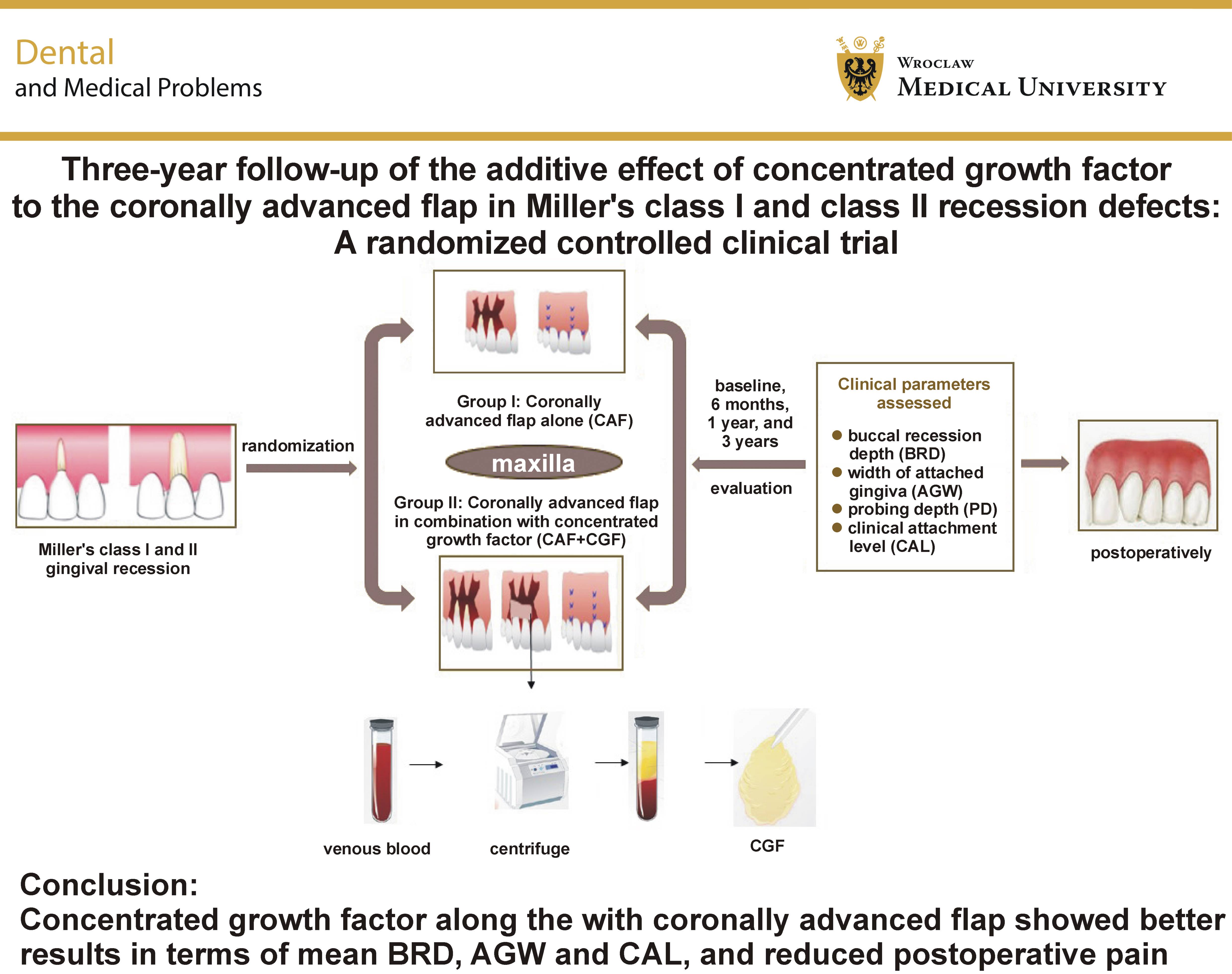
Results. The CAF+CGF group showed better results in terms of mean BRD, AGW and clinical attachment level (CAL), as well as reduced postoperative pain (p < 0.05).
Conclusions. With a 3-year long-term follow-up, the present trial supports the additive effect of CGF in combination with CAF for maxillary gingival augmentation coronal to the recession.
Keywords: gingival recession, root coverage, coronally advanced flap, concentrated growth factor
Introduction
The fundamental goals of traditional periodontal therapy have been the eradication of disease and the preservation of a healthy, functional dentition and its supporting tissues. However, in recent years, the emphasis has shifted increasingly toward esthetic outcomes, extending beyond dental prostheses and tooth color to also include the soft tissues surrounding the dentition. One of the most common esthetic concerns involving periodontal tissues is gingival recession. Gingival recession is characterized by the apical displacement of the free gingival margin from the cementoenamel junction (CEJ), which may contribute to increased root hypersensitivity, caries and attachment loss. Its onset has frequently been associated with dental malposition, traumatic toothbrushing, the frenal pull, and periodontal disease.1
A myriad of studies have shown that various periodontal plastic surgical techniques can effectively treat gingival recession. Many techniques, including free gingival grafts, pedicle flaps with and without grafts, coronally advanced flaps (CAFs), and others, have been studied.2, 3 Numerous graft materials, such as connective tissue grafts (CTGs), xenogeneic collagen matrices, dermal matrices, guided tissue regeneration membranes, allografts, and platelet concentrates, have been used.4, 5 Among these techniques and graft materials, the combination of CAF and CTG has the greatest potential for achieving complete root coverage (CRC).6, 7
Numerous clinical and histological studies have shown that different types of platelet concentrates may be effective in promoting soft tissue repair and periodontal regeneration.8, 9, 10 Concentrated growth factor (CGF) is one of the cutting-edge platelet concentrates. The centrifuge rotation speed fluctuates between 2,400 and 3,000 rpm during the preparation of CGF.11 As a result of this variation in rotation speed, the fibrin matrix produced by centrifugation is larger, more concentrated, and contains higher levels of growth factors than platelet-rich fibrin (PRF).12 Compared with other platelet preparations, CGF has greater viscosity, tensile strength and adhesive properties. In addition to growth factors, CGF contains platelets, leukocytes and CD34+ stem cells, which contribute to the regenerative process.13 Being an autologous fibrin-rich matrix, CGF also contains immunological cells, which may effectively control the inflammation processes.14
Investigations have been conducted to evaluate the effect of CGF on recession coverage, with follow-up periods ranging from 3 months to 1 year.15, 16, 17 Nevertheless, there are no long-term studies evaluating the potential benefits of using CGF in addition to CAF compared with the conventional CAF approach for the treatment of gingival recession. Hence, comparing the impact of CGF in combination with CAF treatment with that of CAF alone in terms of recession coverage was the primary aim of this randomized clinical trial (RCT). Patient-centered outcomes were assessed as secondary objectives.
Material and methods
The Consolidated Standards of Reporting Trials (CONSORT) statement was followed in reporting the present study. This clinical trial was designed as a parallel, randomized, single-center study. Two alternative therapeutic approaches were evaluated in this trial: CAF; and CAF with CGF.
Study setting and population
Between January 2020 and November 2020, patients with Miller’s class I or class II gingival recession in the maxilla who reported to the Department of Periodontology, Saveetha Dental College and Hospitals, Chennai, India, were enrolled in this trial. The study protocol was approved by the institutional ethics committee (IHEC/SDC/PERIO-2001/19/056). Written informed consent was obtained from all participants. The study was conducted in accordance with the principles outlined in the Declaration of Helsinki for research involving human participants.
Eligibility criteria
The inclusion criteria were as follows: individuals aged between 18 and 45 years; systemically healthy individuals; full-mouth plaque score (FMPS) and bleeding score of less than 15%; teeth with a probing depth (PD) ≤3 mm and no evidence of periodontal disease; presence of a clearly identifiable CEJ; and Miller’s class I or class II gingival recession in the maxilla.
The exclusion criteria were as follows: patients with systemic diseases; patients with a history of long-term medication use; smokers; pregnant or lactating women; patients who had undergone periodontal therapy or any mucogingival surgery within the previous 3 months; patients with non-carious cervical lesions.
Randomization
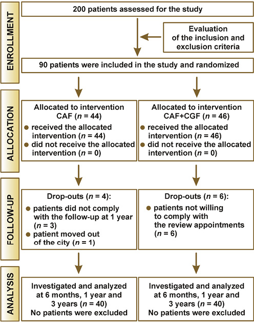
This clinical trial was a monocentric, parallel, randomized, controlled, single-blinded study. Simple randomization was performed by asking each patient to select a chit bearing the type of intervention. The study was conducted in 4 phases: enrollment; allocation; follow-up; and analysis. The sample size was calculated using the G*Power software (https://www.psychologie.hhu.de/arbeitsgruppen/allgemeine-psychologie-und-arbeitspsychologie/gpower), based on data from a previous study.16
Intervention
All surgical procedures were performed by a single operator (C.K.). The experimental procedures were carried out in the Department of Periodontology, Saveetha Dental College and Hospitals, by an experienced operator with extensive expertise in periodontal plastic surgery, including root coverage techniques. All procedures were performed under sterile and adequately equipped conditions, following the predefined surgical protocol.
Groups
The patients were assigned to the groups as follows: group I (control group) – CAF (n = 44); and group II (test group) – CAF+CGF (n = 46).
Surgical technique
All patients were instructed to rinse their mouth with 2% chlorhexidine for 1 min. Prior to the procedure, local infiltrative anesthesia was administered using 2% lidocaine with epinephrine (1:100,000). Following anesthesia, 2 oblique incisions extending beyond the mucogingival junction (MGJ) were made along the line angles of the adjacent teeth. Intrasulcular incisions were then made on the buccal aspect of the teeth with recession. Following subpapillary incisions, the flap was raised in the split-full-split thickness pattern. The flap was then passively displaced coronally to achieve tension-free closure. The surgical site was thoroughly irrigated with saline. Subsequently, the papillae adjacent to the teeth with recession were de-epithelialized. Root debridement was performed using a Gracey curette (Hu-Friedy Manufacturing, Chicago, USA) on the buccal and interdental exposed root surfaces. Root biomodification was then performed using 24% ethylenediaminetetraacetic acid (EDTA) for 1 min, followed by thorough rinsing with saline.
In the test group, approx. 9 mL of the patient’s venous blood was collected into a sterile Vacuette tube without the use of anticoagulants. The tubes were then centrifuged using a one-step centrifugation process (Medifuge; Silfradent, Santa Sofia, Italy) consisting of acceleration for 30 s, followed by centrifugation at 2,700 rpm for 2 min, 2,400 rpm for 4 min and 3,000 rpm for 3 min, followed by deceleration and stopping for 36 s. This process separated the blood into 4 layers: the 1st layer consisting of serum; the 2nd layer consisting of CGF; the 3rd layer consisting of the fibrin buffy coat; and the 4th layer consisting of red blood cells (RBCs). The CGF layer was separated using sterile scissors. The CGF membrane was then obtained using a compressor and immediately placed over the recession area.
The flap was then coronally displaced by 1–2 mm beyond CEJ in both the test and control groups. A sling suture was used to position the flap coronally, using 5-0 Vicryl sutures. Subsequently, interrupted sutures were placed to approximate the releasing incisions.
Data collection
Clinical outcome measurements were recorded at baseline, 6 months, 1 year, and 3 years. All clinical parameters were assessed by a single examiner (A.R.), using a UNC-15 periodontal probe. Clinical outcome measures included buccal recession depth (BRD), the width of attached gingiva (AGW), probing depth (PD), and the clinical attachment level (CAL).
Postoperative pain and potential complications or adverse effects were assessed 10 days after the intervention. Patient discomfort was evaluated using a visual analog scale (VAS).
Statistical analysis
Statistical analysis was performed using IBM SPSS Statistics for Windows, v. 23 (IBM Corp, Armonk, USA). The normality of the data was assessed using the Shapiro–Wilk test. Based on the results, the data followed a parametric distribution. Frequencies and percentages were used to describe qualitative variables, whereas means and standard deviations (M ±SD) were used for quantitative variables. The analysis of variance (ANOVA) was performed to compare differences in the outcome parameters between the groups at different time points.
Results
Baseline features of the patients and the defects
A total of 90 patients received treatment for gingival recession. Forty-four patients received CAF alone, while 46 patients received CAF+CGF. Of the 90 patients, 4 patients in the control group and 6 patients in the test group discontinued the study during the follow-up period (Figure 1).
The mean age of the control group was 42.1 ±11.4 years, with 23 of the 40 participants (57.5%) being female and the remaining 17 (42.5%) being male. Among the 71 maxillary teeth treated with CAF alone, 11 were incisors (15.5%), 19 were canines (26.8%), 36 were premolars (50.7%), and 5 were first molars (7.0%). In the test group, 25 of the 40 participants (62.5%) were female and 15 (37.5%) were male, with a mean age of 45.4 ±7.0 years. A total of 70 maxillary teeth were treated with CAF+CGF, including 12 incisors (17.1%), 22 canines (31.4%), 28 premolars (40.0%), and 8 first molars (11.4%). The demographic characteristics of the study population, including age and gender, are presented in Table 1. There was no statistically significant difference between the 2 groups.
Assessment of the surgical technique and postoperative pain (at 10 days)
The mean duration of the surgical procedure was 69.4 ±6.6 min in the test group and 64.7 ±4.2 min in the control group, with no statistically significant difference between the groups (p = 0.128). The comparison of the VAS scores showed that patients in the control group reported significantly greater postoperative pain than those in the test group (41.2 ±9.4 vs. 32.0 ±1.9; p = 0.032).
Clinical outcomes (at baseline, 6 months, 1 year, and 3 years)
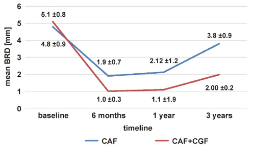
Table 2 depicts the clinical characteristics of the participants in both groups at baseline, 6 months, 1 year, and 3 years in terms of BRD, AGW, PD, and CAL. At baseline, there was no statistically significant difference between the groups for any of the parameters. At 6 months, 1 year, and 3 years of follow-up, there was a statistically significant reduction in BRD, an increase in AGW and improvement in CAL in the test group (p = 0.000). At all time points, there was no statistically significant difference in the PD values between the 2 groups (p > 0.05). Recession coverage was significantly improved as compared to baseline at each follow-up interval in both groups. However, the test group demonstrated better results at all time points (Figure 2).
Discussion
For the treatment of gingival recession, the present RCT compared the use of CGF in combination with CAF with CAF alone. The study evaluated changes in BRD and AGW, and subsequent root coverage, in addition to periodontal clinical parameters, such as PD and CAL.
The literature remains equivocal regarding the impact of platelet concentrates on the outcomes of root coverage procedures. Although recent analyses have reported that platelet concentrates may reduce PD, CAL and recession depth, as well as patient morbidity,18, 19 earlier reviews found that platelet concentrate therapies did not improve root coverage, the width of keratinized tissue or CAL.20, 21 Recent advances in platelet concentrate centrifugation techniques and preparation protocols have raised concerns that these variations may alter the characteristics of the fibrin structure and, consequently, influence therapeutic outcomes.22 However, the literature still lacks a sufficient number of clinical studies evaluating the effect of platelet-associated biomaterials on root coverage outcomes. To the best of our knowledge, this is the first investigation to report the efficacy of CGF in recession coverage with a 3-year follow-up.
In the present trial, with no significant difference in PD and lower VAS scores, the CAF+CGF group outperformed the CAF group in terms of recession coverage, AGW and CAL. Doğan et al. employed CGF for the first time to treat gingival root defects in the maxilla.23 After 6 months, they reported a mean root coverage of 86.67% in the CAF+CGF group. According to their study results, CGF treatment had no additive effect on root coverage. However, it was suggested that the use of CGF with CAF may improve long-term stability by increasing the width and thickness of keratinized tissue and improving CAL.23 The findings of the present study, which are in agreement with those of Doğan et al.,23 demonstrated enhanced AGW and CAL.
Probing depth is considered a direct indicator of periodontal health,24 and was therefore included as an outcome measure in the present study. At baseline, the mean PD was 2.3 ±1.9 mm in the control group and 2.1 ±0.9 mm in the test group. At 6 months, 1 year and 3 years, there was no statistically significant change in PD in either group. These findings are consistent with previous studies that evaluated the use of platelet concentrates, such as CGF and PRF, in the treatment of recession defects.15 When CAL was compared between the 2 groups, the test group demonstrated better results than the control group. This finding is in agreement with previous studies supporting the potential benefit of platelet concentrates in improving CAL following recession coverage procedures.25, 26
Apart from a few limitations, such as not considering buccal recession width (BRW), the height of the interproximal papilla, the gingival biotype, and the thickness of keratinized gingiva, several factors were considered in the present study to determine the effect of CGF on recession coverage and its long-term stability. Non-carious cervical lesions were excluded from the study, as they are known to have a negative impact on root coverage over time. Studies have also emphasized the influence of the tooth position on the outcomes of root coverage procedures.27, 28 More recently, Zucchelli et al. highlighted that the tooth position was crucial in determining the amount of root coverage that could be achieved, as sites in the mandibular arch demonstrated significantly lower mean and complete recession coverage than the treated maxillary recession defects.29 In the present study, only maxillary teeth were included. Collectively, the findings of this long-term trial support the additive effect of CGF in gingival augmentation coronal to the recession.
Conclusions
Within the limitations of the present study, the following conclusion can be drawn: With a 3-year long-term follow-up, the CAF+CGF group showed better results in terms of root coverage and AGW, as well as lower pain scores and reduced patient discomfort, compared with the CAF group.
Trial registration
The trial was registered with the Clinical Trials Registry–India (CTRI) (2019/02/079317).
Ethics approval and consent to participate
The study was conducted in accordance with the principles outlined in the Declaration of Helsinki for research involving human participants. The study protocol was approved by the institutional ethics committee at the Department of Periodontology, Saveetha Dental College and Hospitals, Chennai, India (IHEC/SDC/PERIO-2001/19/056). Written informed consent was obtained from all participants.
Data availability
The datasets supporting the findings of the current study are available from the corresponding author on reasonable request.
Consent for publication
Not applicable.
Use of AI and AI-assisted technologies
Not applicable.