



Abstract
Background. Obesity is a chronic disease resulting from the interaction of genetic, environmental, socio-economic, and behavioral factors, and is associated with the development of numerous comorbidities. Its prevalence is increasing worldwide, making obesity one of the most significant public health challenges.
Objectives. The aim of the present study was to investigate the relationship between obesity and the cortisol levels and periodontal status in children.
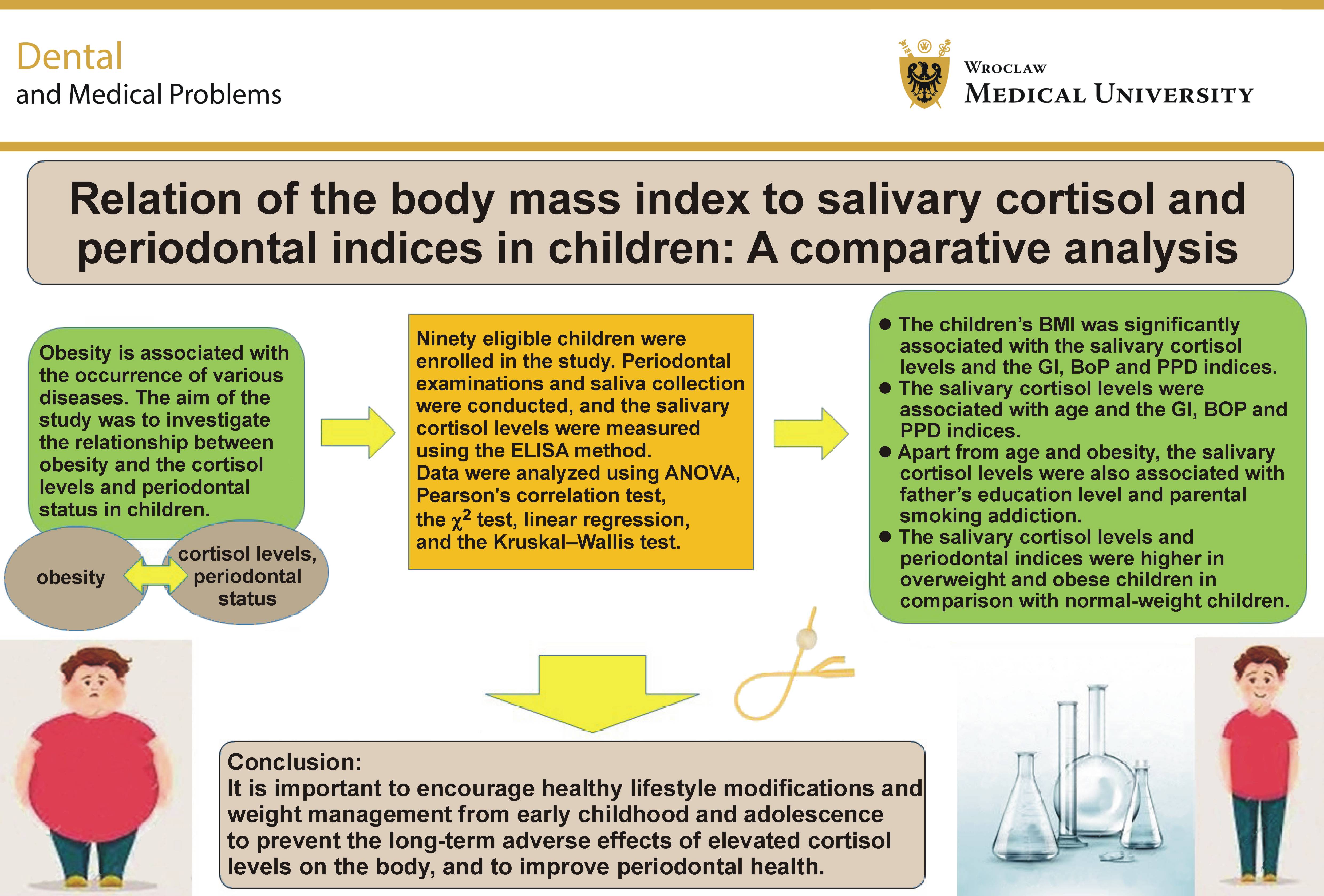
Material and methods. Ninety eligible children aged 8–12 years were enrolled in the study and allocated into 3 groups: normal-weight; overweight; and obesity. After the parents completed a demographic questionnaire, the children underwent periodontal examinations and saliva collection. The salivary cortisol levels were measured using the ELISA method. Data were analyzed using ANOVA, Pearson’s correlation test, the χ2 test, linear regression, and the Kruskal–Wallis test.
Results. The children’s body mass index (BMI) was significantly associated with the salivary cortisol levels and the gingival index (GI), bleeding on probing (BoP) and periodontal probing depth (PPD) (p = 0.010, p < 0.001, p < 0.001, and p = 0.005, respectively). However, BMI was not significantly associated with father’s education, mother’s education, the parental smoking addiction status, total family income, the frequency of mouthwash use per week, toothbrushing frequency per day, or daily study duration (p = 0.599, p = 0.143, p = 0.480, p = 0.875, p = 0.884, p = 0.203, and p = 0.338, respectively). The salivary cortisol levels were also significantly associated with age and GI, BOP and PPD (p = 0.010, p = 0.001, p = 0.018, and p = 0.016, respectively). In the multivariable linear regression analysis, the salivary cortisol levels remained significantly associated with age (β = 0.276; p = 0.004), obesity (β = 0.269; p = 0.006), father’s education level (β = 0.393; p < 0.001), and the parental smoking addiction status (β = 0.238; p = 0.026).
Conclusions. The salivary cortisol levels and periodontal indices were higher in overweight and obese children. Therefore, encouraging children and adolescents to adopt a healthier lifestyle and achieve weight reduction may help reduce the salivary cortisol levels and improve periodontal health.
Keywords: body mass index, periodontal pocket, cortisol, gingival index, periodontal indices
Introduction
Obesity refers to the abnormal or excessive accumulation of adipose tissue in the body.1 It is considered not only an esthetic concern, but also a chronic disease resulting from the complex interaction of genetic, environmental, socioeconomic, and behavioral factors.2 It has been suggested that the genes associated with sweet and bitter taste perception may influence both obesity and oral health.3 In recent years, obesity has replaced smoking as one of the most important lifestyle-related risk factors for premature death because of its association with numerous health problems and chronic diseases.1, 4
According to the World Health Organization (WHO), more than 340 million children and adolescents aged 5–19 years were overweight or obese in 2016.4 Akbari and Mohammadi reported an overall obesity prevalence of 11.4% among Iranian children.5 The diagnosis of obesity in children is usually based on the calculation of the body mass index (BMI). Although BMI has limitations as a direct measure of body fat, it remains the most widely used and practical method for diagnosing obesity in children.6
Cortisol is commonly known as the body’s stress hormone.7 Studies have demonstrated alterations in cortisol secretion, function and metabolism in individuals with obesity.8 Subclinical hypercortisolism increases the risk of osteoporotic fractures, hyperglycemia, hypertension, dyslipidemia, and obesity.9, 10 In addition, hypercortisolism may promote insulin resistance, thereby perpetuating a vicious cycle.11
However, the available evidence is conflicting. Several studies have reported that the mean salivary and serum cortisol levels are lower during the daytime in children with obesity.12, 13, 14 In contrast, other studies have demonstrated a positive association between the cortisol levels and BMI in children.15, 16, 17 Similarly, conflicting findings have been reported regarding the relationship between the serum cortisol levels and BMI in adults.18, 19, 20
Severe periodontal disease is the 11th most prevalent disease worldwide.21 It often begins as gingivitis, which is characterized by gingival bleeding, swelling and pain. If left untreated, gingivitis may progress to periodontitis, resulting in the loss of periodontal attachment, alveolar bone, and ultimately teeth, thereby impairing mastication, esthetics, self-confidence, and quality of life.22
Concern about periodontal disease lies not only in its high prevalence, but also in its well-established association with several systemic conditions, including cardiovascular disease, diabetes mellitus, and adverse pregnancy outcomes, such as low birth weight.23, 24, 25 Perinatal exposures and pregnancy outcomes have also been associated with an increased predisposition to obesity.26
The association between obesity and inflammatory processes has long been recognized.27 Obesity may be considered a risk factor for periodontal disease, as it alters immune function and promotes a chronic low-grade inflammatory state associated with excess adipose tissue.28 Although numerous studies have investigated periodontal health in adults with obesity,29, 30 only limited studies have been conducted in children and adolescents, and these suggest a possible association between obesity and periodontal health.2, 28, 31 Given the increasing prevalence of overweight and obesity among children, understanding the early periodontal changes associated with obesity is of considerable importance.32, 33
Considering the insufficient number of studies and the conflicting findings regarding the relationship between obesity and periodontal health in children and adolescents, further research in this field is warranted. Therefore, the aim of the present study was to investigate the relationship between overweight and obesity, the salivary cortisol levels, and the periodontal status in children.
Material and methods
Study design and participants
This cross-sectional comparative descriptive study was conducted from 2021 to 2022 in 3 downtown schools in Qom, Iran. The study protocol was developed in accordance with the Strengthening the Reporting of Observational Studies in Epidemiology (STROBE) statement.
According to a study by Putignano et al., considering the average salivary cortisol levels in 2 groups of individuals with obesity and normal weight, with an alpha level of 5% and a beta level of 10%, the required sample size was calculated as 30 participants per group.34 Therefore, a total of 90 participants were included in this study, using the non-random convenience sampling method.
The inclusion criteria were children aged 8–12 years with total dmft and DMFT (decayed, missing and filled teeth in primary and permanent dentition, respectively) scores <3. The exclusion criteria comprised a history of any systemic disease (such as epilepsy, diabetes, hypothyroidism, cancer, psychological disorders, precocious puberty, or short stature), severe periodontitis, the use of any medication, a history of surgery, trauma, or other stressful events within the previous 3 months, participation in professional sports activities, living with only one parent (due to parental divorce or death), saliva sampling during the menstrual period in female participants, and resistance or the lack of cooperation during saliva collection and clinical examinations.
After obtaining informed consent from the children’s parents, eligible children were enrolled in the study and equally divided into 3 groups: 30 children with normal weight; 30 children with overweight; and 30 children with obesity. The groups were matched based on age and sex. The participants were also divided into 2 groups according to sex, including 45 males and 45 females. Each sex group consisted of 15 children with obesity, 15 children with overweight and 15 children with normal weight.
Measurements and the questionnaire
The children’s weight was measured while wearing light clothing (such as a T-shirt and pants) and without shoes or belts, using a digital scale (SBS 4414; Sinbo Precision Industry, Shenzhen, China) with an accuracy of 100 g. Height was measured using a wall-mounted stadiometer with an accuracy of 0.5 cm. The body mass index was then calculated by dividing weight in kilograms by the square of height in meters.
Based on the BMI values and the standard BMI charts developed by WHO,6, 35 the 90 eligible children were included in the study and equally divided into 3 groups – normal weight, overweight, and obesity – with 30 participants in each group. According to the WHO standards for children and adolescents aged 2–20 years, a BMI percentile of 85–97% was considered overweight, while a BMI above the 97th percentile was considered obesity.6, 35
The children and their parents were instructed on proper toothbrushing and flossing techniques 1 month before data collection. The parents were also encouraged to carefully supervise their children’s oral hygiene practices during this period.
Then, the designed questionnaire was completed by the children’s parents, and the children’s demographic characteristics, parental smoking status, daily activity status, medical and dental history, frequency of mouthwash use per week, toothbrushing frequency per day, and daily study duration in hours were recorded.
Collection of saliva samples
Before saliva collection, the participants were instructed to refrain from toothbrushing, eating, drinking, and vigorous physical activities for 1 h. During this period, they were allowed to relax through activities such as reading, writing and drawing.15 Due to circadian variations in cortisol concentrations, saliva samples were collected at noon (12:00). Saliva samples were obtained using the spitting method.36 After rinsing the mouth with water and swallowing saliva once, each participant was instructed to allow saliva to accumulate in the floor of the mouth, and then transfer it into a Falcon tube 1–2 times per minute for 5 min while sitting with the head and neck positioned at a 45-degree angle.37
Oral examinations
All oral examinations were performed by a trained dentist. To avoid bias related to the incomplete eruption of some permanent teeth, periodontal indices were assessed only on first permanent molars and maxillary and mandibular central and lateral incisors. Primary teeth were not included in the periodontal evaluation.2
Gingival index
The gingival index (GI) was scored from 0 to 3 according to the Löe and Silness criteria as follows38:
– score 0: no bleeding on probing (BoP); normal gingiva without signs of inflammation;
– score 1: presence of non-bleeding gingival changes; mild inflammation of the marginal gingiva detected during a clinical examination (the marginal gingiva is slightly redder than normal, with slight edema, and colorless gingival fluid may be observed at the entrance of the sulcus);
– score 2: bleeding of the marginal gingiva after stimulation with a dental probe or air spray; moderate gingival inflammation (the gingiva appears red or bluish-red and shiny, with the edema of the marginal gingiva);
– score 3: spontaneous gingival bleeding; severe gingival inflammation (the gingiva is markedly red or bluish-red, enlarged, and swollen).
Bleeding on probing
To evaluate BoP, a Williams probe (Smart Instrument, model MS5201; Hu-Friedy Manufacturing, Chicago, USA) was gently inserted into the sulcus of all teeth up to the base of the pocket or sulcus without applying additional pressure. After 10–15 s, the number of teeth exhibiting gingival bleeding was recorded.39
Periodontal probing depth
To assess periodontal probing depth (PPD), a Williams probe (Smart Instrument, model MS5201) was inserted into the sulcus or periodontal pocket with gentle force while maintaining contact with the tooth surface and keeping the probe parallel to the root surface (the long axis of the tooth). The probe was advanced until the tip reached the base of the pocket and encountered increased resistance, indicating the limit of further penetration. The distance between the base of the pocket (the point where the probe stopped) and the gingival margin was measured and recorded.40
Measurements were performed at 6 sites for each tooth (mesiobuccal, mid-buccal, distobuccal, distolingual, mid-lingual, and mesiolingual). A pocket depth greater than 3 mm was considered indicative of an inflammatory condition.2 The mean PPD values for each tooth were then calculated and recorded.41
Salivary cortisol levels
The cortisol levels can be measured using 3 types of samples: blood; saliva; and urine. The salivary cortisol levels have a direct and strong correlation with the serum cortisol levels and follow the circadian pattern of serum cortisol secretion. Due to the simplicity and non-invasive nature of saliva collection, as well as the ease of saliva transport and storage, the salivary cortisol measurement is widely used in pediatric research. In addition, blood sampling can be stressful for children and may result in elevated serum cortisol levels.42, 43
The salivary cortisol levels were measured using a salivary cortisol kit (DKO020; DiaMetra, Spello, Italy) based on the enzyme-linked immunosorbent assay (ELISA) method.44
Bias
The risk of bias was minimized by excluding children with total dmft and DMFT scores >3 from the study, providing oral hygiene instructions to the participants and their parents, and assessing the periodontal status using reliable methods (GI, BoP and PPD). The salivary cortisol levels were analyzed in a specialized laboratory in Qom, Iran.
Statistical analysis
Data were analyzed using IBM SPSS Statistics for Windows, v. 26 (IBM Corp., Armonk, USA). Descriptive and analytical statistics were applied. The Kolmogorov–Smirnov test and the Q–Q plot were used to assess the normality of data distribution. Differences between the groups were analyzed using the one-way analysis of variance (ANOVA) and the Kruskal–Wallis test according to the distribution of data (normal and not normal, respectively). The χ2 test was used for categorical variables, and Pearson’s correlation test was used to assess correlations between variables. The multivariable linear regression analysis was performed to assess the associations between the salivary cortisol levels (the quantitative dependent variable) and the independent variables while controlling for potential confounding factors.
Results
Characteristics of the participants
In this study, 90 eligible children, including 45 males and 45 females, were enrolled. The mean age was 10.00 ±1.48 years in the obese and normal-weight groups, and 10.16 ±1.39 years in the overweight group. There were no significant differences among the 3 groups regarding age and sex (p = 1.000).
Correlation between BMI and the demographic status
The BMI status was not significantly associated with the paternal or maternal education level (χ2 test; p = 0.599 and p = 0.143, respectively). The relationship between the children’s BMI status and total family income was also non-significant (χ2 test; p = 0.875).
In addition, 50% of parents of normal-weight children reported smoking addiction, compared with 36.7% of parents of children from the overweight and obesity groups. Therefore, parental smoking was more prevalent among the parents of normal-weight children than among those of overweight and obese children. However, no significant association was found between the children’s BMI status and the parental smoking addiction status (χ2 test; p = 0.480).
The BMI status was not significantly associated with the frequency of mouthwash use per week, toothbrushing frequency per day or daily study duration in hours (Kruskal–Wallis test; p = 0.884, p = 0.203 and p = 0.338, respectively) (Table 1).
Correlation between BMI and the salivary cortisol levels and periodontal indices
The mean salivary cortisol levels in the normal-weight, overweight and obesity groups were 2.42 ±0.74, 2.93 ±1.32 and 3.68 ±2.26 ng/mL, respectively. Thus, the mean salivary cortisol levels were highest in children with obesity, followed by overweight children and normal-weight children. There was a significant association between the BMI status and the salivary cortisol levels (one-way ANOVA; p = 0.010) (Table 2).
The mean GI values in the normal-weight, overweight and obesity groups were 0.42 ±0.13, 0.51 ±0.16 and 0.61 ±0.15, respectively. Thus, the mean GI was highest in children with obesity, followed by overweight children and normal-weight children.
The mean BoP values in the normal-weight, overweight and obesity groups were 17.83 ±7.20%, 21.50 ±7.61% and 26.00 ±6.82%, respectively. Accordingly, the mean BoP was highest in children with obesity, followed by overweight children and normal-weight children.
The mean PPD values in the normal-weight, overweight and obesity groups were 1.52 ±0.22, 1.60 ±0.24 and 1.72 ±0.23 mm, respectively. Similarly, the mean PPD was highest in children with obesity, followed by overweight children and normal-weight children.
There was a significant association between the children’s BMI status and GI, BoP and PPD (one-way ANOVA; p < 0.001, p = 0.001 and p < 0.005, respectively) (Table 2).
The pairwise comparisons of the salivary cortisol levels among the study groups showed a significant difference only between the obesity and normal-weight groups (post hoc test; p = 0.003). No significant differences were observed between the obesity and overweight groups, or between the overweight and normal-weight groups (post hoc test; p = 0.069 and p = 0.209, respectively) (Table 3).
The pairwise comparisons of GI showed significant differences between the obesity and overweight groups, the obesity and normal-weight groups, and the overweight and normal-weight groups (post hoc test; p = 0.011, p < 0.001 and p = 0.017, respectively) (Table 3).
For BoP, significant differences were observed between the obesity group and both the overweight and normal-weight groups (post hoc test; p = 0.018 and p < 0.001, respectively). However, no significant difference was found between the overweight and normal-weight groups (post hoc test; p = 0.052) (Table 3).
For PPD, a significant difference was observed only between the obesity and normal-weight groups (post hoc test; p = 0.001). No significant differences were found between the obesity and overweight groups, or between the overweight and normal-weight groups (post hoc test; p = 0.054 and p = 0.166, respectively) (Table 3).
According to Pearson’s correlation analysis, there were significant positive correlations between GI and BoP (r = 0.948; p < 0.001), and between GI and PPD (r = 0.680; p < 0.001). A significant positive correlation was also observed between BoP and PPD (r = 0.674; p < 0.001). In addition, the salivary cortisol levels were positively correlated with GI (r = 0.340; p = 0.001), BoP (r = 0.250; p = 0.018) and PPD (r = 0.253; p = 0.016) (Table 4).
Based on the results of the univariable linear regression analysis, the salivary cortisol levels were significantly and positively associated with BMI (β = 0.116; p = 0.001), obesity (β = 1.221; p = 0.005), GI (β = 3.325; p = 0.001), and age (β = 0.311; p = 0.010). However, no significant association was observed between the salivary cortisol levels and the overweight status (β = 0.474; p = 0.263) (Table 5).
Based on the results of the multivariable linear regression analysis, the salivary cortisol levels were significantly and positively associated with age (β = 0.276; p = 0.004), obesity (β = 0.269; p = 0.006), father’s education level (β = 0.393; p < 0.001), and the parental smoking addiction status (β = 0.238; p = 0.026) (Table 6). These findings indicate that obesity remained independently associated with the salivary cortisol levels after adjusting for age, father’s education level and the parental smoking addiction status.
Discussion
Correlation between the salivary cortisol levels and age
In this study, the salivary cortisol levels were measured to investigate physiological changes in cortisol metabolism during childhood and adolescence.
The present study found that the salivary cortisol levels increased significantly with increasing age in children. According to Kiess et al., the cortisol levels are age-dependent, such that after the age of 6 years, they are significantly correlated with maturation stages.16 Törnhage45 and Larsson et al.46 also reported higher salivary cortisol levels in older children. This positive association between age and increased cortisol levels has been reported in other studies as well.47 However, these findings are inconsistent with those of Kjölhede et al.12 and Knutsson et al.48 The discrepancies between the present findings and those of the aforementioned studies may be attributed to differences in the sampling location and time, the methods used to measure salivary cortisol, and the age range of the study populations.
Correlation between the salivary cortisol levels and obesity
The present study found that the salivary cortisol levels were positively and significantly associated with increasing BMI and obesity. Chu et al. also reported that obese children had higher morning salivary and urinary cortisol levels.15 Several other studies have likewise demonstrated a positive relationship between the salivary, serum and urinary cortisol levels and obesity.16, 17, 43, 49 These studies suggest that cortisol concentrations increase long before a child becomes obese.15 Yu et al. also found that the salivary nocturnal cortisol levels were positively correlated with weight gain, abdominal fat distribution (the waist-to-height ratio (WHtR)), and body fat percentage in all participants,43 consistent with the findings of the present study. However, the present findings are inconsistent with those reported by Kjölhede et al., who found lower morning and evening salivary cortisol levels in overweight and obese children aged 6–12 years,12 as well as with the studies by Hillman et al.13 and Chalew et al.,14 which reported lower plasma cortisol levels in obese children. Hillman et al. further showed a negative association between the daytime cortisol levels and BMI z-score and central obesity, but a positive association during the night.13 The discrepancies between the present findings and those of these studies may be attributed to differences in the type of cortisol measured (e.g., plasma rather than salivary cortisol), the cortisol measurement methods and the timing of saliva sample collection.
Cortisol concentrations are not elevated in obese adults with peripheral fat distribution, whereas individuals with central obesity exhibit increased cortisol levels.50 Unfortunately, due to the limitations of the present study, fat distribution was not assessed using waist circumference or other anthropometric measures. Therefore, based on the present findings, no definitive conclusions can be drawn regarding fat distribution and its relationship with the cortisol levels in children. Further studies are needed to investigate this association.
Correlation between obesity and the periodontal status
According to several studies, a high BMI may be a risk factor for periodontal disease in adolescents and young adults, as assessed by the plaque index (PI), BoP, PPD, clinical attachment loss (CAL), and alveolar bone loss.51
Several hypotheses have been proposed to explain biological interactions between obesity and periodontal disease, including alterations in inflammatory and immune responses, impaired glucose tolerance, dyslipidemia, changes in host immunity, increased macrophage activation, impaired microvascular function, physiological responses, psychosocial stress, and the secretion of pro-inflammatory cytokines (tumor necrosis factor-alpha (TNF-α), interleukin-1 (IL-1), IL-6, and IL-8), adipokines (leptin, adiponectin, resistin, and plasminogen activator inhibitor-1 (PAI-1)), and other bioactive substances, such as reactive oxygen species (ROS) from adipose tissue and C-reactive protein (CRP).52, 53 However, the precise molecular and cellular mechanisms underlying these interactions remain unclear, and further studies are needed to elucidate these mechanisms, which may contribute to the development of preventive and therapeutic strategies.
According to some reports, periodontal disease in obese individuals is associated with alterations in the composition of the oral microbiota.54, 55 However, most studies investigating the relationship between obesity and periodontitis have primarily considered immunological and metabolic alterations as the main predisposing factors for periodontal disease.
Several mechanisms have been proposed to explain this relationship. Adipocytes may contribute to a systemic pro-inflammatory state through the release of various inflammatory mediators.56, 57 Some studies have shown that obesity is associated with adverse postoperative outcomes, including infectious complications and impaired wound healing.58, 59 Therefore, the altered immune responses and impaired wound healing associated with obesity may influence the clinical response to periodontal treatment. However, Suvan et al.30 and Chaffee and Weston60 report that the evidence regarding the association between BMI, overweight or obesity and the clinical response to periodontal treatment remains limited.
In the present study, the GI, BoP and PPD indices were used to assess the periodontal status. The findings showed that obesity was positively and significantly associated with all 3 indices.
Although the association between obesity and periodontal disease has been widely investigated in adults, it has been less extensively studied in the pediatric population.
De Castilhos et al. reported a statistically significant association in young adults between obesity and GI and calculus, both of which are indicators associated with periodontal disease; however, no significant association was observed between obesity and periodontitis.61 On the other hand, Suvan et al. confirmed the association between increasing BMI and the severity and extent of periodontitis,62 consistent with the findings of the present study.
Other researchers have shown that obesity in children is associated with increased PI, gingival inflammation and BoP, and that BMI is significantly correlated with periodontal indices, including PI, BoP, PPD, CAL, the oral hygiene index (OHI), and the community periodontal index (CPI).2, 41, 51, 63, 64, 65 Martens et al. likewise demonstrated a positive association between overweight and obesity and the prevalence of periodontal disease.53 They further reported that obesity was associated with less frequent daily toothbrushing and flossing, a higher prevalence of dental caries, a lower parental education level, a higher socioeconomic status, and less healthy dietary habits.53
The increased BoP observed in obese participants may be attributed to elevated levels of pro-inflammatory cytokines in gingival crevicular fluid (GCF).31
Vaziri et al. reported that the mean PPD was higher in obese than in non-obese participants; although this difference was statistically significant, it was not considered clinically significant.41 Similarly, Modéer et al.31 and Scorzetti et al.63 found that participants in the obese groups had a significantly higher prevalence of PPD ≥ 4 mm.
In a study by Vallogini et al., the findings were inconsistent with those of the present study and the aforementioned studies.66 The obese group showed lower visible PI values and gingival bleeding index (GBI) scores, as well as a better periodontal status in terms of PPD. In their study, obese pediatric participants were recruited from a children’s hospital, whereas non-obese participants were recruited from a school.66 Therefore, the discrepancies between the findings of that study and the present study may be attributed to differences in the sampling location, as participants recruited from a hospital setting may have received specific dietary and oral hygiene counseling, potentially resulting in improved periodontal parameters.
Kim et al. showed that individuals with a BMI higher than 25 (classified as overweight or obese) had a lower risk of periodontitis as compared to individuals of normal weight, whereas those with abdominal obesity had a significantly higher risk of periodontal disease.67 These findings suggest that metabolic syndrome may play an important role in periodontal inflammation.68, 69, 70
Reeves et al. concluded that the onset of periodontitis may be associated with weight gain and increased waist circumference.71 They found that among individuals aged 17–21 years, each 1-kilogram increase in body weight was associated with a 6% increase in the risk of periodontal disease, while each 1-inch increase in waist circumference was associated with a 5% increase in the risk of periodontitis. In contrast, no significant association was observed between these parameters and periodontal disease among younger children aged 13–16 years.71
The findings of most of the aforementioned studies indicate an adverse effect of obesity on periodontal health in young individuals, which is likely due to a combination of factors, including metabolic and inflammatory changes, inadequate attention to oral and dental hygiene practices and preventive measures, an unbalanced diet, and irregular dental examinations.72, 73
Correlation between the salivary cortisol levels and the periodontal status
The findings of this study showed that the salivary cortisol levels were positively and significantly associated with the GI, BoP and PPD indices. Various studies have investigated the role of chronic stress as an underlying factor in the development of oral and dental health problems. Based on substantial scientific evidence, stress and psychological disorders may cause individuals to neglect oral and dental hygiene, thereby accelerating plaque accumulation and the progression of periodontal disease.74 Academic stress has also been reported as a risk factor for gingival inflammation, through increased levels of IL-1β in GCF, as well as reduced oral hygiene.75, 76, 77
It has been hypothesized that stress induces behavioral changes, such as overeating and an increased preference for high-fat diets, which may contribute to immunosuppression through increased cortisol production.74 Furthermore, chronic stress can alter the salivary flow rate, pH and chemical composition.74, 78 Rai et al. demonstrated that stress-related biomarkers, including the salivary cortisol concentration, were increased in children with dental caries.79 In the present study, children with dmft/DMFT scores above 3 were excluded due to the potential association between the cortisol levels and dental caries.80, 81, 82 Therefore, the influence of this confounding variable was reduced in the current study, which represents one of its strengths.
It appears that periodontal disease and stress have a bidirectional relationship. Several studies have reported that the chronic elevation of the cortisol levels may be a potential risk factor for the onset or severity of periodontal disease in adults.74, 83, 84
Consistent with the findings of the present study, Schmidt et al. showed that the salivary cortisol levels were significantly higher in adolescents with periodontitis than in those without periodontitis.85 Individuals with elevated cortisol levels appear to be at a higher risk of developing periodontitis. In contrast, Panagiotou et al. found no association between stress and dental caries or periodontal disease.86 One possible explanation for this discrepancy may be the lack of adherence by parents and participants to the scheduled saliva collection times at home.
Considering the complex nature of periodontal disease and numerous confounding factors that influence its development, comprehensive longitudinal studies are recommended in the future to further clarify and confirm the potential relationship between periodontal disease, BMI and the cortisol levels.
Limitations
The difficulty in obtaining participants’ cooperation during oral examinations or saliva collection under calm conditions resulted in the need for repeated sampling.
Conclusions
The findings of the present study showed that:
– children’s BMI was positively associated with the salivary cortisol levels and the GI, BOP and PPD indices, indicating that overweight and obese children have a poorer periodontal status compared with children of normal weight;
– the salivary cortisol levels, as an indicator of the serum cortisol levels, were higher in overweight and obese children than in children of normal weight.
These findings highlight the importance of encouraging healthy lifestyle modifications and weight management from early childhood and adolescence to prevent the long-term adverse effects of elevated cortisol levels on the body, and to improve periodontal health.
Ethics approval and consent to participate
The study was conducted in accordance with the Declaration of Helsinki after obtaining approval from the Ethics Committee of Qom University of Medical Sciences, Iran (No. IR.MUQ.REC.1400.231). All methods were carried out in accordance with the relevant guidelines and regulations. Written informed consent was obtained from the parents or legal guardians of all participating children.
Data availability
The datasets supporting the findings of the current study are available from the corresponding author on reasonable request.
Consent for publication
Not applicable.
Use of AI and AI-assisted technologies
Not applicable.