



Abstract
Emerging evidence suggests that viral integration into non-coding and genomically vulnerable regions may represent one of several mechanisms contributing to oncogenesis, warranting further investigation through long-read whole-genome sequencing (WGS).
Keywords: HBV, EBV, oncogenic viruses, instability, cancer evolution
Oncogenic viruses are no longer viewed merely as isolated infectious causes of cancer, but as context-dependent drivers and cofactors that shape tumor evolution through viral persistence, host genome integration, genomic instability, and the disruption of coding and non-coding regulatory landscapes. This modern understanding, however, emerged from a much simpler early idea: that cancer might, at least in some cases, be caused by a transmissible biological agent.
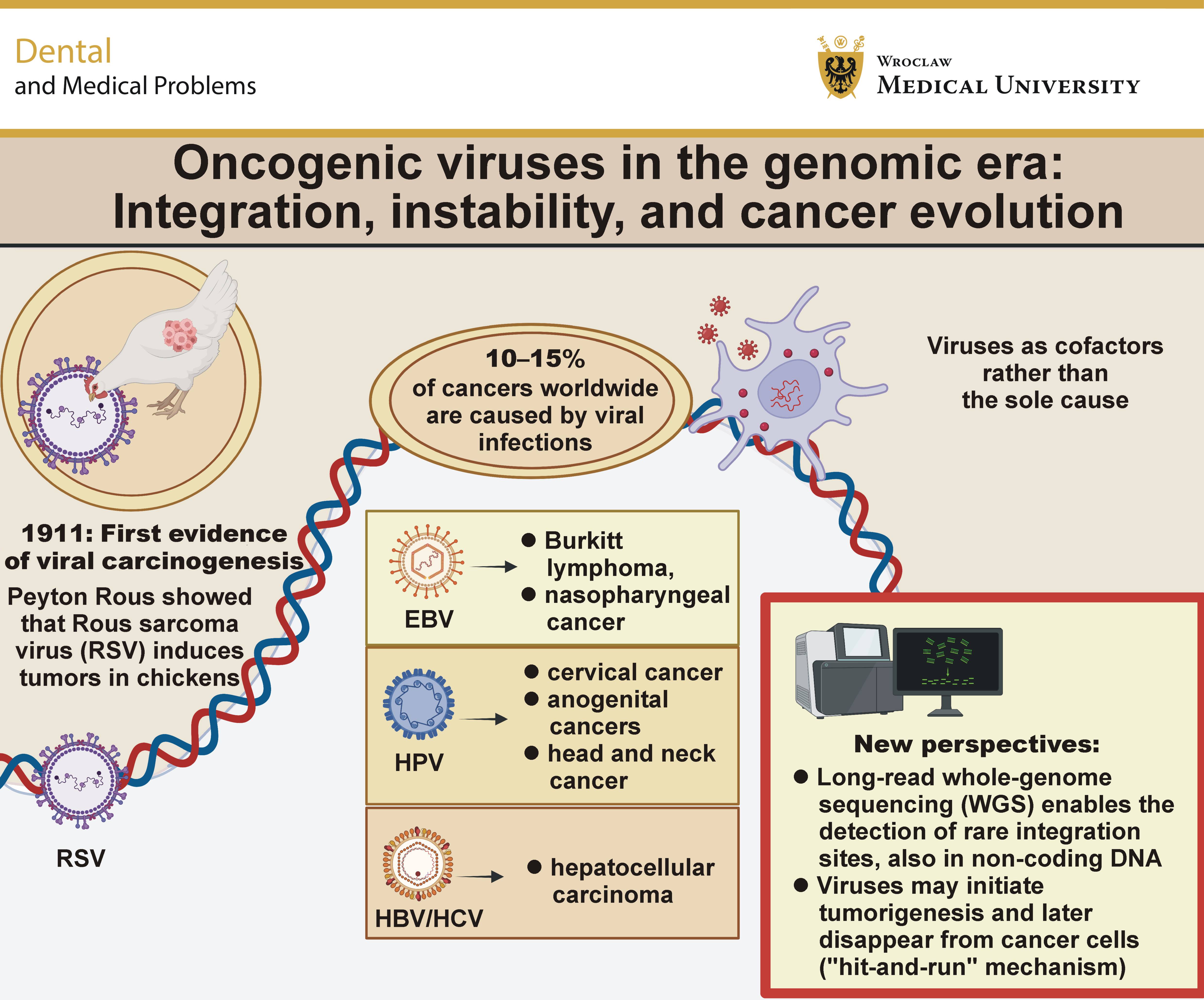
At the beginning of the 20th century, cancer lacked a unifying causal framework. In 1911, Peyton Rous demonstrated that a transmissible agent, Rous sarcoma virus (RSV), could induce tumors in chickens, providing the first experimental evidence that cancer could arise from a defined transmissible biological agent.1
This hypothesis gained momentum in the mid-20th century with advances in virology and molecular biology. Oncogenic viruses were progressively identified, beginning with the discovery of Epstein–Barr virus (EBV) in Burkitt lymphoma cells in 1964, followed by the demonstration of its ability to immortalize human lymphocytes in 1967, and the recognition of hepatitis B virus (HBV) as the causative agent of serum hepatitis in 1965.2 Over time, several human tumor viruses were firmly established, including human herpesvirus 8 (HHV-8; Kaposi sarcoma), human papillomavirus (HPV; cervical and other anogenital cancers), HBV and hepatitis C virus (HCV) (hepatocellular carcinoma), EBV (Burkitt lymphoma, nasopharyngeal carcinoma, and selected lymphomas), and human T-cell lymphotropic virus 1 (adult T-cell leukemia/lymphoma).3, 4, 5, 6 Collectively, these oncogenic viruses are now estimated to account for 10–15% of cancers worldwide.2, 7
These discoveries translated into tangible clinical benefits – prophylactic vaccines against HPV and HBV became the first effective tools for cancer prevention. Their impact is particularly evident for HPV-associated malignancies. A recent global analysis of cervical, penile, vulvar, vaginal, laryngeal, oropharyngeal, oral, and anal cancers estimated 831,204 HPV-attributable cancer cases and 422,935 deaths in 2022.8 In oral oncogenesis, however, the role of HPV appears more heterogeneous than in cervical or oropharyngeal cancer. Human papillomavirus has been detected by genetic testing in 0.0–74.5% of oral squamous cell carcinoma (OSCC) cases, with a weighted mean odds ratio of 17.1 for the detection of HPV-16 or HPV-18 in patients with OSCC.5
However, the early expectation that cancer might be explained by a single viral cause gradually gave way to a more complex model. Most cancers arise through the accumulation of genetic alterations driven by defective DNA repair and environmental exposures. Viruses remain important, but as components of a broader oncogenic landscape, acting as initiators, cofactors or catalysts rather than universal drivers. This broader cofactor-based view is supported by systematic evidence from colorectal cancer, where gut microbiota dysbiosis may contribute to carcinogenesis through chronic inflammation, carcinogenic metabolites and altered host immune responses.9
Today, in the era of genomics, attention has returned to the ways in which viral DNA integrates into the host genome: which chromatin contexts are permissive; how integration sites intersect with genomic instability; and how defects in DNA repair pathways influence these processes. The original search for a singular cause has thus evolved into a deeper insight – viruses are not the universal explanation for cancer, but they provide a powerful lens through which the mechanisms of oncogenesis can be understood.
Among human oncogenic viruses, EBV has emerged as a paradigmatic model linking viral persistence to malignant transformation. Epstein–Barr virus infects more than 90% of the global population and establishes lifelong latency, yet only a small fraction of infected individuals develop malignancy, underscoring the importance of host, environmental and genomic modifiers.10 Epstein–Barr virus is etiologically associated with a broad spectrum of lymphoid and epithelial malignancies, including Burkitt lymphoma, Hodgkin lymphoma, nasopharyngeal carcinoma, and EBV-associated gastric carcinoma, collectively accounting for approx. 1% of all cancers worldwide.11 Despite this well-established association, the mechanisms by which EBV contributes to oncogenesis remain incompletely understood and likely extend beyond a single unifying pathway.10
Traditionally, EBV-driven tumorigenesis has been attributed to latent infection programs, in which viral gene products, such as EBNA1, LMP1 and LMP2, modulate host signaling pathways, promote cell survival and induce epigenetic reprogramming.12 However, accumulating evidence suggests that viral–host genome interactions represent an additional, yet underappreciated, layer of oncogenic regulation.
Although EBV genomes are typically maintained as episomes, integration into the host genome has been repeatedly documented in both lymphoid and epithelial malignancies, where it may disrupt gene expression, alter chromatin architecture and contribute to genomic instability.11 Notably, integration events have been identified near oncogenes such as REL and BCL11A in Burkitt lymphoma, suggesting a potential role in the deregulation of key oncogenic pathways.13
Moreover, EBV integration has been linked to regions of chromosomal fragility and instability, raising the possibility that viral insertion may both exploit and exacerbate vulnerable genomic contexts.14 These observations are consistent with emerging genomic evidence showing that integration sites are enriched in regions prone to DNA damage and microhomology-mediated repair, implicating defective DNA repair pathways as key determinants of viral insertion and its oncogenic consequences.11
Intriguingly, the “hit-and-run” hypothesis further expands this framework by proposing that EBV may initiate oncogenic transformation through transient genomic or epigenetic alterations, even if viral genomes are subsequently lost from tumor cells.10
Building on this framework, recent advances in high-throughput genomic technologies are beginning to redefine our understanding of viral–host interactions in cancer. Conventional approaches, largely based on targeted sequencing or short-read platforms, have provided important insights into viral presence and integration, but remain limited in resolving complex structural events, repetitive regions and deeply intronic insertions. In contrast, whole-genome sequencing (WGS), particularly when incorporating long-read sequencing technologies, now enables a more comprehensive and unbiased interrogation of the cancer genome, including previously inaccessible genomic regions.
These technologies are particularly relevant in the context of EBV, where integration events are relatively rare, heterogeneous, and frequently located outside protein-coding regions. Emerging data indicate that viral DNA often integrates within intronic or intergenic regions, including loci harboring long non-coding RNAs (lncRNAs), regulatory elements, or chromatin domains involved in transcriptional control. Such events may not directly disrupt protein-coding genes, but can nonetheless alter transcriptional programs through enhancer hijacking, epigenetic remodeling, or the modulation of three-dimensional (3D) chromatin architecture.
Our recent work provides an illustrative example of this paradigm.15 Using high-resolution genomic approaches in lymphomas arising in the context of Nijmegen breakage syndrome, we identified multiple EBV-associated genomic alterations within non-coding regions, including 3 distinct loci characterized by deep intronic integration signals. Notably, these regions were enriched for lncRNA elements, suggesting that EBV may preferentially target regulatory genomic compartments rather than canonical oncogenes.15 This observation aligns with a broader shift in cancer genomics, in which non-coding regions are increasingly recognized as critical drivers of oncogenesis.
Importantly, similar patterns have been observed beyond EBV-associated malignancies. In hepatocellular carcinoma, HBV integration has been reported within non-coding regions, including loci associated with regulatory RNAs, where it may influence gene expression and promote tumorigenesis through cis-regulatory effects.16 These parallels suggest that viral integration into the non-coding genome may represent a conserved oncogenic mechanism across distinct virus–tumor systems.
Taken together, the integration landscape revealed by modern genomic technologies supports a model in which viral insertion is not merely a stochastic event, but rather one shaped by chromatin accessibility, DNA repair dynamics, and higher-order genome organization. By enabling the precise mapping of integration breakpoints and their genomic context, long-read WGS provides critical tools to dissect these processes at unprecedented resolution. Ultimately, this integrative approach may bridge the gap between viral infection and malignant transformation, offering new insights into how alterations within the non-coding genome contribute to cancer development.
Ethics approval and consent to participate
Not applicable.
Data availability
Not applicable.
Consent for publication
Not applicable.
Use of AI and AI-assisted technologies
During the preparation of this work, the authors used ChatGPT (OpenAI) for language editing and stylistic refinement. The authors reviewed and edited the content, and take full responsibility for the final version of the manuscript.