



Abstract
Background. Clinical use of mineral trioxide aggregate (MTA) is often associated with several challenges, including a prolonged setting time, poor handling characteristics and compromised physical properties in the presence of tissue fluids. Mineral trioxide aggregate, when used as a perforation repair or root-end filling material, needs adequate strength to withstand the forces acting on it. Recently, several researchers have attempted to overcome these challenges through the use of various additives.
Objectives. To evaluate the physical and antimicrobial properties of MTA reinforced with gold nanoparticles (AuNPs).
Material and methods. Sixteen customized putty molds (6 mm in height x 4 mm in diameter) were filled with the materials assigned to 2 groups: control group, MTA (n = 8); and experimental group, MTA + AuNPs (n = 8). Compressive strength (CS) was evaluated using a universal testing machine, while setting time was assessed using a flat-ended indenter. The antimicrobial properties of MTA and MTA + AuNPs were evaluated using the agar diffusion method against Enterococcus faecalis, Escherichia coli, Pseudomonas aeruginosa, Staphylococcus aureus, and Candida albicans. The data was statistically analyzed using an unpaired t-test.
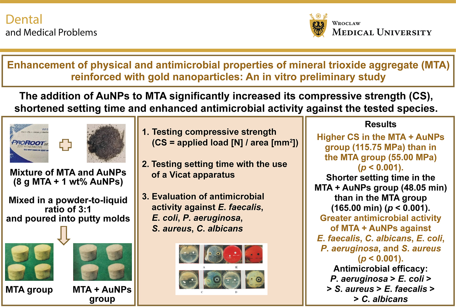
Results. Compressive strength was significantly higher in the MTA + AuNPs group (115.75 ±2.22 MPa) than in the MTA group (55.00 ±5.35 MPa) (p < 0.001). The MTA + AuNPs group also demonstrated a significantly shorter setting time than the MTA group (48.05 min vs. 165.00 min, respectively). The unpaired t-test revealed a significantly greater antimicrobial activity against all tested microorganisms in the MTA + AuNPs group than in the MTA group (P. aeruginosa > E. coli > S. aureus > E. faecalis > C. albicans).
Conclusions. Incorporation of AuNPs into MTA enhanced CS, shortened setting time and increased antimicrobial activity against the tested microorganisms.
Keywords: compressive strength, mineral trioxide aggregate, gold nanoparticles, setting time, antimicrobial efficacy
Introduction
Mineral trioxide aggregate (MTA), owing to its superior biocompatibility and cementogenic properties, has shown successful outcomes when used for perforation repair, pulp capping, apexification, or as a retrograde filling material.1 Hydration of this silicate-based biomaterial with distilled water leads to the formation of calcium silicate hydrate and calcium hydroxide in the crystalline phase of set MTA, followed by the formation of hydroxyapatite (Ca10(PO4)6(OH)2). An extended setting time, poor handling characteristics and compromised physical characteristics in the presence of tissue fluids are the main challenges of MTA, especially when used as a perforation repair or root-end filling material, where adequate strength is required to withstand the forces acting on the material.2 Recently, a number of researchers have attempted to address these issues by utilizing specific additives (calcium chloride, bioactive glass, TiO2 particles) and by using alternative hydration liquids (calcium chloride and calcium gluconate).3
In recent years, nanotechnology has emerged as a promising approach for enhancing the physical properties of various dental materials.4 A greater surface-to-volume ratio and a greater quantity of atoms at their grain peripheries make nanoparticles (NPs) unique. The physical and chemical properties of materials become quite different when the size and number of atoms are greatly reduced.5, 6 Nanoparticles have the ability to be easily arranged in various configurations due to their high surface-to-core ratio, thus making them amenable to manipulation and enabling diverse applications. Nanoparticles have found their place in various aspects of dental sciences, including the use of titanium dioxide or zirconium dioxide NPs for improving the physical properties of denture resin, the addition of bioglass, zirconia and glass ceramics to endodontic sealers, or the addition of nanosilver to root canal disinfectants, to name a few.7
A nanoparticle that has recently garnered significant interest is the gold nanoparticle (AuNP). Being one of the oldest dental restorative materials, gold is known for its superior antibacterial properties and high biocompatibility.8 When converted to an NP form, its properties are further enhanced. Gold NPs have high chemical stability, photostability and low toxicity.9 The use of organic complexes of Au(I) and Au(III) ions as a mode of antibiotic delivery enhances the bactericidal effect of the antibiotics.10 Many studies have investigated the use of AuNPs in dentistry, including applications in AuNP-coated dental floss, AuNP-modified dental aligners, AuNP-stabilized dental implants, and tumor screening.11, 12
The present study aimed to evaluate the potential positive effects of the incorporation of AuNPs on the physical and antimicrobial properties of MTA. The null hypothesis was that the addition of AuNPs would not enhance the antimicrobial efficacy, increase the compressive strength (CS) or reduce the setting time of MTA.
Material and methods
Synthesis of citrate-capped AuNPs
Gold nanoparticles (30 nm in diameter) were produced by refluxing 0.625 mL of 1% aqueous trisodium citrate with 50 mL of 0.25 mM chloroauric acid (HAuCl4) until the solution turned wine red. Trisodium citrate acted as both a reducing and a stabilizing agent. The resulting AuNP solution was collected and lyophilized for further use. The size of the AuNPs was confirmed by placing 2.0 μL of the AuNP solution at a concentration of 0.1 mg/mL−1 in purified water (Milli-Q® H2O; Merk Millipore, Darmstadt, Germany) on a carbon-coated grid and allowing it to dry. The sample was then examined with the use of a transmission electron microscope (Leo 912 AB; Carl Zeiss, Oberkochen, Germany) at 120 kV (×40,000 magnification).13
Preparation of a mixture of MTA and AuNPs
Sixteen customized cylindrical molds made of addition silicone putty, with a height of 6 mm and an internal diameter of 4 mm, were prepared (Advancing Standards Transforming Markets (ASTM) standard E384).14 Mineral trioxide aggregate (8 g) and AuNPs corresponding to 1% of the MTA weight (0.08 g) were weighed using an electronic weighing scale (Wensar, Chennai, India). The materials were combined and mixed with a stainless-steel spatula on a glass slab using saline as the liquid phase at a powder-to-liquid ratio of 3:1. The resulting MTA–AuNP mixture constituted the experimental material.
For comparison of the physical properties, the specimens were allocated to 2 groups:
• control group: MTA alone (n = 8);
• experimental group: MTA containing 1% AuNPs by weight (n = 8).
Compressive strength testing
Four samples from each group were used for CS testing. The mixed cement was packed into the molds and stored in an incubator at 37°C for 4 days. After setting, the specimens were removed from the molds and visually inspected for voids and chipped edges.
Compressive strength was evaluated with a universal testing machine (Heico, New Delhi, India). A compressive load was applied parallel to the long axis of the molds at a crosshead speed of 1 mm/min until the fracture occurred. The maximum load required to fracture each specimen was recorded. Compressive strength was calculated using the following formula:
CS = applied load [N] / area [mm2]
Setting time testing
Setting time was evaluated in accordance with ISO 9917-1. A flat-ended indenter measuring 1.0 mm in diameter and carrying 400 g load was carefully applied vertically to the surface of the tested material. The procedure was repeated every 60 s. The final setting time was noted when the indenter no longer produced an indentation on the material.15
Antimicrobial activity testing
Antimicrobial activity was evaluated against 4 bacterial species: Enterococcus faecalis (ATCC 29212); Escherichia coli (ATCC 25922); Pseudomonas aeruginosa (ATCC 15692); and Staphylococcus aureus (ATCC 29213), as well as 1 fungal species, Candida albicans (ATCC 10231). Antimicrobial efficacy was assessed using a double-layer agar diffusion method. Plates containing fresh cultures were prepared for all microorganisms tested. Two wells, each measuring 4 mm in depth and 6 mm in diameter, were prepared in each plate using a sterile copper band and filled with the respective test materials. The plates were maintained at room temperature for 2 h to allow pre-diffusion of the materials and were subsequently incubated at 37°C for either 24 h or 72 h. The microbial inhibition zones surrounding the set materials were measured using a caliper with a precision of 0.1 mm.
Statistical analysis
An independent samples t-test was used to compare the mean values of the physical and antimicrobial properties between the 2 groups. A two-sided (two-tailed) test with a significance level of α = 0.05 was used. A p-value <0.05 was considered statistically significant.
Results
Table 1 and Table 2 present the mean and standard deviation values for the physical properties and growth inhibition zone diameters against the different microorganisms tested in the MTA and MTA + AuNPs groups. For both physical properties evaluated, the unpaired t-test demonstrated a significant effect of material type (with and without AuNPs) on setting time and CS. Compared with the control group, the experimental group showed significantly higher CS and lower setting time values (p < 0.001). The unpaired t-test also revealed significant differences between MTA and MTA + AuNPs in antimicrobial activity against E. faecalis, C. albicans, E. coli, P. aeruginosa, and S. aureus (p < 0.001). Overall, the antimicrobial activity ranked in descending order as: P. aeruginosa > E. coli > S. aureus > E. faecalis > C. albicans.
Discussion
The first known use of AuNPs dates back to 1971.16 Gold nanoparticles are characterized by chemical stability, compatibility and antibacterial activity, making them a potential additive to dental cements, particularly because they can retain their nanometric dimensions in the presence of an appropriate stabilizer.8 In the present study, AuNPs were produced using a reduction method with sodium citrate as the reducing agent. This approach is widely employed due to its versatility and the ease with which particle size and shape can be controlled. To prevent NP aggregation, citrate was used as a capping agent to stabilize the NPs.17
The physical parameters evaluated in the present study were setting time and CS, while antimicrobial efficacy was assessed against the bacterial species (E. coli, E. faecalis, S. aureus, P. aeruginosa) and the fungal species (C. albicans). Gram-positive bacteria are predominant in primary root canal infections, while E. faecalis and yeasts such as C. albicans are more commonly found in retreatment cases.18
High CS is a critical property of any dental restorative material, as the restoration must withstand masticatory forces that may reach 284 N.19 The results of the present study showed a significant increase (110.5%) in mean CS in the experimental group compared with the control group (115.75 ±2.22 MPa vs. 55.00 ±5.35 MPa, respectively; p < 0.001) (Table 1). High CS is particularly desirable when MTA is used as a perforation repair material, for pulp capping, or as an apical plug in apexogenesis. It has been reported that the initial CS of MTA is 40 MPa and may increase to 67 MPa within 3 weeks.19 The CS of calcium silicate-based biomaterials reflects the extent to which hydration and maturation occur during cement setting. The increase in CS observed after the incorporation of AuNPs may be attributed to accelerated hydration, reduced cement porosity, increased density of particle interfaces, and the ability of the NPs to act against compressive forces.20, 21
Similar outcomes with the use of NPs have been reported in previous studies. Samiei et al. observed a substantial increase in the CS of MTA following the incorporation of 1% by weight TiO2 NPs, while Elsaka et al. noted improved CS when glass ionomer cement (GIC) was reinforced with TiO2 NPs.22, 23 Reinforcement of GIC with 3% TiO2 NPs improved CS; however, higher concentrations of 5% and 7% adversely affected surface microhardness. This effect may be attributed to the smaller particle size and larger surface area of TiO2 NPs compared with glass particles, resulting in an insufficient amount of the polyacrylic ionomer matrix to effectively bind the increased quantity of TiO2 NP powders and consequently weakening the interfacial bonding between the particles and the ionomer matrix. Interestingly, another study by Samiei et al. reported contrasting results, with a noteworthy reduction in the CS of MTA following the incorporation of 2% by weight Ze–Ag–Zn NPs.24 These findings suggest that both NP size and the amount incorporated have an effect on the physical properties of the biomaterial. As no previous studies have evaluated the effect of AuNP incorporation on the properties of MTA, a concentration of 1% AuNPs by weight was selected based on findings obtained with other nanoparticles. Higher AuNP concentrations might adversely affect the adequacy of MTA constituents required to maintain its inherent bioactive and physical properties.
A long setting time of almost 3 h has long been considered a drawback of MTA.25 This is particularly relevant when MTA is used as a root-end filling material following apical surgery or as a perforation repair material, because the unset material may be susceptible to contamination or washout. Clinically, a setting time of up to 25–30 min is desirable, as faster setting reduces the likelihood of contamination and provides greater initial strength, thereby reducing the risk of material washout. The results of the present study showed that the mean setting time was significantly shorter in the MTA + AuNPs group than in the MTA group (48.05 ±0.40 min (range: 47.50–48.40 min) vs. 165.00 ±1.63 min (range: 163–167 min), respectively; p < 0.001).
A smaller particle size of the powder reactant provides a larger surface area per unit mass, which catalyzes the reaction. Thus, the setting reaction is influenced by the size of the NPs incorporated into the material.26 The nanostructure and composition of the unreacted powder facilitate rapid hydration and the formation of a nanoporous calcium aluminate silicate hydrate microstructure, thereby promoting both rapid setting and an effective bioactive response.27
Evaluation of the antimicrobial properties showed that MTA reinforced with AuNPs produced significantly larger zones of inhibition against all tested microorganisms than MTA alone. The antimicrobial activity ranked in descending order as: P. aeruginosa > E. coli > S. aureus > E. faecalis > C. albicans.
Previous studies have similarly demonstrated the potential of nanotechnology to improve the antimicrobial efficacy of MTA. In a study conducted by Samiei et al. in 2013, the antimicrobial properties of MTA and MTA containing silver NPs (MTA/SN) were tested against E. faecalis, P. aeruginosa, S. aureus, and C. albicans. For E. faecalis, C. albicans and P. aeruginosa, the microbial inhibition zones produced by MTA/SN were significantly larger than those produced by MTA alone.28 The results of the present study are also consistent with those reported by Akbari et al., who observed increased antimicrobial efficacy of MTA with and without silver NPs against E. faecalis, S. aureus and C. albicans.29
In the present study, the antimicrobial effect of AuNPs differed between gram-positive and gram-negative bacteria. The difference may be related to differences in bacterial membrane structure, particularly the thickness of the peptidoglycan layer, which is 50% higher in gram-positive than gram-negative bacteria.29
The significant difference between the 2 groups may also be related to direct interactions between AuNPs and bacterial cells, potentially resulting in membrane penetration and disruption of cellular function. The high surface-to-volume ratio of NPs may enhance their antibacterial activity by promoting intracellular generation of reactive oxygen species.30 Gold nanoparticles incorporated into MTA exerted antimicrobial effects by altering membrane potential and reducing adenosine triphosphate synthase activity, thereby disrupting bacterial metabolism. Another significant advantage of NPs is that, unlike antibiotics, they are less likely to promote bacterial resistance, as NPs directly interact with bacterial cell walls, leading to cell death.31, 32
The present investigation was limited by its in vitro design and relatively small sample size. Further research should evaluate different concentrations and particle sizes of AuNPs. A clinical study will be required to validate the results obtained in the present study.
Conclusions
The findings of the study suggest that the addition of AuNPs to MTA increases CS, reduces setting time, and enhances antimicrobial activity against the tested species. Reinforcement of MTA with AuNPs appears to improve material properties and may therefore have potential clinical relevance. Further studies evaluating different AuNP concentrations are warranted.
Ethics approval and consent to participate
Approval from the Institutional Ethics Committee was obtained prior to the commencement of the study (Ref. No. 90th ECM II BThesis/P9).
Data availability
The datasets generated and/or analyzed during the current study are available from the corresponding author on reasonable request.
Consent for publication
Not applicable.
Use of AI and AI-assisted technologies
Not applicable.