Abstract
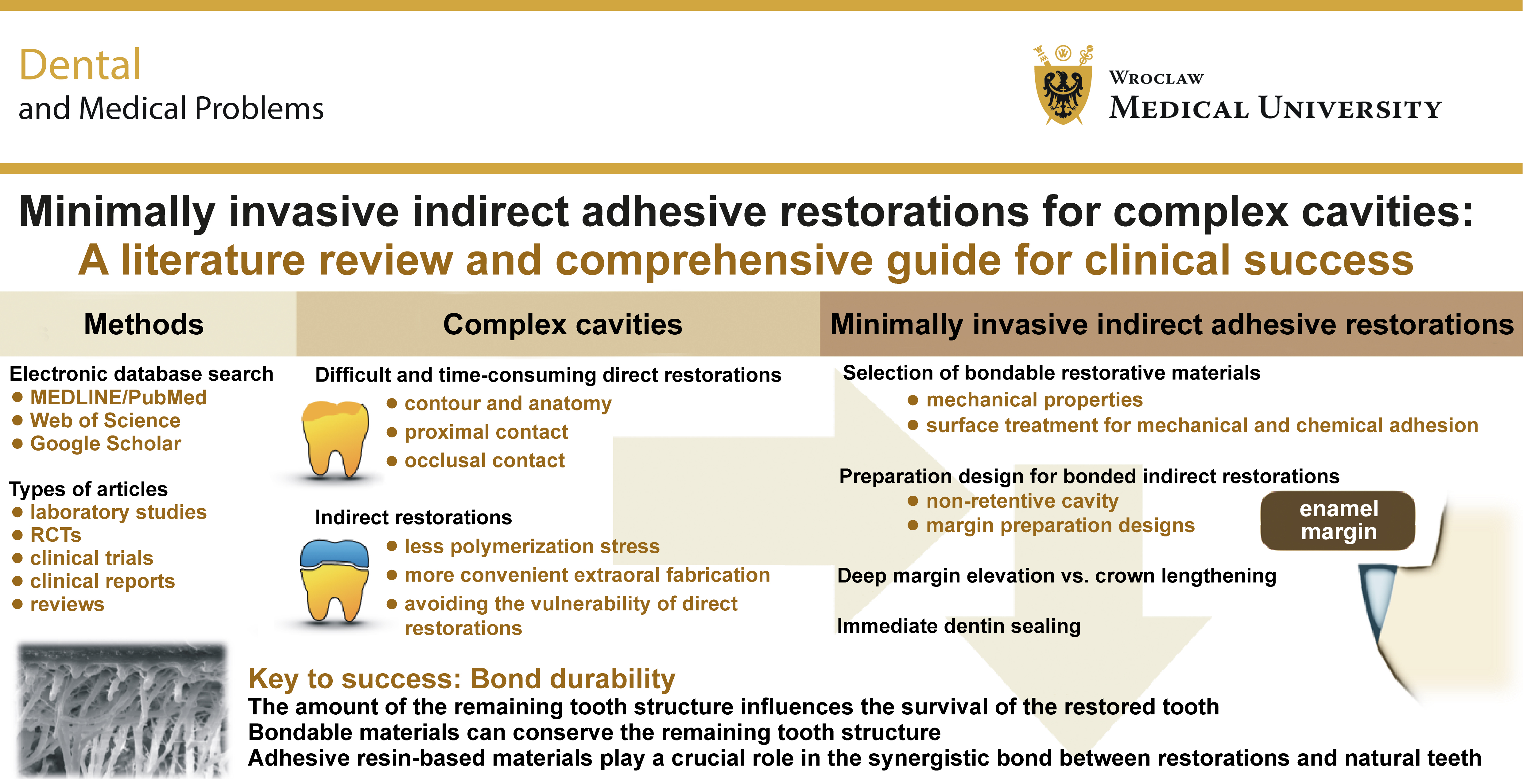
A complex cavity is so named because it results from damage to the enamel–dentin complex, and such cavities can cause patients considerable difficulties in daily life. The reconstruction of damaged areas with synthetic restorative materials requires careful consideration of the unique functional properties of natural enamel and dentin, whose inherent characteristics remain superior to those of any synthetic restorative material currently available. The objective of this article was to organize the available information on indirect adhesive restorations for complex cavities, with particular emphasis on the factors and techniques that require consideration during clinical procedures.
An electronic search was performed in MEDLINE/PubMed, the Web of Science and Google Scholar. The articles included in this study comprised randomized controlled trials (RCTs), clinical trials, clinical reports, reviews, and laboratory studies.
The amount of the remaining natural tooth structure directly affects the longevity of the restored tooth. The remarkable properties of natural enamel and dentin have long been recognized, and bondable restorative materials have therefore been continuously developed with the aim of preserving these natural tissues and reinforcing the remaining tooth structure through effective adhesion. Even when a large portion of the tooth structure has been destroyed, conservative treatment can often still be performed, allowing the benefits of these advanced techniques to be achieved. All aspects of the procedure need to be considered, including material selection, cavity design and preparation, cavity and restoration surface preparation, and the cementation procedure. Additional techniques, including immediate dentin sealing (IDS) and the controversial deep margin elevation technique, are also addressed in this article.
Keywords: indirect adhesive restorations, bondable restorative materials, non-retentive adhesive restorations, complex cavity, minimally invasive restorations
Introduction
A common dental problem that brings patients to the dentist is the presence of a large cavity. Large cavities are often associated with troublesome food retention, uncomfortable tooth sensitivity or a precarious sharp edge. A large cavity can result from a variety of causes, such as active dentin caries beneath fragile enamel, secondary caries beneath a previous restoration, or a partial fracture of the natural tooth structure.1, 2
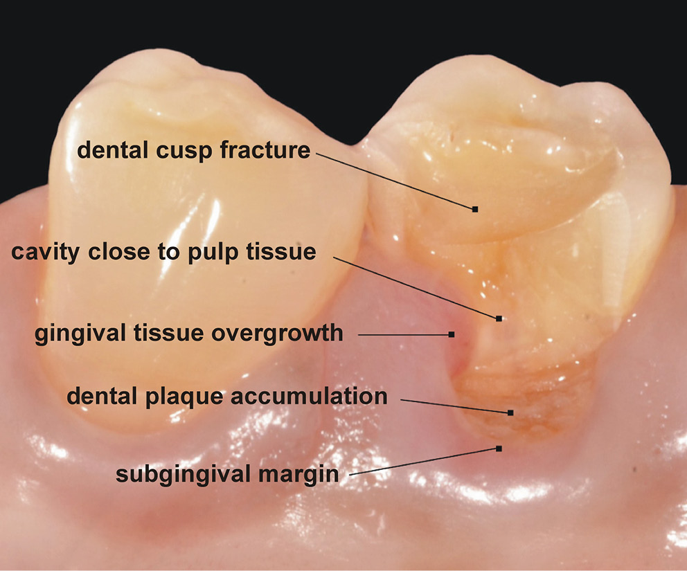
A large cavity often involves 2 tooth surfaces, and then it is referred to as a compound cavity. When the damage is extensive and involves 3 or more surfaces, it is referred to as a complex cavity.3 The restoration of a complex cavity is complicated by the fact that it often involves subgingival margins, including gingival overgrowth, or a cavity extending close to the pulpal tissue (Figure 1). In addition to the mechanical aspect of withstanding chewing forces, this situation also involves the biological aspect of ensuring that the tooth can function properly during mastication. There is also an esthetic aspect, as all patients naturally desire a natural-looking restoration.4
Materials for dental restorations have been intentionally developed to mimic the properties of the natural tooth structure for reconstruction. However, enamel and dentin are often destroyed simultaneously, and both need to be replaced, even though their properties are quite different. Enamel is brittle and stiff, with a high elastic modulus of approx. 80 GPa, whereas dentin is more flexible, with an elastic modulus of approx. 20 GPa.5 This represents a highly effective combination created by nature, in which the strong outer shell of enamel and the resilient inner layer of dentin work together to prevent enamel from shattering when the tooth is subjected to fatigue loading.6 To date, there are still no restorative materials that can adequately substitute for the natural enamel–dentin combination when it is damaged.
Conservative dental treatment, or conservative dentistry, is an intellectual concept aimed at preserving as much of the unique natural tooth structure as possible, since the properties of all artificial materials fall short of those of the original tooth structure. Unbondable restorative materials require the otherwise unnecessary removal of a sound tooth structure to create a retentive form that holds the restoration in place through macromechanical retention.7 Consequently, the remaining tooth structure becomes more susceptible to fracture, as unbondable materials cannot replace the natural reinforcement provided by the tooth structure that has been removed. Thus, removing any part of the tooth for the purpose of creating macromechanical retention inherently weakens the remaining natural tooth structure.7, 8, 9 In contrast, by definition, bondable materials adhere to an intact tooth structure through micromechanical retention. For this reason, the intact portion of the tooth should be preserved as much as possible to provide a large tooth surface area for effective adhesion.10 These bondable materials can also reinforce the remaining tooth structure, thereby reducing the risk of subsequent fracture.8, 9, 11
A comprehensive search for the existing reviews on dental adhesive indirect restorations was conducted using MEDLINE/PubMed, the Web of Science and Google Scholar. This search revealed that most reviews focused exclusively on either cavity preparation techniques or the treatment of cavity surfaces before cementation. However, comprehensive reviews integrating all relevant procedures and the rationale behind them are lacking. This review aims to address this gap by collecting and organizing the available information on dental adhesive indirect restorations, with particular emphasis on the factors and techniques that need to be considered during the clinical process, as these can significantly affect the success of treatment.
Methods
An electronic database search, with no date restrictions, was performed using MEDLINE/PubMed, the Web of Science and Google Scholar. The articles included in this study comprise randomized controlled trials (RCTs), clinical trials, clinical reports, reviews, and laboratory studies. Only articles published in English were included. Titles and abstracts were screened as the initial screening step. The selected articles were retrieved, read in full, and then included in this review. An additional search for relevant information was performed by reviewing the references of the selected articles.
Bondable restorative materials
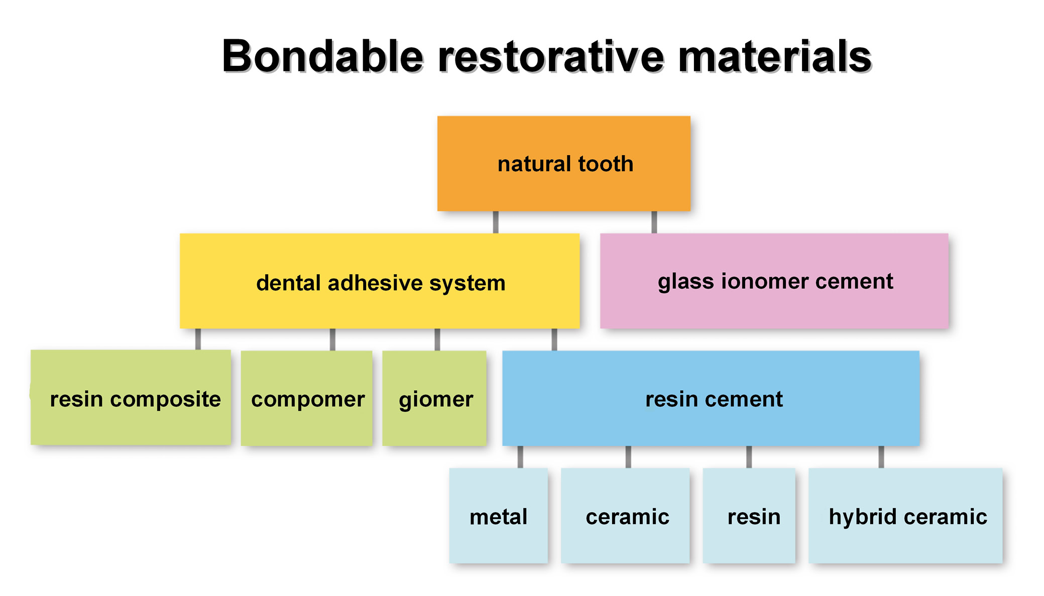
There are essentially 2 types of bondable materials used with the natural tooth structure: glass ionomer cement (GIC); and dental adhesives.12 Glass ionomer cement initially establishes hydrogen bonds with the tooth through the free carboxyl groups of the cement binding to the bound water on the tooth surface.13 These hydrogen bonds are gradually replaced by the ionic bonds formed between the carboxylate groups of poly(acrylic acid) in GIC and the calcium ions of hydroxyapatite, which also results in the formation of an ion-exchange layer at the tooth–GIC interface.14, 15 Dental adhesives are currently the most widely used bondable materials and provide reliable micromechanical adhesion to the tooth structure.16 Dental adhesives are used in 2 main ways – for direct and indirect restorations. Direct restorations are made of conventional resin-based materials, including resin composites, compomers and giomers, which bond directly to the dental adhesive. In contrast, indirect restorations are made of materials such as metal, ceramic, resin, or hybrid ceramic, and require an intermediate layer of resin cement to establish a bond with the dental adhesive. Thus, dental adhesives are versatile materials that can bond to almost all types of dental restorative materials (Figure 2).
When attempting to restore a complex cavity using the direct technique, it is difficult to create good contours of the proximal surfaces and the precise occlusal anatomy corresponding to the opposing tooth. Direct restorations are also time-consuming, and they provide good functionality for only a medium term, 3–5 years, in a complex cavity.17, 18, 19 They frequently encounter bond degradation, leading to marginal leakage, broken restorative materials, or ineffective restoration anatomy and contour.20, 21 For long-term success, bonded indirect partial restorations are suggested as a better alternative.
These more durable restorations are considered minimally invasive for large defects in posterior teeth.1, 22, 23, 24 The amount of the remaining intact natural tooth structure influences the survival of the restored tooth by helping to prevent catastrophic failure.2, 25 Bonded indirect partial restorations can also overcome some of the limitations of direct restorations. Indirect restorations with a cement thickness of less than 200 µm generate less polymerization stress than direct resin composite restorations with a thickness of 2 mm.26 They also provide more accurate proximal contacts and occlusal relationships with the neighboring teeth as a result of more convenient extraoral fabrication. Bonded indirect partial restorations offer an opportunity to preserve and strengthen the remaining natural tooth structure, and Figure 3 shows various designs typically used to achieve this.1, 25, 27, 28 The synergistic bond between bondable restorative materials and the natural tooth structure, mediated by adhesive resin-based cement, plays a crucial role.2, 25, 27, 29
Today, resin cements are by far the most commonly used adhesive cements. Adhesive cements must bond to a variety of substrates, including enamel, dentin, ceramic, metal, and resin.30 Resin cements can be classified according to their polymerization mechanism as light-cured, chemically cured or dual-cured, or according to their adhesive strategy as total-etch, self-etch or self-adhesive.31, 32 The ability of light to penetrate all areas of the cement and activate its photo-initiators is vital to the longevity of light-cured resin cement; therefore, thin, translucent restorations are recommended when using light-cured resin cement. Light-cured resin cement has greater color stability than chemically cured or dual-cured resin cements, and provides a convenient working time before setting.31 Dual-cured resin cement is polymerized by both chemical and light activation. This type of cement is indicated when the restoration is thick or opaque, such that light cannot adequately penetrate it. However, dual-cured resin cement still requires light activation to achieve a high degree of polymerization.33, 34
Restorative materials for non-retentive indirect restorations
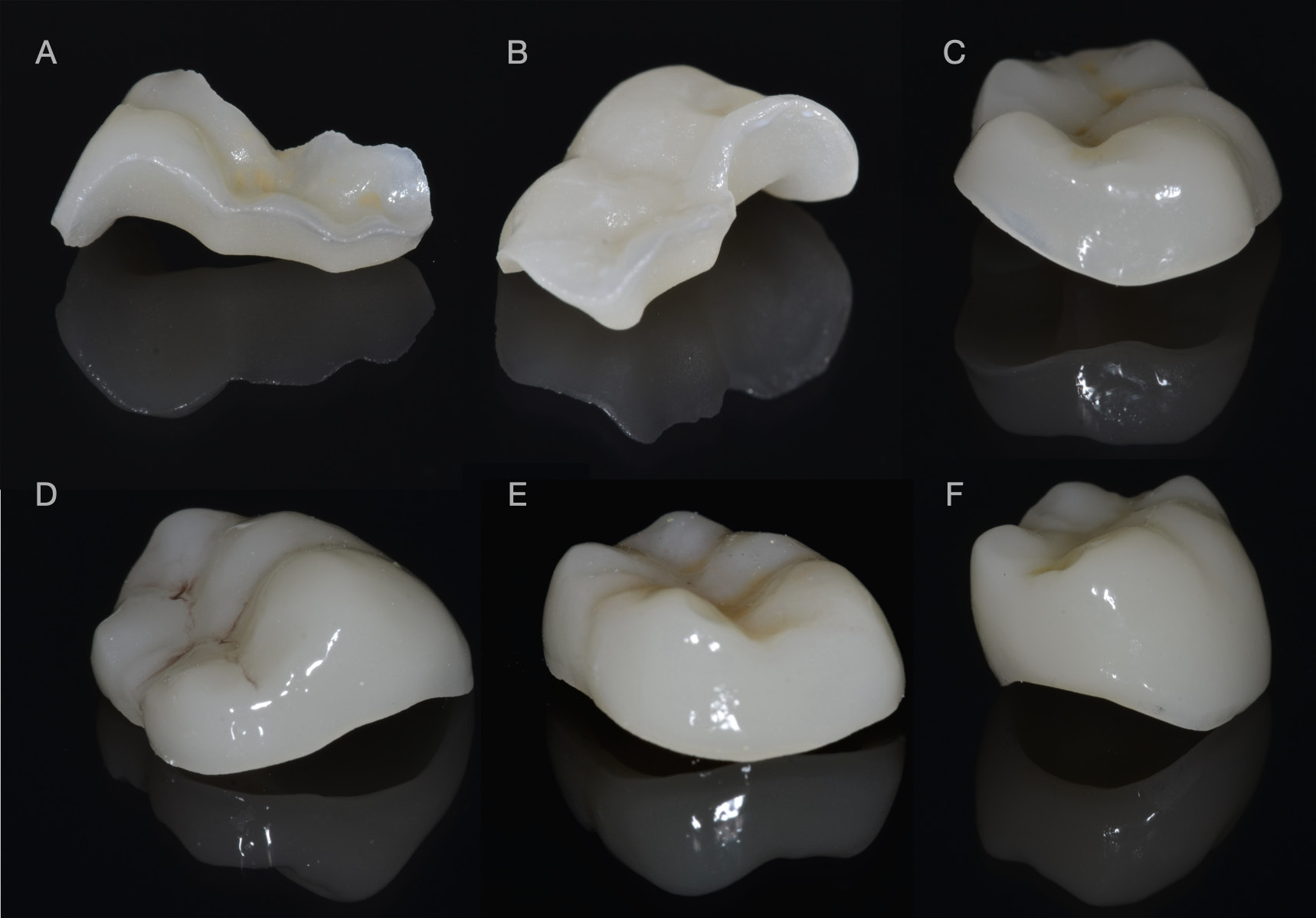
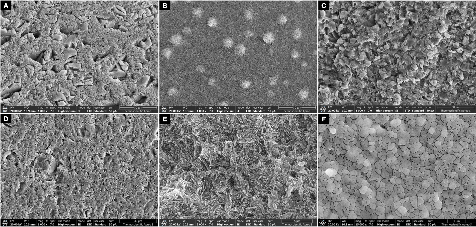
This review investigates dental materials commonly used for tooth-colored bonded indirect restorations, as shown in Figure 4. These materials include indirect resin composite, polymer-infiltrated ceramic network (PICN), feldspathic ceramic, lithium disilicate glass ceramic, and zirconia. The retention of bonded indirect restorations relies on adhesive bonding to the tooth structure, and the quality of the bond is critical to a long-term success.35 Among tooth-colored bonded indirect restorative materials, etchable ceramics, such as feldspathic ceramics, leucite-reinforced glass ceramics and lithium disilicate glass ceramics, provide satisfactory bond strength to the natural tooth structure.22, 30, 36 However, feldspathic ceramics (flexural strength of 60–70 MPa; fracture toughness of 0.70–1.30 MPa·m½)37, 38 and leucite-reinforced glass ceramics (flexural strength of 107–160 MPa; fracture toughness of 1.28–1.38 MPa·m½)39, 40 exhibit a higher frequency of fracture than stronger lithium disilicate glass ceramics (flexural strength of 370–460 MPa; fracture toughness of 2.80–3.50 MPa·m½).23, 41, 42 Therefore, highly etchable lithium disilicate glass ceramics are recommended for restoring complex posterior cavities using the bonded indirect technique.43
Lithium disilicate glass ceramic
Lithium disilicate glass ceramic provides high adhesive strength to the tooth structure through both micromechanical and chemical bonding mechanisms. Microroughness is created on the intaglio surface of this etchable ceramic by applying 4.5% hydrofluoric acid for 20 s.44 However, aggressive treatment with either a higher concentration or prolonged application of hydrofluoric acid can negatively affect mechanical performance, resulting in reductions in flexural strength and bonding potential, and can also compromise the long-term success of the restoration, particularly in low-thickness restorations.45, 46, 47, 48 After creating an irregular surface, silane is applied to promote a chemical reaction that forms strong siloxane linkages between the silica in the ceramic and the methacrylate groups in resin-based materials.49, 50
Self-etching ceramic primers were developed as a single-component alternative to the hydrofluoric acid and silane treatment described above. The principal advantage of self-etching ceramic primers is that they simplify the technique and reduce technique sensitivity by decreasing the number of clinical steps.51 The self-etching ceramic primer is applied to the intaglio surface of the glass-based ceramic material for 20 s using an agitating microbrush, and is then allowed to react for an additional 40 s. When the bond strength achieved with self-etching ceramic primer treatment is compared with that of the conventional procedure (hydrofluoric acid followed by silane), the conventional procedure provides a higher bond strength across all tested ceramic types, including leucite-based ceramic, lithium disilicate glass ceramic, zirconia-reinforced lithium disilicate glass ceramic, and hybrid ceramic.52 In addition, the bond strength of the materials treated with the self-etching ceramic primer decreases significantly after simulated aging.52, 53
The currently available lithium disilicate glass ceramics have different crystal structures. Rod-shaped and platelet-shaped crystals are common characteristics, but each material contains a different proportion of these 2 crystal morphologies.54 After heat treatment, lithium disilicate glass ceramic exhibits reduced porosity and increased density due to the tight interlocking of the disilicate crystals. The mismatch in thermal expansion coefficients between the lithium disilicate crystals and the glassy matrix generates compressive stress around the crystals, thereby impeding subsequent crack propagation within the material.42 Regarding friction and wear, lithium disilicate glass ceramic has abrasiveness similar to that of enamel.55 It also exhibits excellent biocompatibility, including a favorable soft-tissue response, low plaque retention, and good adhesion and proliferation of human epithelial cells and gingival fibroblasts when the ceramic surfaces are properly polished.56, 57
Indirect resin composite
Indirect resin composite restorations represent improvement over direct restorations in terms of physical properties, such as hardness, wear resistance, color stability, polymerization shrinkage, and biocompatibility, as they are subjected to heat, visible-spectrum light, vacuum, and pressure during fabrication (flexural strength of 120–160 MPa; fracture toughness of 0.80–1.20 MPa·m½; modulus of elasticity of 8.5–12.0 GPa).58, 59 They also have a higher percentage of filler particles (75–92% by weight) than direct resin composite materials (50–65% by weight).58, 60, 61 Although these procedures produce restorations that are smaller than the prepared cavities because of polymerization shrinkage, the shrinkage is still less than that of direct resin composites. The resulting spaces are ultimately compensated for by the cementation layer.58 Indirect resin composites have lower resistance to compressive stress than ceramics, but they have greater tensile strength and better stress distribution.58 Therefore, they are still used for the restoration of large defects.62 Common indications for indirect resin composites include long-term semi-permanent restorations, such as large restorations in young patients or in teeth undergoing orthodontic treatment.
Indirect resin composite undergoes polymerization with a high degree of conversion, leaving few unreacted monomers available for bonding to the adhesive resin.63 Therefore, indirect resin composite requires mechanical surface preparation. Creating a microrough surface on the indirect resin composite by sandblasting is recommended.64 Before applying the adhesive resin, the mechanical bond to the microrough surface can be enhanced by establishing a chemical bond through the application of a silane coupling agent. The silane coupling agent binds to the silica-based fillers in the indirect resin composite and also increases the wettability of its surface.65
The intraoral repair of indirect resin composite restorations with resin-based materials is less complicated than the repair of ceramic restorations because of the compatibility between the materials.66 The substrate, indirect resin composite, is prepared in the same manner by sandblasting, followed by a direct restoration using a dental adhesive and a resin composite.67, 68
Hybrid ceramic
Hybrid ceramic materials are commercially available as polymer-infiltrated ceramic networks (PICNs), consisting of an 86% interconnected silicate glass-ceramic skeleton, with leucite as the major phase and zirconia as the minor phase, and 14% resin. Some microcracks have been found between the different phases of this material.69 When the fracture toughness of PICN is compared with that of other tooth-colored restorative materials, PICN, at 1.5 MPa·m½, has a lower fracture toughness than almost all types of ceramics, except feldspathic ceramic, which has a fracture toughness of approx. 1 MPa·m½. In comparison, lithium disilicate glass ceramic has a fracture toughness of 2.37 MPa·m½, while zirconia has a fracture toughness of 4.94 MPa·m½.70 Polymer-infiltrated ceramic networks and indirect resin composites have desirable low abrasiveness toward the opposing teeth.71, 72 To create surface irregularities for bonding, the application of 4.5% hydrofluoric acid for 60 s is recommended, as silicate glass ceramic is the major component of PICN and forms its framework.73
Polymer-infiltrated ceramic network has a lower elastic modulus than other ceramic materials. Materials with a low elastic modulus tend to absorb stress and distribute it through the deformation of the restoration rather than transferring it to the underlying resin cement and tooth structure.74, 75 These low-elastic-modulus materials have demonstrated a higher rate of failure, such as displacement or fracture. Nevertheless, they have also been shown to protect the remaining natural tooth structure, particularly against catastrophic fractures.76, 77
Edge chipping is an inherent disadvantage of glass-ceramic materials. The development of PICN has improved this limitation of glass ceramics. However, the edge-chipping resistance of PICN (120 N/mm) remains lower than that of nanohybrid resin composites (275 N/mm), feldspathic ceramics (179 N/mm), and leucite-reinforced glass ceramics (169 N/mm).71
Zirconia
The zirconia used in this study was yttria-stabilized zirconia (YSZ), a biocompatible material widely used as a biomaterial. Yttria-stabilized zirconia can be classified according to its yttria content. Yttria stabilizes the tetragonal phase of zirconia at room temperature, reduces the coefficient of thermal expansion, and affects the grain size of zirconia.78 The classification is often expressed as the mole percentage of yttria in tetragonal zirconia polycrystal (TZP).79, 80 Three-mol% yttria-stabilized tetragonal zirconia polycrystal (3Y-TZP) is an early-developed form of YSZ with excellent mechanical properties (flexural strength of 1,200–1,500 MPa; fracture toughness of 3.50–4.50 MPa·m½; modulus of elasticity of 210.0 GPa), but relatively low translucency. An increase in the yttria content results in the zirconia material containing both tetragonal and cubic phases, classified as partially stabilized zirconia (PSZ). Eight-mol% yttria-stabilized tetragonal zirconia polycrystal (8Y-TZP) has a stable cubic phase at room temperature, and is commonly referred to as cubic stabilized zirconia (CSZ).79, 80
There are 3 main phases of zirconia: monoclinic at room temperature; tetragonal above approx. 1,170°C; and cubic above approx. 2,370°C. The mechanical properties of the monoclinic phase are relatively poor, whereas those of the tetragonal phase are superior. Incorporating dopants, such as yttria, into the starting powder can partially stabilize the tetragonal phase at room temperature, resulting in improved strength and fracture resistance. Under external stress, the metastable tetragonal phase can transform back to the monoclinic phase. This stress-induced transformation is accompanied by the expansion of the zirconia crystals and alterations in their shape, which generates compressive stress around the transformed regions and help impede crack propagation. These changes in the crystal structure increase the energy-absorption capacity and resistance to damage of the material, a phenomenon known as transformation toughening.81, 82
Although stabilization with yttria and transformation toughening reduce the risk of fracture in zirconia, the material remains susceptible to an aging process known as low-temperature degradation. The mechanical properties, particularly strength, of aged zirconia decrease as a result of the spontaneous transformation of the tetragonal phase into the monoclinic phase. During usage, the zirconia lattice develops multiple oxygen vacancies associated with the substitution of trivalent yttrium ions for tetravalent zirconium ions. These oxygen vacancies facilitate the penetration of water into the zirconia lattice.83 The interaction with water destabilizes the tetragonal phase and induces its transformation into the monoclinic phase. This transformation is accompanied by an increase in grain volume, which generates stress at the zirconia surface, and causes grain uplift and the dislodgement of individual grains. Microcracks subsequently develop, allowing further penetration of water into the deeper regions of the material. As the transformation progresses into the bulk of the material, these microcracks can propagate and eventually lead to fracture. In contrast, the cubic phase does not undergo the same stress-induced tetragonal-to-monoclinic transformation; therefore, zirconia with a higher cubic-phase content is less susceptible to low-temperature degradation.84, 85
Zirconia contains no silica in its composition, and therefore cannot be effectively chemically etched, making chemical adhesion alone insufficient for achieving a strong bond.86 The principal method used to enhance the bonding ability of zirconia is the creation of microroughness on the intaglio surface by airborne-particle abrasion with alumina, followed by the application of a phosphate methacrylate monomer, particularly 10-methacryloyloxydecyl dihydrogen phosphate (MDP), to the treated surface to promote chemical bonding.87, 88 An alternative technique is tribochemical silica coating followed by the application of silane. However, tribochemical silica coating has been questioned, as the silica layer formed on the surface may be unstable over time.88
Similar to other restorative materials bonded using adhesive techniques, zirconia exhibits a higher bond strength when bonded to tooth enamel than when bonded to dentin.89 However, the bond strength of zirconia is significantly lower than that of glass-based ceramics, particularly when bonded to dentin, which constitutes much of the substrate in complex cavities.44, 90, 91, 92, 93, 94 Although zirconia has greater fracture resistance than silica-based ceramics, its bonding potential to dentin is limited. For minimally invasive indirect adhesive restorations of complex cavities, tooth preparation commonly results in a non-retentive cavity predominantly involving dentin. Consequently, zirconia is not recommended for non-retentive cavity preparations that rely primarily on adhesion to dentin.
Preparation design for the bonded indirect restorations of complex cavities
General features of the cavity
Catastrophic fractures, whether partial or complete, are the main cause of failure in the ceramic restorations of posterior teeth.95 The preparation design has a significant influence on stress distribution within the restoration when it is subjected to occlusal loading. A proper preparation design can reduce mechanical stress, and thereby decrease the risk of restoration fracture.96, 97 Although indirect resin composites are less brittle than ceramics, the principles of tooth preparation are the same for both materials.
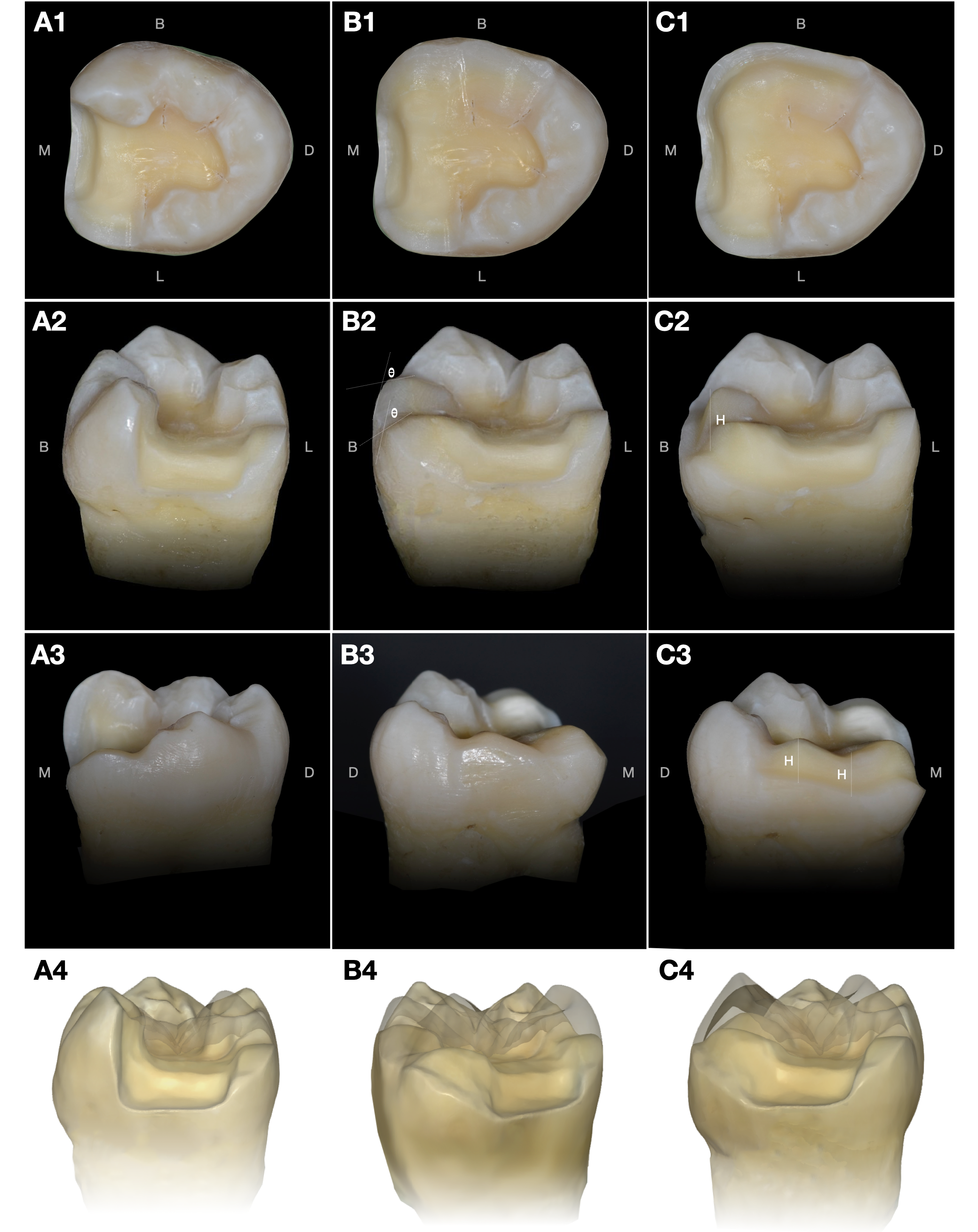
After the removal of defects, the cavity commonly presents with intradentin undercuts and enamel that is unsupported by dentin. Unsupported enamel is no longer considered usable and should be removed initially to allow the adequate visualization of the underlying dentin undercuts. However, all intact, sound enamel without cracks or demineralized areas should be preserved as much as possible, since enamel provides an excellent bonding substrate for adhesive systems. Undercuts within the intact dentin should be blocked out with a bondable resin composite, thereby creating an appropriate path of insertion for the restoration (Figure 5).2, 98, 99, 100, 101
The most important physical feature of the prepared cavity is that all internal edges and line angles must be rounded. A smooth, rounded transition should be created from the pulpal wall to the para-pulpal walls, and from the para-pulpal walls to the approximal cavity walls. Sharp transitions between the cavity walls should be avoided because, as can generate wedging forces between the cement and the restoration, potentially increasing the risk of fracture.95
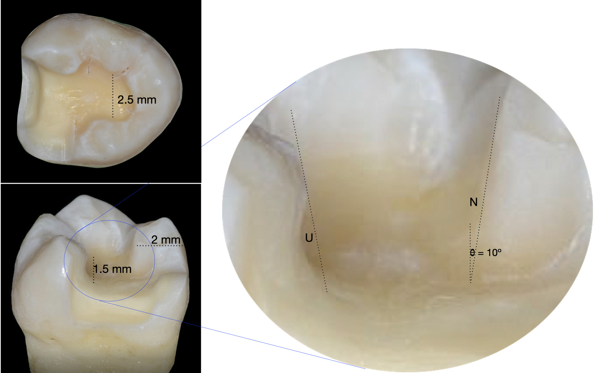
Parallel walls are not required for adhesive bonding, since retention is micromechanical on the surface of enamel and dentin.95 The degree of divergence of the cavity walls affects the susceptibility of ceramics to fracture. When the cavity walls have a large degree of divergence, the restored tooth is more vulnerable to fracture. The cavity walls should not diverge more than 10° (Figure 5).102
Even though the thickness of the restoration has no substantial effect on its fracture resistance when using the adhesive technique, the thickness of a brittle material should not be neglected when it is used in a stress-bearing area.103 Different types of materials from different manufacturers require different ideal thickness for function: lithium disilicate ceramic – 1.5 mm (min. 1.0 mm); PICN – 1.5 mm (min. 1.0 mm); 3Y-TZP – 1.0 mm (min. 0.5 mm); 4Y-TZP and 5Y-TZP – 1.5 mm (min. 1.0 mm). Commonly, the thinnest region of a posterior restoration is at the fissure areas, which should have a minimum thickness of 1.5 mm (Figure 5).104 If there is insufficient clearance space in the fissure area to receive the restoration, a round instrument is recommended to deepen the fissure area. It has been shown that a flat pulpal wall is not required for adhesive restorations.95
If the preparation design involves an intracoronal cavity, the width of the cavity should be at least 2.5 mm, and the thickness of the remaining cusp should be no less than 2 mm (Figure 5).2, 95 A compromised tooth, whether endodontically treated or cracked, should have a thickness of the remaining wall of no less than 3 mm.105, 106 If the preparation cannot meet this requirement, the cusp of the affected wall should be shortened. The proximal margin should be extended without contacting the adjacent tooth. This approach enables clear identification of the margin with either a conventional impression or a digital scan, and facilitates the removal of excess adhesive cement and the finishing of the margin after the restoration is delivered.107
The efficacy of the bond between the restorative material and the remaining tooth structure is vital to the longevity of the restoration. Phosphoric acid-etched enamel provides significantly greater shear bond strength (50–90 MPa) than dentin (45–70 MPa).108 It is recommended to preserve as much intact enamel as possible, as it is more stable over time than dentin.109 This is because enamel is not subject to the enzymatic degradation associated with dentin, which is mediated by matrix metalloproteinases (MMPs) and cysteine cathepsins (CCs).110, 111 The restoration margins are among the most critical areas, as they are particularly susceptible to dental caries, which is a common cause of indirect restoration failure. A non-beveled preparation should be used at the margins of ceramic and resin-based restorations, since adequate material thickness contributes to their strength.112 Therefore, a continuous enamel margin without a bevel and a preparation margin angle of 90° is recommended to maximize bonding effectiveness and marginal durability, thereby contributing to the longevity of the restoration.95, 97
Margin preparation design
Placing the preparation margin on enamel is a reliable approach for ensuring effective bonding with the dental adhesive system. In addition to its effect on bond strength, the marginal design influences the clinical success of the restoration in several other ways. The preparation margin must be sufficiently strong to withstand chewing forces and sufficiently smooth to allow proper seating of the restoration, and facilitate cleaning and plaque removal. Importantly, the restoration margin should fit as closely as possible to the preparation finishing line to minimize the amount of exposed cement, and thereby reduce microleakage and the risk of secondary caries.
When the remaining wall thickness of a compromised tooth is less than 3 mm, the cusp associated with the thin wall should be reduced.2, 95, 105, 106 Importantly, it is recommended that compromised teeth, including endodontically treated and cracked teeth, have all cusps restored and receive restorations with full occlusal coverage.113, 114, 115 The required restoration thickness in a reduced-cusp area depends on the type of material used. Lithium disilicate glass ceramics should be at least 1.5-millimeter thick, whereas feldspathic ceramics and leucite-reinforced glass ceramics should be at least 2-millimeter thick in reduced-cusp areas (Figure 6).105
A shoulder margin preparation with a rounded internal line angle (a radial shoulder) is recommended for resin composite or ceramic restorations. A shoulder margin is a common margin design that can be placed at any location on the prepared tooth and has a width of approx. 1 mm (max. 1.5 mm). A bevel preparation (contra-bevel preparation) is an alternative margin design for areas of cuspal reduction when an esthetic transition between the tooth and the restoration is desired.106 A bevel preparation with an inclination of 45° or greater is performed to avoid unsupported enamel and provide an adequate bulk of the restorative material at the margin (Figure 6).25
The height of the axial walls determines the path of insertion of the restoration, and contributes to its stability and reliability during delivery. The height of the axial walls has no effect on the fracture resistance of the restoration.25 However, an axial wall height of at least 2 mm is recommended for compromised teeth to strengthen the remaining tooth structure by providing a ferrule effect (Figure 6).116
The biologically oriented preparation technique (BOPT) was introduced because gingival recession is one of the most common complications associated with restorations in proximity to the periodontal tissues. This procedure is intended to enhance periodontal tissue stability and promote the coronal migration of the marginal gingival tissue.117 The technique combines tooth preparation without a defined finishing line with the immediate placement of a provisional restoration. A vertical tooth preparation is performed to eliminate the anatomical emergence profile without creating a finishing line.118 Simultaneously, the gingival sulcus is de-epithelialized (gingitage), allowing tissue regeneration.119 The provisional restoration must have an emergence profile that accurately mimics that of the natural tooth. The immediate placement of the provisional restoration helps preserve the biological space, the cervical contour of the tooth, and the blood clot formed following the de-epithelialization of the gingival sulcus. Moreover, the provisional restoration accelerates the tissue-healing process, allowing the surrounding tissues to adapt their form and position to the new restoration profile. The thickened gingival tissue subsequently remodels over time to conform to the definitive restoration.120
Deep margin elevation (coronal margin relocation)
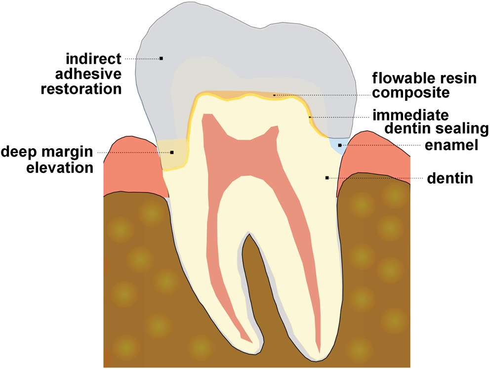
Deep margin elevation is the process of repositioning a subgingival finishing line to a supragingival margin by applying directly bondable restorative materials beneath the margin of an indirect restoration.121, 122 Several benefits can result from deep margin elevation. For example, it facilitates access to otherwise difficult to visualize areas during impression taking, intraoral scanning and restoration delivery.123, 124, 125, 126 The deep margin elevation technique is generally performed in conjunction with immediate dentin sealing (IDS) in the same area to reduce marginal leakage and enhance bond strength (Figure 7).127
In the deep margin elevation procedure, dental adhesives and resin composite materials are used to elevate the prepared margin by no more than 2 mm. Glycerin gel coating is recommended during the final polymerization step, followed by finishing and polishing. A bitewing radiograph should then be taken to evaluate the quality and adaptation of the resin composite elevation.123
In some situations, deep margin elevation can substitute for surgical crown lengthening, thereby avoiding potential anatomical complications, such as furcation exposure or the loss of periodontal attachment.128 However, the decision to use deep margin elevation as an alternative depends on the individual patient, as the invasion of the biological width has also been reported following this procedure.129, 130 Surgical crown lengthening is generally required when the distance between the restoration margin and the alveolar bone crest is less than 2 mm. When this distance is greater than 2 mm, deep margin elevation can be performed, as sufficient space remains to preserve connective tissue attachment. When the distance is less than this threshold, surgical crown lengthening is required to create adequate space for connective tissue attachment.131
Even conservative treatment using an adhesive restoration can sometimes require a preparation with a subgingival margin extending onto the root dentin. This is because the proximal margin is frequently located beyond the cementoenamel junction (CEJ) as a result of extensive dental caries. This location can make the restoration preparation and cementation procedures difficult, so deep margin elevation is commonly recommended in such circumstances (Figure 7). However, this area lacks enamel, and dentin and cementum do not provide bonding as effectively as enamel.100, 122, 132 Thus, although many studies have found no difference in the overall marginal integrity of restorations with or without deep margin elevation, some studies have demonstrated more favorable outcomes with deep margin elevation in other aspects, such as sealing ability, bond strength and fracture resistance.124, 125, 126, 133, 134, 135, 136, 137, 138 Currently, there is insufficient evidence to support the routine use of this technique, since its clinical outcomes can be unpredictable. Nevertheless, deep margin elevation can be a useful alternative when surgical crown lengthening is contraindicated or presents anatomical challenges, such as furcation involvement, root proximity or root concavity.139
Immediate dentin sealing
The traditional procedure for indirect restorations requires at least 2 appointments. During the 1st appointment, the tooth is prepared, followed by an immediate conventional impression or digital scan, and temporary luting of a provisional restoration. During the 2nd appointment, the definitive restoration is cemented onto the tooth. Specifically, the adhesive resin used during the cementation procedure is applied to dentin that is no longer freshly cut, as the freshly cut dentin was exposed to both the impression material and the temporary luting cement during the 1st appointment. This process is therefore referred to as delayed dentin sealing (DDS).140, 141, 142 Residues from the impression material and the temporary luting cement can remain on the dentin surface and infiltrate the dentinal tubules. This can interfere with hybridization by the adhesive resin and reduce bond strength.142
The IDS procedure involves applying an adhesive resin to freshly cut dentin immediately after tooth preparation (Figure 7). Although a wide variety of adhesive systems can be used for IDS, only three-step etch-and-rinse and two-step self-etch adhesive systems have been shown to improve the bond strength between freshly cut dentin and the restoration by creating a stable hybrid layer.141, 143 In contrast, when two-step etch-and-rinse or one-step self-etch adhesive systems are used for IDS, they do not provide advantages over the DDS technique.144 Ideally, the adhesive resin should bond to the uncontaminated dentin to create an effective hybrid layer.143, 145 The IDS procedure allows the adhesive resin to penetrate deeply into dentin and polymerize before cementation, resulting in a well-developed hybrid layer. This differs from conventional adhesive cementation, in which the adhesive resin is applied when the restoration is seated to avoid incomplete seating of the restoration.146, 147 During seating, pressure can cause the collagen fibers within dentin to collapse, resulting in an imperfect hybrid layer and a lower bond strength as compared to the separate polymerization of the adhesive resin and the resin cement.148, 149, 150, 151 A well-developed hybrid layer can protect against bacterial infiltration and dentin sensitivity while enhancing bond strength.142, 152, 153 Effective IDS can also reduce microleakage through the gaps between dentin and the restoration, and promote better adaptation of the restoration to dentin.145, 148, 154
Either a three-step etch-and-rinse adhesive system or a two-step self-etch adhesive system is recommended for IDS. Moreover, an adhesive resin containing fillers provides a uniform film thickness of approx. 88 μm, which is advantageous for creating a high-quality hybrid layer.155 If an unfilled adhesive resin must be used, a flowable resin composite is particularly recommended. When a flowable resin composite is applied over the thin film of an adhesive resin, the hydrophobic properties of both resins improve the integrity of the adhesive interface and make it more receptive to resin cement, particularly chemical- or dual-cured cements, thereby avoiding adhesion incompatibility.156
When performing IDS, the adhesive resin must undergo a second polymerization under glycerin gel, which functions as an air-blocking agent, and thereby reduces the oxygen-inhibited layer. This step is followed by rinsing with an air-water spray. Importantly, any excess adhesive resin on enamel should be removed with a dental bur to complete the IDS procedure.143 Although glycerin is used to reduce the thickness of the oxygen-inhibited layer, an approx. 40-micrometer layer can still remain on the surface.157, 158 The hydrophilic monomer hydroxyethyl methacrylate (HEMA), which is a component of adhesive resins, is also present in the oxygen-inhibited layer. This can cause the residual inhibition of the polymerization of elastomeric impression materials.159 The platinum salt that functions as the polymerization catalyst in vinyl polysiloxane can be scavenged by and react with the oxygen-inhibited layer, resulting in the inhibition of vinyl polysiloxane polymerization.158 Polyether impression materials, which are hydrophilic and have greater stiffness and lower tear resistance than vinyl polysiloxane, contain cationic initiators that can react with monomer free radicals in the oxygen-inhibited layer. This can result in ionic copolymerization, allowing the superficial surface of the polyether to adhere to the adhesive resin surface and causing the superficial layer of the impression to tear easily during removal.159
The oxygen-inhibited layer of IDS can also affect the provisional restoration. The resin-based materials used for the provisional restoration can bond to the adhesive resin of IDS during fabrication through the oxygen-inhibited layer.160 Therefore, the oxygen-inhibited layer should be removed with either pumice or ethanol before taking the impression and fabricating the bis-acryl provisional restoration under a heavy separating medium.159, 161
At the appointment for the delivery of the definitive restoration, the cavity surface should be air-abraded,143 after which the appropriate adhesive technique for the resin cement can be performed. It is important to note that various adhesives used for IDS and/or definitive cementation may contain acidic monomers, and the interactions between different acidic monomers can affect bond strength. For example, a low shear bond strength has been reported when phosphate methacrylate interacts with carboxylic methacrylate.162 Even when 2 adhesives contain phosphate methacrylate monomers, the bond strength between them can be low, since they contain different types of phosphate functional monomers.162 To avoid compromised bond strength caused by the interactions between acidic monomers, the safest approach is to avoid combining different types of acidic monomers.162 When two adhesives contain the same acidic monomer, this interaction is not a concern.
Durability of the bonded indirect restorations of complex cavities
The fracture resistance of a large complex cavity is significantly reduced by the loss of a substantial portion of the tooth structure. After the placement of a bonded restoration, the fracture resistance substantially increases.2 Although a bonded restoration can reinforce the remaining tooth structure, the durability of the adhesive bond remains a concern. The flexure of the tooth during chewing contributes to the mechanical degradation of the adhesive interface, which occurs in combination with hydrolytic and enzymatic degradation.163 The subsequent loss of adhesion, marginal leakage, and an increased flexure of the cavity walls can ultimately lead to the fracture of the remaining tooth structure.164 Although the bond strength to enamel is greater and more reliable than that to dentin, the fatigue resistance of the enamel bond is substantially lower than that of the dentin bond.164, 165, 166 Enamel fatigue failure often initiates and propagates within enamel as a crack, without adhesive failure, in the form of enamel cohesive failure.164
The fracture of an etchable ceramic restoration used as a bonded indirect restoration in a complex cavity is a particular concern in patients who clench or grind their teeth (bruxism). The failure rate of lithium disilicate glass ceramic restorations in patients with bruxism has been found to be not significantly different from that in patients without bruxism. However, feldspathic ceramics and leucite-reinforced glass ceramics have been associated with higher failure rates in patients with bruxism.167, 168, 169
The thickness of lithium disilicate glass ceramic restorations affects the tensile stress distribution within the restoration. Thin restorations develop higher tensile stress, which increases their risk of fracture.170 The thickness of the cement layer also influences stress at the adhesive interface. As cement layer thickness increases, greater stress is generated at the adhesive interface as a result of the polymerization shrinkage of the adhesive resin cement.171 Thus, both the thickness of the restoration and the thickness of the cement layer can influence the longevity of the restoration.
The degree of conversion of the adhesive resin cement is another important factor that should be considered. The effects of the translucency and thickness of lithium disilicate glass ceramic restorations on the degree of conversion of light-cured adhesive resin cement have been investigated. The translucency of the restorations had no significant effect on the degree of conversion, whereas lithium disilicate glass ceramic restorations thicker than 1 mm significantly reduced the degree of conversion of the adhesive resin cement.172 Since posterior restorations made of lithium disilicate glass ceramic generally require a minimum thickness of 1.5 mm,2, 25, 173 dual-cured or chemically cured adhesive resin cement is recommended instead of light-cured resin cement. In this way, adequate polymerization can be achieved despite the thickness of the restoration.
Conclusions
Minimally invasive indirect adhesive restoration is a conservative procedure for reconstructing large areas of destroyed natural tooth structure. This technique is recommended for complex cavities that are difficult to restore directly with conventional fillings. Since the technique allows the intact natural tooth structure to be preserved as much as possible, the cavity is prepared in a non-retentive form and does not require the creation of a major mechanical retentive form.
Ethics approval and consent to participate
Not applicable.
Data availability
All the data generated and/or analyzed during this study are included in this published article.
Consent for publication
Not applicable.
Use of AI and AI-assisted technologies
Not applicable.