



Abstract
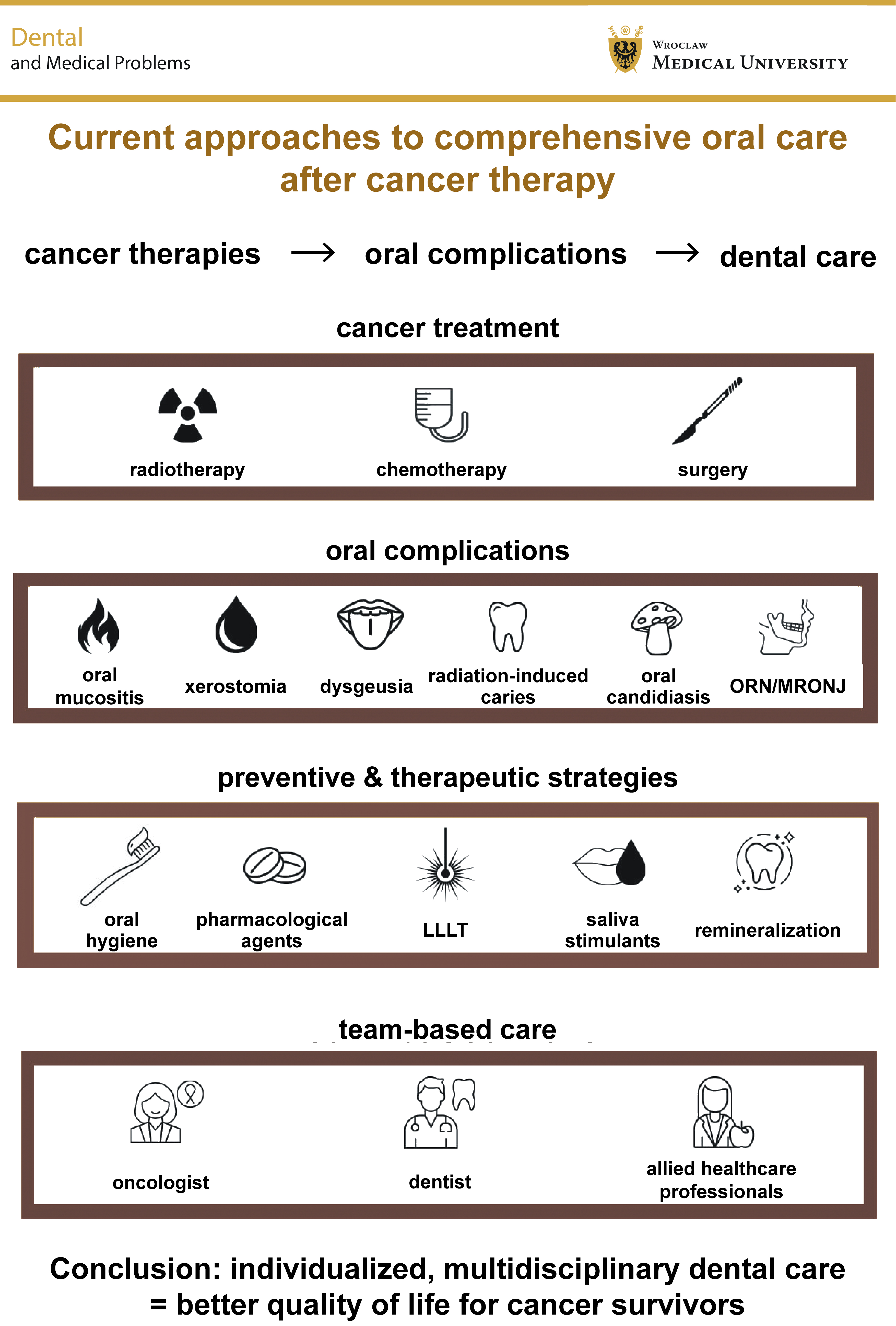
Cancer therapies, including radiotherapy, chemotherapy and surgical interventions, are essential components of modern oncology, but are frequently associated with long-term, multifactorial oral complications that can impair patients’ oral function and quality of life (QoL). This narrative review integrates current evidence, clinical guidelines and expert consensus to provide a comprehensive overview of contemporary approaches to oral management following cancer treatment.
The aim of this review was to synthesize and critically evaluate current dental strategies for the comprehensive care of individuals after oncologic therapy, identify the most common oral complications, including oral mucositis (OM), xerostomia, dysgeusia, radiation-induced caries, oral candidiasis, osteoradionecrosis (ORN), and medication-related osteonecrosis of the jaw (MRONJ), and highlight the areas requiring further research.
A literature search was conducted across PubMed, Wiley Online Library, the Web of Science, Google Scholar, and ScienceDirect from March to October 2025, focusing on clinical trials, reviews and international guidelines. Preventive measures, including remineralization protocols, rigorous oral hygiene practices and salivary stimulants, were emphasized as the crucial components of supportive care. The emerging therapeutic modalities, such as low-level laser therapy (LLLT) and novel pharmacological agents, were critically assessed with regard to their clinical effectiveness.
Overall, the evidence emphasizes the need for an interdisciplinary, patient-centered approach involving oncologists, dental specialists and allied healthcare professionals to minimize morbidity and optimize clinical outcomes. Multidisciplinary dental care remains indispensable for preventing and managing oral complications, and improving the long-term QoL of cancer survivors.
Keywords: oral mucositis, xerostomia, osteoradionecrosis, post-cancer dental care, interdisciplinary oncology
Introduction
Cancers are among the leading causes of death worldwide, particularly among middle-aged and older adults, contributing to chronic pain, reduced quality of life (QoL), and substantial costs associated with the treatment of advanced disease. Although traditionally classified as non-communicable diseases, a growing body of evidence has shown that some cancers are closely linked to viral infections, particularly human papillomavirus (HPV). Human papillomavirus plays a significant role in the development of cervical, laryngeal and oral cancers, with HPV-positive cases accounting for 20–40% of oral cancers.1
Cancers of the lip, oral cavity and pharynx are among the most common malignant tumors of the head and neck region, accounting for more than 400,000 new cases worldwide each year. Recent studies have indicated that their incidence is unevenly distributed geographically and shows a global upward trend. As medical and social costs continue to rise, oral and pharyngeal cancers in younger populations are becoming an increasingly significant public health concern.2 Dental complications in individuals who have survived childhood cancer are often irreversible, as they most commonly affect the permanent dentition.3
Post-treatment dental care represents a vital component of comprehensive oncologic management, particularly in patients undergoing therapy for head and neck malignancies. Current evidence supports the integration of dental professionals into multidisciplinary oncology teams, demonstrating improved oral health outcomes among head and neck cancer survivors. Patients managed within such coordinated frameworks exhibit significantly better periodontal health and oral hygiene as compared to those referred solely to general dental services.4 Researchers conducted a dose–response meta-analysis including 25 studies to further clarify and assess the association between tooth loss and cancer risk.5 They demonstrated that each additional loss of 10 teeth was associated with a 9% increase in cancer risk. They also estimated that tooth loss was associated with a 3–31% increased risk of various malignancies, including cancers of the head and neck, esophagus, stomach, colon, pancreas, lung, hematopoietic system, and bladder, in a clear dose-dependent manner.5
Oncologic therapies – including radiotherapy, chemotherapy and surgery – are associated with both acute and chronic oral complications, highlighting the need for long-term dental monitoring and intervention.6 Acute radiation-induced toxicity often develops early during treatment and may persist for several weeks after therapy, whereas late complications may arise weeks or even years later.7
Oral cavity and oropharyngeal cancers are recognized worldwide as major health concerns, given their substantial impact on patients’ QoL.8 The primary risk factors for oropharyngeal cancer (OPC) include tobacco smoking, alcohol consumption, betel chewing, and HPV infection, which may occur individually or in combination. Unlike tobacco-related cancers of the middle pharynx, whose incidence has declined in many high-income countries, the incidence of HPV-associated OPC has risen sharply over recent decades.9
The incidence of OPC in the United States has risen steadily among men since the 1970s. Notably, both the incidence and annual number of OPC cases now exceed those of cervical cancer, making OPC the most common HPV-related malignancy in the United States. Unlike cervical cancer, OPC cannot currently be screened for, as precancerous lesions cannot be reliably identified, and the existing diagnostic tools are insufficient for detecting early-stage, localized tumors.10
In the context of post-treatment care, it is important to note that patients with HPV-related oral cancer generally have better prognoses, and greater sensitivity to radiotherapy and chemotherapy. The projected increase in HPV-associated cancers also suggests continued improvement in patient survival, reflecting advances in treatment and the overall QoL.1
Oropharyngeal squamous cell carcinoma (OPSCC), which includes malignancies of the tonsils, base of the tongue, soft palate, and uvula, is now one of the most rapidly increasing cancers in incidence across high-income countries.11 Over the past several decades, the epidemiology of OPSCC in the United States has undergone notable changes, with important clinical and public health implications for patients after cancer treatment.12 The standard therapeutic approach for OPSCC generally includes the surgical resection of the tumor, definitive radiotherapy, or combined chemoradiotherapy. Among the treatment-related side effects that adversely affect patients’ perceived QoL and functional performance – and therefore require multidisciplinary care, including dental support – are xerostomia, odynophagia and oral thrush.11
Interdisciplinary collaboration improves functional outcomes and helps preserve oral health.4, 13, 14 As cancer survival increases, greater emphasis is being placed on maintaining oral function and comfort. Although many cancer patients exhibit oral health profiles similar to those of the general population, a substantial proportion present with deteriorating restorations, periodontal disease, and other complications exacerbated by cancer therapy, thereby affecting QoL.7 Emerging evidence suggests that early radiographic and biochemical changes around implants, such as subtle alterations in crestal bone and elevated levels of pro-inflammatory cytokines, may precede overt clinical signs. This highlights the need for more sensitive monitoring in post-cancer patients, whose reduced regenerative capacity and increased susceptibility to oral complications make early detection essential for maintaining long-term function and comfort.15 Furthermore, the potential application of stem cell-based therapies with tissue-specific differentiation capacity offers a promising adjunct to peri-implant and post-oncologic oral rehabilitation. Systematic reviews indicate that mesenchymal stem cells (MSCs) can enhance osteoblastic differentiation, promote new bone formation, and improve implant osseointegration through molecular signaling and interactions with biofunctionalized implant surfaces. Although the evidence remains heterogeneous and largely preclinical, these emerging approaches highlight a potential future direction for managing complex tissue defects in cancer survivors.16
Dentists play an essential role in early diagnosis and prevention through oral hygiene promotion, fluoride use and salivary care.6 Patients experiencing oral mucositis (OM), dry mouth, gingival sensitivity, and gingival pain exhibit markedly greater taste alterations and reduced QoL, while functional impairment, such as trismus and dysphagia, further contributes to malnutrition and diminished well-being. These findings underscore the need for early rehabilitation, dietary modification, and coordinated multidisciplinary care.17, 18 Artificial intelligence (AI), including neural networks (NNs), shows growing promise as a surveillance tool. Many studies report that NNs can identify early oral cancer with accuracies exceeding 85%, offering valuable support to clinicians, particularly in settings with limited access to oral pathology expertise. Risk factor and behavioral assessments should complement AI-based evaluations to enhance diagnostic precision.19
Although guidelines are available for healthcare professionals caring for patients undergoing oncologic treatment, minimizing common side effects, such as mucositis, xerostomia and radiation-induced caries, depends largely on patients’ active involvement in oral self-care, supported by appropriate education. Although these self-care protocols are often supported by a low level of evidence, their implementation remains essential for maintaining oral health.20
Methods
This narrative review synthesizes current evidence and clinical practice guidelines to provide an integrated overview of contemporary approaches to oral management in oncology patients. Its aim was to summarize and critically evaluate modern dental strategies for the comprehensive care of individuals after cancer treatment, highlight major oral complications, and identify the areas requiring further investigation.
Studies were included based on their relevance to post-cancer oral complications, current dental management protocols, peri-implant tissue health, early diagnostic approaches, and the emerging regenerative or stem cell-based therapies. A total of 74 publications were included. The literature comprised clinical practice guidelines, systematic reviews and meta-analyses, narrative or scoping reviews, randomized controlled trials (RCTs), observational studies, epidemiological studies, preclinical/in vitro studies, and case reports or case series. Although no formal risk-of-bias assessment was performed, study selection followed the principles of transparency and relevance consistent with the Scale for the Assessment of Narrative Review Articles (SANRA) recommendations, with a focus on clinical applicability and contribution to contemporary dental oncology practice.
Literature search was conducted in PubMed/MEDLINE, Wiley Online Library, the Web of Science, Google Scholar, and ScienceDirect between March and October 2025. Given the narrative design of this review, study selection was guided by thematic relevance and the contribution of each source to the understanding of contemporary concepts and clinical practice, rather than by rigid, predefined systematic criteria.
Oral mucositis
The oral mucosa, periodontal tissues and dentition are among the regions most commonly affected by radiation-induced toxicity due to their high cellular turnover and essential roles in oral health and function.7 These structures are particularly susceptible to ionizing radiation, which can compromise tissue integrity and lead to substantial clinical complications.
Oral mucositis is a frequent and debilitating adverse effect of oncologic therapies. It initially presents as mucosal erythema and atrophy, and may progress to ulceration. One of the earliest signs is leukoedema – an ill-defined, diffuse, milky-white opalescence, most often observed on the buccal mucosa, which typically disappears upon stretching.7 Erythema may appear within the first week of fractionated radiotherapy (2 Gy/day)7 and within 4–5 days after the initiation of chemotherapy.21 The onset of OM reflects epithelial aplasia resulting from cytotoxic injury to rapidly proliferating basal cells, generally occurring 7–14 days after chemotherapy. Early symptoms include burning or tingling sensations, and increased sensitivity to mechanical and chemical stimuli.7 Pain, dysgeusia, dysphagia, and odynophagia commonly interfere with nutrition and hydration.22 Lesions frequently involve the lips, buccal mucosa, ventrolateral tongue, and floor of the mouth, typically resolving within 2–4 weeks after the completion of therapy, although complete healing may require up to 3 months.7
Chlorhexidine and other biologically active rinses provide antimicrobial and anti-inflammatory benefits, but have limited effects on the molecular pathways underlying OM.13 Basic oral care remains fundamental to reducing secondary infections and minimizing mechanical trauma. Symptomatic management is central, particularly in chemotherapy-induced OM, with compounded analgesic rinses (“Magic Mouthwash”) containing diphenhydramine, viscous lidocaine, bismuth subsalicylate, or corticosteroids commonly used for palliation.7
Preventive oral hygiene reduces the risk of OM, although its direct effect on the underlying mechanisms of mucosal injury remains unclear.13 Chlorhexidine and benzydamine may be used for antiseptic and anti-inflammatory purposes; however, their use may be associated with adverse effects, including mucosal burning, taste alterations, and staining.23
Chlorhexidine is not recommended for the prevention of OM in patients receiving radiotherapy for head and neck cancer, although it remains useful for gingivitis and plaque control.13 Diagnosis relies primarily on clinical history, symptoms and lesion distribution.24 Chemotherapeutic agents most commonly associated with OM include doxorubicin, bleomycin, fluorouracil, and methotrexate.25
For the World Health Organization (WHO) grade I–IV OM, weekly low-level laser therapy (LLLT) is recommended until complete recovery.21 Prophylactic photobiomodulation (PBM) initiated 2–3 weeks before cancer treatment, combined with education on oral hygiene, nutritional guidance and risk-factor modification (e.g., tobacco and alcohol use), is strongly advised.26
Analgesic rinses, such as doxepin and methylene blue, may be effective in managing oral mucositis-related symptoms. Methylene blue oral rinse has demonstrated rapid and clinically meaningful analgesic effects in cancer-related oral mucositis, with improvement in oral function when used alongside standard therapy. Its safety profile appears favorable, with no serious adverse events reported, and only mild, transient effects, such as brief oral burning or temporary discoloration of the lips and teeth, which resolve spontaneously. Owing to its demonstrated efficacy, safety, and limited adverse effects, methylene blue represents a promising supportive option for refractory oral mucositis-associated pain.27 Topical doxepin rinse also appears to be an effective analgesic option for managing oral mucositis-related pain, providing clinically meaningful reductions in symptom intensity. Although generally well tolerated, its use may be accompanied by mild, transient adverse effects, such as oral burning, unpleasant taste, or drowsiness, the latter likely related to partial systemic absorption. Patient acceptability is relatively high, with many individuals expressing willingness to continue therapy when adequate pain relief is achieved. Overall, doxepin represents a useful adjunct to supportive care, offering a symptomatic benefit with an acceptable safety profile.28
Benzydamine is indicated for the prevention of OM in patients receiving moderate-dose radiotherapy (<50 Gy), while a 0.2% morphine oral solution may provide both analgesic and anti-inflammatory effects.29 Topical morphine has been shown to reduce pain intensity, shorten the duration of severe symptoms, and improve functional outcomes, such as swallowing and oral opening. Its safety profile appears favorable, with predominantly mild and transient local adverse effects, most commonly brief burning, dryness or itching, and minimal systemic toxicity. Reported adverse reactions are infrequent and generally do not limit tolerability. Palifermin significantly reduces the incidence, duration and severity of OM, but may cause transient taste disturbances. However, according to the Multinational Association of Supportive Care in Cancer and International Society of Oral Oncology (MASCC/ISOO) guidelines, the available evidence is insufficient or inconsistent to support formal clinical recommendations for these agents. Oral cryotherapy using ice chips is widely employed in chemotherapy-induced OM to reduce mucosal exposure to chemotherapeutic agents through vasoconstriction.7, 30, 31
Key recommendations are summarized in Table 1.
Xerostomia
Despite efforts to limit radiation exposure to healthy tissues, the salivary glands, the oral mucosa and maxillofacial bones are often unintentionally affected, resulting in functional and structural changes with long-term effects on QoL.7 Radiotherapy and certain cytotoxic agents commonly impair salivary gland function, causing hyposalivation and xerostomia.26, 32 This predisposes patients to dental caries, fungal infections and bacterial overgrowth due to the disruption of the oral microbiota and reduced salivary defense mechanisms.33 It has been noted that oncologists frequently underestimate the importance of salivary function, whereas they should incorporate its assessment into routine patient evaluation.17 Hyposalivation may develop early, sometimes after only a few 2-Gy fractions. Radiation doses >30 Gy are associated with irreversible glandular injury, with severity correlating with the cumulative dose.26 Although the compensatory hypertrophy of the spared glands may occur months later, the total salivary output may decrease by 50–60% within the first week when all major glands are irradiated.7
Management involves supportive and pharmacological strategies. Ice chips, adequate hydration (≥1.5 L/day), mucosal lubricants, and salivary stimulation with xylitol-containing products are considered first-line measures. Saliva substitutes are generally more effective than water for managing oral dryness.34 In severe cases, muscarinic receptor agonists, such as pilocarpine and cevimeline, may be prescribed.26, 35, 36, 37 Corticosteroids may benefit patients with immune-mediated xerostomia, although they should be used with caution. Anethole trithione may be considered as an adjunctive therapy. Patients should avoid alcohol, caffeine, and acidic or spicy foods,26, 29, 36 and may also benefit from saline/bicarbonate rinses, humidifiers, and moisturizing gels or sprays.34, 38
Complementary therapies under investigation include acupuncture, omega-3 fatty acid supplementation, vitamins C and E, green tea polyphenols, and aloe vera, although the evidence supporting their use remains limited. Photobiomodulation and transcutaneous electrical nerve stimulation (TENS) show emerging potential, whereas hyperbaric oxygen therapy (HBOT) has limited supporting evidence and homeopathy lacks the evidence of efficacy.35
Topical pilocarpine (1–2%) effectively stimulates salivary secretion, with lower concentrations (e.g., 0.1%) also demonstrating benefit. A 2% topical formulation may provide salivary stimulation comparable to a 5-mg oral dose, with fewer systemic effects. Novel delivery systems, including pilocarpine lozenges, aerosols, hydrogels, and nanofiber carriers, are under development. Localized mucosal tablet formulations may optimize therapeutic outcomes while minimizing systemic exposure.39 However, pilocarpine is effective only when functional salivary tissue is preserved. Evidence from controlled clinical studies indicates that its therapeutic benefit is modest, with only minimal increases in the salivary flow reported across trials. Local administration (e.g., ophthalmic drops applied intraorally) is associated with fewer adverse effects than systemic tablet formulations, which frequently cause cholinergic side effects, such as sweating, rhinorrhea, nausea, and excessive salivation. Bethanechol demonstrates comparable efficacy with a more favorable side-effect profile, suggesting that it may be a better-tolerated alternative in some patients.40
Lipids act as coating and lubricating agents in the oral cavity by forming a protective layer that reduces friction. Unlike other macromolecules that rely on water retention, lipids interact with oral surfaces primarily through weak dispersion forces, while longer lipid chains provide greater viscosity and longer-lasting adhesion to the mucosa. Studies indicate that plant-based oils, such as sunflower, canola, coconut, and olive oils, may serve as saliva substitutes by improving oral comfort, reducing xerostomia and enhancing the salivary flow. Olive oil may additionally exhibit anti-inflammatory and antibacterial properties.41, 42
Sodium hyaluronate (hyaluronic acid – HA), a naturally occurring polysaccharide with mucoadhesive and water-binding properties, is considered a safe and effective therapeutic option, providing viscosity and lubrication comparable to that of natural saliva. It is biocompatible and can be degraded by hyaluronidase. However, attention should be given to its molecular weight: high–molecular-weight HA may inhibit the antimicrobial activity of lysozyme and peroxidase, potentially increasing the risk of infection. Therefore, low–molecular-weight HA (1,000–4,000 kDa) is recommended for use in artificial saliva formulations.41, 43
Since the salivary film forms a protective coating over the oral mucosa, its reduction in hyposalivation leaves the epithelium more exposed and susceptible to chemical irritants. A diminished mucosal/salivary film compromises lubrication and barrier function, increasing the risk of irritation in response to topical agents. Therefore, when high-concentration fluoride products are considered, the condition of the oral mucosa should be carefully assessed in patients with hyposalivation, as the absence of an adequate salivary coating may increase their sensitivity to these agents.44, 45, 46
Key recommendations are summarized in Table 2.
Taste alterations
Chemotherapy is known to disrupt gustatory function and reduce appetite. Although taste alteration is a common treatment-related toxicity, it is frequently underreported by patients and may be underestimated by clinicians, as it does not pose an immediate threat to survival. Greater severity of taste alteration has been observed in women than in men, and among individuals with breast cancer as compared to those with gastrointestinal, pulmonary, genitourinary, or hematologic neoplasms. Patients who reported a good or very good appetite exhibited substantially lower levels of taste alteration than those with poor or moderate appetite. No significant associations were identified between taste alteration and either patient age or the number of chemotherapy cycles when analyses were stratified by the appetite level.17
Dysgeusia
Dysgeusia, a common complication in patients undergoing head and neck radiotherapy (HNRT), often coexists with xerostomia due to the integral role of saliva in taste perception. The condition is dose-dependent, with disturbances in taste sensation typically occurring at radiation doses of approx. 30 Gy. At this threshold, patients may experience a near-complete loss of fundamental taste modalities – namely salty, sweet, sour, and bitter – secondary to damage to gustatory epithelial cells.21, 47 While mild dysgeusia is generally manageable, more severe forms can significantly impair appetite, reduce oral intake, and negatively affect the nutritional status and QoL.7
Partial recovery of taste function usually begins within 20–60 days following the cessation of treatment, with full restoration frequently occurring over a period of 2–4 months.7 Chemotherapy can also contribute to transient dysgeusia, often presenting as a metallic taste due to the diffusion of cytotoxic agents into saliva. Drugs such as cyclophosphamide, methotrexate and 5-fluorouracil may alter taste either by affecting cranial nerves (VII, IX and X), damaging the oral mucosa, or directly impacting taste bud cells.22, 26
Although clinical data regarding the efficacy of zinc supplementation are variable, zinc has been proposed to improve taste perception by stabilizing the regulatory proteins involved in taste bud maintenance.7 Zinc supplementation also appears to offer therapeutic benefits in reducing the incidence, duration and severity of OM, likely due to its roles in immune function, epithelial repair and antioxidant defense. Its safety profile is generally favorable, with minimal adverse effects reported when used at appropriate doses. However, variability in zinc formulations, dosing strategies, and routes of administration limits the comparability of the existing studies and underscores the need for standardized, long-term clinical trials.48 Vitamin D supplementation has also been explored as a potential therapeutic option for mitigating taste alterations. In chronic or persistent cases, dietary counseling and simple nutritional intervention remain important for improving patient outcomes.7
Supportive strategies, such as slow, mindful mastication, can enhance flavor perception and stimulate the salivary flow, which may be particularly beneficial in patients with concurrent xerostomia. Additionally, rotating food types during meals may help prevent sensory adaptation and promote dietary diversity, thereby supporting adequate nutrition and overall oral intake.7
According to some researchers, future cancer treatment strategies and post-therapy supportive care should prioritize the development of microbiota-modulating interventions and personalized therapeutic approaches to enhance oncologic treatment efficacy. Given that gut microbiota dysbiosis has been associated with chronic inflammation, the production of carcinogenic metabolites and altered immune responses, and considering that inflammatory conditions, including those affecting the oral cavity, are frequently observed in patients undergoing chemotherapy and radiotherapy, dental practitioners should also consider systemic microbiological interactions when aiming to improve patient outcomes.49
The principal recommendations are summarized in the Table 3.
Radiation-induced caries
It refers to an aggressive and rapidly progressing form of dental decay that commonly develops following HNRT. Although ionizing radiation can alter the structural integrity of dental hard tissues, the underlying pathogenesis of radiation-induced caries is largely analogous to that of caries associated with other hyposalivation-related conditions. Clinically, it resembles caries observed in patients with xerostomia, but more frequently involves atypical tooth surfaces, such as cusp tips and cervical margins. The disease also exhibits a higher recurrence rate and more rapid progression than conventional caries.26
The elevated risk is primarily attributable to radiation-induced enamel demineralization, shifts toward a cariogenic oral microbiota, poor oral hygiene, and high-carbohydrate dietary patterns.50 Xerostomia, commonly induced by radiotherapy, often precedes the onset of radiation-induced caries, which may manifest as early as 3 weeks after treatment and rapidly compromise dental integrity. Radiation further weakens tooth structure, rendering enamel more vulnerable to acid-induced breakdown. Contributing factors include frequent consumption of sugar-laden beverages, which facilitates bacterial adhesion and enhances enamel demineralization.22 Under conditions of reduced salivary flow, Streptococcus mutans may proliferate, further increasing the risk of caries in irradiated patients.
Effective prevention requires rigorous oral care protocols, including routine dental evaluations and continuous fluoride therapy. Although custom fluoride trays remain the most efficacious delivery method, alternatives such as fluoride varnishes, rinses and high-fluoride toothpaste are also recommended. Fluoride application should be maintained for as long as hyposalivation persists. When trays are not used, topical fluoride gels or brush-on formulations, combined with dietary modifications, are essential.50 Additional strategies include daily sodium fluoride rinses,26 quarterly dental visits and regular professional cleanings.
Nutritional counseling should emphasize reduced consumption of fermentable carbohydrates, decreased snacking frequency and substitution with non-cariogenic sweeteners. Xylitol-containing products may be beneficial due to their ability to inhibit cariogenic bacteria and stimulate the salivary flow. Advanced preventive care may include fluoride varnishes for prolonged enamel protection and calcium phosphate- or hydroxyapatite-based agents to promote remineralization. Probiotic therapy may also help support a balanced oral microbiome and reduce pathogenic bacterial colonization.33
In advanced cases, restorative interventions, such as composite restorations, crowns or dental implants, may be required. To preserve enamel integrity, patients should use non-abrasive toothpaste and avoid acidic foods and beverages.29, 51 Long-term oral health maintenance depends on patient education focused on oral hygiene practices, caries prevention and healthy dietary habits.52
The principal recommendations are summarized in the Table 4.
Oral candidiasis
It is a frequent opportunistic infection in oncology patients, particularly following chemotherapy and radiotherapy, due to treatment-induced immunosuppression and hyposalivation.53 The most common form, pseudomembranous candidiasis, presents clinically as white, curd-like plaques that can be gently scraped off, typically revealing an erythematous, eroded or bleeding mucosal surface underneath. Another notable subtype, hyperplastic candidiasis, manifests itself as thickened, non-removable white plaques. This variant closely resembles leukoplakia, necessitating careful differential diagnosis because of its potential for malignant transformation.7
Oral candidiasis is associated with burning, pain, dysphagia, dysgeusia, and mucosal discomfort. A minimum 2-week course of antifungal therapy is generally required for resolution.54 The condition is predominantly caused by Candida albicans, and commonly affects the tongue, hard palate and buccal mucosa, often coexisting with angular cheilitis, erythema and mucosal bleeding. Predisposing factors include local and systemic immunosuppression, xerostomia, prolonged use of antibiotics or corticosteroids, immunosuppressive agents, poor denture hygiene, high-sugar diets, and tobacco use.29, 36
Diagnostic confirmation is achieved through clinical evaluation supported by microbiological testing, including fungal cultures.26, 33, 36, 55 Although a Cochrane meta-analysis reported insufficient evidence to definitively support or oppose the use of antifungal agents in cancer-associated candidiasis,7 treatment typically begins with topical antifungal agents, such as nystatin (suspension or lozenges), clotrimazole or miconazole. In refractory or systemic cases, systemic antifungal agents, such as fluconazole or itraconazole, may be necessary. Adjunctive management includes meticulous oral hygiene, mechanical tongue cleaning and antiseptic rinses, such as chlorhexidine or sodium bicarbonate solutions.
The principal recommendations are summarized in the Table 5.
Denture stomatitis
In cases of denture stomatitis resistant to conventional therapies, tea tree oil (Melaleuca alternifolia), due to its antifungal properties, may be used alongside the COE-COMFORT™ tissue conditioner. The microwave disinfection of complete dentures is another effective adjunctive method. If these measures fail, temporary discontinuation or replacement of the denture is recommended. Chronic or recurrent oral candidiasis should prompt medical evaluation to reassess diagnosis and modify treatment accordingly.26, 33, 36, 54, 55
Osteoradionecrosis
Osteoradionecrosis (ORN) is a serious complication of HNRT, characterized by radiation-induced hypoxia, hypocellularity, and reduced vascularity within osseous tissues.36 These alterations impair tissue regeneration and compromise mucosal integrity, thereby predisposing patients to bone necrosis and sequestration.7 The mandible is the most frequently affected site, owing to its dense bone structure and relatively limited blood supply.
Clinically, ORN presents with exposed necrotic bone, chronic pain, mucosal ulceration, fistula formation, and, in severe cases, pathological fractures, often in the absence of overt infection. Radiographic findings typically include diffuse radiolucencies with ill-defined borders and, in advanced stages, radiopaque sequestra.36, 56 Osteoradionecrosis is commonly classified according to its clinical course, anatomical extent, severity of symptoms, and response to HBOT. It may significantly impair QoL, particularly in advanced cases.
Multiple risk factors contribute to the development of ORN, including poor oral hygiene, pre-radiotherapy dental or surgical procedures, high radiation doses or fraction sizes, tumor proximity to bone, and direct bone invasion. The condition is more prevalent in males over 55 years of age, particularly those with a history of tobacco and alcohol use. Although most cases occur within 3 years after radiotherapy, delayed onset is also possible. Prevention relies on comprehensive oral evaluation and the stabilization of oral health before and after radiotherapy. Reducing the need for invasive dental procedures and minimizing oral infections and inflammation are critical components of ORN prevention.50 Management involves a combination of conservative and surgical strategies, including debridement, wound care, antibiotic therapy, and, when indicated, HBOT.8
The principal recommendations are summarized in the Table 6.
Medication-related osteonecrosis of the jaw
Medication-related osteonecrosis of the jaw (MRONJ) is associated with the antiresorptive agents (e.g., bisphosphonates and denosumab) and antiangiogenic therapies used in cancer care. These drugs impair osteoclast function, reduce bone turnover and increase the risk of osteonecrosis.57 Medication-related osteonecrosis of the jaw can lead to progressive jawbone destruction and considerable morbidity, particularly with intravenous formulations used in cancer treatment as compared to oral formulations used for osteoporosis.
Diagnostic criteria include a history of antiresorptive or antiangiogenic therapy, non-healing exposed bone in the maxillofacial region persisting for ≥8 weeks, the absence of previous radiotherapy to the jaws, and the absence of jaw metastases.57 Evidence suggests that quarterly dental evaluations and preventive protocols, such as prophylactic antibiotics and primary soft-tissue closure following tooth extraction, may reduce the incidence of MRONJ more effectively than reactive care. The benefit of platelet-rich growth factor (PRGF) application following tooth extraction remains inconclusive.58
Studies have shown no significant advantage of adding HBOT to standard MRONJ treatment, including antiseptics, antibiotics and surgery, highlighting the need for further research.57 A comparative study by Mozzati et al. reported no cases of MRONJ with either minimally invasive or more invasive tooth extraction protocols in patients receiving oral bisphosphonates, suggesting that there is insufficient evidence to favor one surgical approach over the other.59
Neurotoxicity
Chemotherapy-related neurotoxicity can result in persistent, diffuse pain localized to the head and neck region. This pain may mimic odontogenic pain, complicating diagnosis. Despite the presence of symptoms, clinical and radiographic examinations typically reveal no dental or mucosal abnormalities, although the thickening of the periodontal ligament (PDL) may be observed in vital teeth.21 Symptom fluctuation and the absence of clear clinical findings present diagnostic challenges in dental settings.
Trismus
Trismus, defined as a restricted ability to open the mouth, is a common complication following oral cancer surgery and radiotherapy. It typically results from postoperative fibrosis and scar contracture, leading to a decreased interincisal distance, often measuring less than 35 mm between the maxillary and mandibular incisors.7
Trismus may develop early during radiotherapy, particularly in patients with tumors involving the nasopharynx, palate or maxillary sinuses. If left unaddressed, the condition can lead to a significant reduction in mandibular mobility, impairing essential functions, such as mastication, speech and oral hygiene.22 Reduced jaw opening is frequently observed approx. 9 weeks after the completion of radiotherapy. Denture-wearing patients are particularly affected, as trismus may prevent the proper insertion of prostheses and complicate the fabrication of new appliances because of restricted intraoral access.7
The management of trismus emphasizes the early initiation of therapeutic exercises, including jaw stretching and the use of assistive devices, such as bite dilators or tongue depressors, aimed at preserving the range of motion.54 While short-term improvement can be achieved, long-term benefits require consistent adherence to these exercises. In chronic cases, the development of dense fibrotic tissue renders passive stretching less effective, underscoring the importance of early detection and intervention to prevent irreversible functional impairment.22
The principal recommendations are summarized in the Table 7.
The effective prevention and management of oral complications related to oncologic treatment require an interdisciplinary approach involving dental and medical professionals. Dental care is critical for reducing the risk of complications and enhancing therapeutic outcomes and QoL.
Pre-radiotherapy dental management is particularly important for patients with head and neck cancer. According to the Canadian Dental Oncology Network (CDON) consensus guidelines, developed using a modified Delphi method, prophylactic tooth extraction may be considered at radiation dose thresholds of 70 Gy for the maxilla and 60 Gy for the mandible.60 Management protocols are tailored to anatomical location and the radiation dose, with all patients – regardless of dentition – recommended for referral to a dental oncology clinic for pre-treatment evaluation. The recommended healing interval between tooth extraction and radiotherapy is 7–14 days.60 Risk assessment should consider factors such as infectious foci, the oral health status, the type of tumor, and the overall prognosis. Fluoride prophylaxis, oral hygiene education and routine dental follow-up are essential components of comprehensive care aimed at reducing post-radiotherapy complications.19, 60
Following anticancer treatment, including chemotherapy, long-term dental care remains essential for controlling infection, restoring function and improving esthetics. Treatment planning should be conducted collaboratively with oncologists.21, 61 Key elements include consistent daily oral hygiene under the supervision of a dentist or dental hygienist, as well as routine follow-up visits – monthly for the first 3 months, every 3 months during the first year, and every 6 months thereafter for at least 3 years.34 Dental prophylaxis involves the regular removal of plaque and calculus,33, 36, 51 and caries prevention through the use of high-fluoride toothpaste, chlorhexidine rinses (for at least 3 weeks every 3 months) and routine fluoride applications.29
Fluoride prophylaxis is especially critical for patients after radiotherapy to prevent radiation-induced caries. Recommended strategies include:
– high-fluoride toothpaste (5,000 ppm F⁻): daily use to promote remineralization;
– sodium fluoride rinses: 0.05% daily or 0.2% weekly;
– 5% sodium fluoride varnish: every 3 months, particularly in patients with xerostomia;
– 1.23% fluoride gel: applied using trays by a dentist or the patient.36
Xylitol-containing products are especially recommended, as xylitol has been shown to inhibit the growth of S. mutans. Patients should be advised to choose water over sugary or acidic beverages, as acidic drinks can contribute to irreversible dental erosion. Additionally, frequent vomiting, commonly associated with cytostatic therapy, may further contribute to enamel degradation through repeated exposure to gastric acid.33
Post-chemotherapy procedures and prosthodontics
After chemotherapy, treatment-related side effects should be appropriately managed, and regular dental follow-up is particularly important during the first few months after treatment. Invasive procedures, such as tooth extractions or oral surgery, should be delayed for at least 1 year when clinically feasible. If urgent intervention is required, antibiotic prophylaxis should be initiated 48 h before the procedure and continued for 7–15 days afterward. Hyperbaric oxygen therapy may also be considered to reduce the risk of complications.29
The use of dentures should generally be avoided for 1 year after treatment. If necessary, prosthetic rehabilitation should be postponed for 4–6 months following chemotherapy. Special caution is advised in patients treated with bisphosphonates, particularly intravenous formulations, as implant placement carries a risk of MRONJ.61 Multidisciplinary care is crucial for managing oral complications and improving patients’ QoL.
Orthodontic considerations in oncology patients
Orthodontic treatment decisions should account for the patient’s general health, caries risk and adherence to oral hygiene practices.62 Treatment planning should consider the applied forces, the anticipated treatment duration and the pre-existing root damage, which may be exacerbated during therapy.63 In patients receiving antiresorptive medications, tooth extractions should be approached with caution because of the risk of MRONJ, which has been reported to range from 0.2% to 6.7%.64 Bisphosphonates may impair tooth movement and prolong treatment duration. Orthodontic treatment is generally contraindicated during and shortly after intravenous bisphosphonate therapy.
Given the adverse effects of oncologic therapies, such as mandibular retrognathia, reduced craniofacial dimensions and increased tooth mobility, orthodontic treatment should be simplified. Lower-force mechanics are recommended to minimize the risk of root resorption, and mandibular treatment should be avoided where possible.65
Orthodontic treatment may be safely resumed 2 years after the completion of chemotherapy or radiotherapy, owing to prolonged alterations in bone metabolism and potential treatment-related discomfort, including nausea, xerostomia, taste alterations, and mucosal sensitivity. However, when treatment has been limited to surgery alone without adjunctive therapies, there is no requirement to delay orthodontic intervention.66
Teeth whitening is generally safe when performed correctly, but may cause transient side effects, such as dentinal hypersensitivity and soft-tissue irritation. Therefore, whitening procedures should be approached cautiously, particularly in post-oncological patients. Patient eligibility should be based on a thorough clinical evaluation of the caries status, non-carious lesions, periodontal health, xerostomia, soft-tissue condition, and tooth sensitivity. To minimize potential risks, lower concentrations of carbamide peroxide applied over longer periods are preferred over high-concentration hydrogen peroxide products.67
Immunotherapy-related oral complications and their management
Immune-related adverse events (irAEs) primarily affect the oral mucosa and the salivary glands, often leading to severe symptoms. Mucosal changes may present as lichenoid lesions, erosions and ulcers, accompanied by intense pain and hypersensitivity that can impair oral intake and contribute to malnutrition. Salivary gland dysfunction is also common, presenting as xerostomia, dysphagia, and difficulties with speaking, often accompanied by thick or sticky saliva. Among these side effects, xerostomia is the most frequently reported and typically develops early during treatment. Mucosal disturbances, including inflammation, ulceration and pain – such as those associated with lichenoid and vesiculobullous reactions – generally develop over the following months. Dysgeusia occurs less frequently, but may also develop early and can significantly affect daily functioning. Most oral irAEs arise within the first months of immunotherapy. Due to their chronic nature, symptoms such as dry mouth, taste disturbances and mucosal disease can substantially impair QoL, limiting food intake, speech and sleep.68, 69, 70
The early initiation of treatment is crucial to avoid the interruption of anticancer therapy. Standard management includes systemic corticosteroids (e.g., prednisone 40–60 mg/day) and topical steroids, such as dexamethasone 0.05% rinse or clobetasol 0.05% gel, often used at higher-than-standard concentrations. Adjunctive approaches include PBM, which can effectively alleviate mucosal pain, and, in cases of salivary gland dysfunction, sialogogues (e.g., pilocarpine), moisturizing agents, and intraductal salivary gland irrigation, which can significantly improve the salivary flow.68
The rapid identification and comprehensive management of these complications are essential for maintaining the continuity of immunotherapy and improving patients’ QoL.
The key recommendations addressing the complications discussed above throughout the manuscript are presented in Table 8.
Limitations
This review is subject to several limitations. As a narrative review, it does not follow a systematic methodology for comprehensive evidence synthesis, which may introduce selection bias. The available evidence is characterized by substantial heterogeneity, including differences in study design, patient populations, cancer types, treatment modalities, radiation doses, outcome definitions, and follow-up duration. This heterogeneity limits the direct comparability of findings and may contribute to inconsistent or conflicting results.
Despite advances in supportive oral care, several aspects of post-cancer dental management remain unresolved, reflecting broader limitations in the current evidence base. In particular, the routine use of chlorhexidine for OM prevention remains controversial; although effective for plaque control and infection prevention, international guidelines do not support its prophylactic use for mucositis, and some studies report increased mucosal irritation. Similarly, the long-term use of high-concentration fluoride in patients with severe xerostomia remains debated. While fluoride prophylaxis is essential for preventing radiation-induced caries, a reduced salivary flow and the impairment of the salivary film may increase mucosal sensitivity, and current guidelines provide limited guidance regarding optimal formulations and dosing in this setting.
Moreover, many of the included studies focus on specific cancer types or treatment settings, which may limit the generalizability of their findings to the broader population of cancer survivors. The relative scarcity of long-term follow-up data – particularly regarding chronic oral complications, implant outcomes and the emerging therapies – further limits conclusions about sustained effectiveness and late adverse effects.
Finally, this review was limited to English-language publications. Given the rapidly evolving nature of oncologic therapies and dental management strategies, new evidence may emerge after the completion of this review, potentially influencing future recommendations.
Conclusions
The long-term management of oral health in cancer survivors is a critical component of multidisciplinary oncology care. As cancer therapies advance and survival rates increase, addressing the persistent and often debilitating oral complications of treatment becomes increasingly important. These complications include OM, xerostomia, dysgeusia, radiation-induced caries, ORN, candidiasis, and functional impairment, such as trismus and dysphagia.
Comprehensive dental care, integrated into cancer management, plays a key role in mitigating these complications. Early dental intervention – particularly before radiotherapy – is essential for risk assessment, preventive care, and reducing post-treatment morbidity. Continued surveillance and personalized strategies, such as high-fluoride therapy, salivary stimulation, nutritional guidance, and patient education, are vital for preserving oral function and QoL.
When interpreting the available clinical guidelines, it becomes clear that recommendations for post-treatment oral care in cancer survivors remain heterogeneous and address different yet complementary aspects of management. The CDON guidelines, although primarily focused on pre-radiotherapy decision-making, acknowledge that patients who have undergone radiotherapy remain at increased long-term risk of dental complications, including radiation-induced caries and ORN.60, 71
The MASCC/ISOO and European Society for Medical Oncology (ESMO) guidelines focus predominantly on the prevention and management of acute oral complications during active treatment, with limited guidance on structured dental follow-up after treatment completion. Consequently, practical recommendations for the prevention and management of chronic oral sequelae in cancer survivors remain largely underdeveloped.72, 73
In contrast, the NCCN Head and Neck Cancer guidelines integrate oral health into survivorship care, particularly for patients treated with radiotherapy. They emphasize regular dental evaluations, intensified preventive strategies, and the careful planning of invasive dental procedures in previously irradiated tissues, thereby recognizing the persistent and cumulative nature of treatment-related oral morbidity.74
This discrepancy reflects a broader gap between the guidelines addressing acute treatment-related toxicity and the long-term oral healthcare needs of cancer survivors, underscoring the need for dedicated, survivorship-focused dental care recommendations to support coordinated, long-term oral health management. The comparison of key recommendations across major guideline frameworks is summarized in Table 9.
Taken together, these documents further highlight the absence of a unified, interdisciplinary, long-term oral care protocol for patients treated for head and neck cancer. Moreover, substantial gaps persist in the existing evidence base for dental care in oncology patients, including the limited availability of high-quality studies on PBM and the low level of the evidence supporting complementary interventions, such as acupuncture or omega-3 acid supplementation. The effectiveness of xerostomia treatment (e.g., pilocarpine and bethanechol) is only moderate, and the lack of standardized dosing protocols makes comparisons difficult. Regenerative therapies using stem cells are still in early research stages, and AI models require larger, clinically validated studies. Clear evidence is also lacking for the efficacy of HBOT, TENS, and other supportive methods. Data on managing dysgeusia (e.g., zinc supplementation), preventing radiation-induced caries (probiotics, plant oils, fluoride regimens) and treating oral candidiasis remain insufficient. The management of ORN and MRONJ likewise requires more robust comparative studies. Additionally, research linking the microbiota status to treatment-related complications is scarce, and many preventive recommendations rely on low-quality evidence.
Future research should aim to establish standardized, evidence-based dental care protocols specifically tailored to patients undergoing oncologic treatment. Priority areas include evaluating the cost-effectiveness of preventive interventions, determining the long-term outcomes of salivary gland preservation strategies, and assessing how different oral care approaches influence treatment tolerance and post-treatment recovery. Further studies are also needed to refine minimally invasive techniques, optimize radiotherapy planning to protect oral structures, and explore novel agents that support mucosal healing and salivary function. Strengthening the evidence base in these domains will help guide more consistent clinical practice, and ultimately improve the QoL of cancer patients.
Ethics approval and consent to participate
Not applicable.
Data availability
Not applicable.
Consent for publication
Not applicable.
Use of AI and AI-assisted technologies
Not applicable.