Abstract
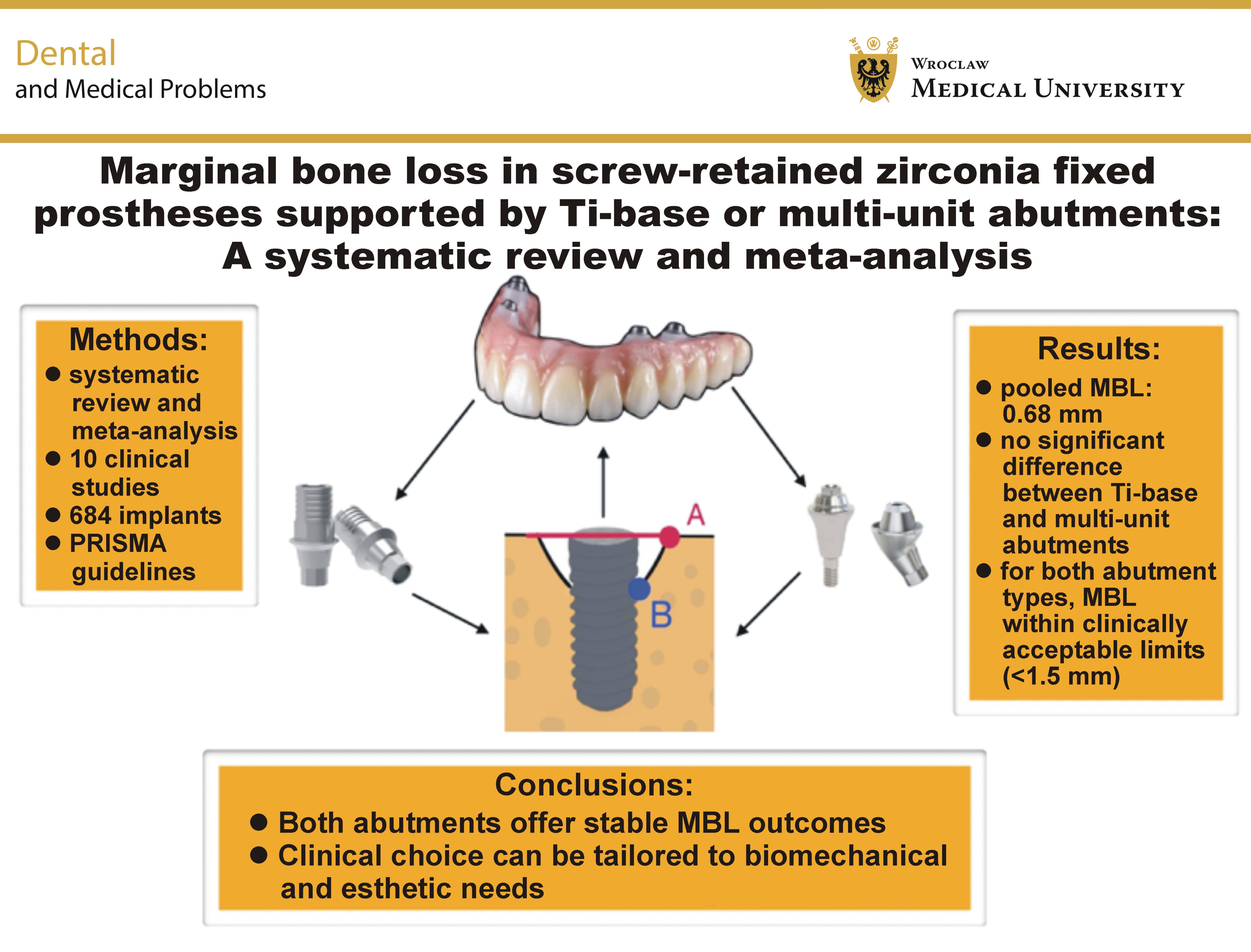
This systematic review aimed to evaluate the marginal bone loss (MBL) associated with screw-retained zirconia fixed dental prostheses (FDP) supported by either titanium-base (Ti-base) or multi-unit abutments.
A comprehensive literature search was conducted in accordance with the PRISMA 2020 guidelines, using PubMed, Scopus, Web of Science, and the Cochrane Library, covering publications up to May 2024. The inclusion criteria focused on clinical trials and cohort studies reporting quantitative MBL outcomes for zirconia screw-retained prostheses supported by either Ti-base or multi-unit abutments, with a minimum follow-up of 6 months. Data extraction and risk-of-bias assessments were performed independently by 2 reviewers. A meta-analysis was conducted using a random-effects model to pool the MBL values.
Of the 752 articles screened, 10 studies met the inclusion criteria, comprising 684 implants in 412 patients. The mean MBL ranged from 0.19 to 1.26 mm over follow-up periods of 12 to 60 months. The pooled mean MBL was 0.68 mm (95% CI: 0.54–0.81 mm; I2 = 47%). Subgroup analyses revealed no significant differences between the Ti-base- and multi-unit-supported restorations.
Screw-retained zirconia FDP supported by Ti-base or multi-unit abutments are associated with minimal MBL within clinically acceptable thresholds. Long-term prospective studies are warranted to confirm these findings and further assess biological and technical complication rates.
Keywords: marginal bone loss, fixed prosthesis, Ti-base abutment, multi-unit abutment, zirconia prosthesis
Introduction
Marginal bone loss (MBL) remains one of the most clinically relevant parameters for assessing the long-term success of dental implants and prosthetic rehabilitation. Progressive peri-implant bone loss can lead not only to esthetic and biological complications, but may ultimately compromise implant stability and result in implant failure. The etiology of MBL is multifactorial and includes mechanical stress, microbial colonization, peri-implant inflammation, prosthetic design, and the type of abutment connection used.1, 2
With the increasing use of metal-free prosthetic materials, zirconia has emerged as a favorable alternative to metal-based frameworks due to its excellent mechanical strength, chemical stability and biocompatibility.3, 4 Screw-retained zirconia fixed dental prostheses (FDP) are now widely used for both single-unit and full-arch restorations. The zirconia superstructure is commonly supported by titanium-base (Ti-base) abutments or multi-unit abutments (MUAs), which serve as intermediaries between the implant and the prosthetic superstructure. Multi-unit abutments are frequently used to compensate for implant angulation and facilitate the fabrication and insertion of screw-retained prostheses. However, this configuration introduces an additional prosthetic screw and, consequently, a secondary mechanical interface that may be susceptible to loosening or fracture under functional loading. Moreover, the coronal surface of MUAs may provide limited retention, particularly in terms of the available bonding area for cement-retained superstructures, potentially affecting the long-term stability of the restoration. These mechanical and prosthodontic considerations have prompted increasing interest in alternative abutment designs, such as Ti-bases, which provide a more streamlined connection and allow direct bonding to the zirconia prosthetic component.
The type of abutment may influence the degree of MBL observed post-restoration. Titanium-base abutments are characterized by a single-piece connection at the implant level and typically rely on the adhesive cementation of the zirconia component onto the Ti base. In contrast, MUAs position the restorative platform above the peri-implant tissue level and provide a more prosthetically driven, screw-retained solution, particularly useful in full-arch rehabilitation and in achieving a passive fit.5, 6
Several clinical studies have reported the MBL outcomes associated with both Ti-base and multi-unit abutments. Vatenas et al., in a randomized clinical trial (RCT) with a 1-year follow-up, observed significantly lower MBL in patients restored with multi-unit-supported zirconia prostheses as compared to those restored with prostheses directly connected at the implant level.1 Similarly, Lind et al. reported a case series in which switching from direct zirconia-to-implant connections to multi-unit-supported restorations resulted in the reversal of early, non-plaque-induced bone loss, suggesting a potential biomechanical and biological protective effect.2
Despite these findings, the available literature remains heterogeneous, with considerable variability in follow-up periods, loading protocols and MBL assessment methods. Moreover, most studies have employed observational or retrospective design, with only a limited number of RCTs available. These limitations contribute to ongoing uncertainty regarding the actual impact of abutment selection on MBL.
Therefore, the objective of this systematic review and meta-analysis was to comprehensively evaluate the MBL associated with screw-retained zirconia prostheses supported by either Ti-base or multi-unit abutments in single- and multiple-implant restorations. The analysis aimed to determine whether abutment design has a statistically and clinically significant effect on MBL outcomes.
Methods
Protocol and registration
This systematic review was conducted in accordance with the Preferred Reporting Items for Systematic Reviews and Meta-Analyses (PRISMA) 2020 guidelines, ensuring transparency, reproducibility and methodological rigor. The review protocol was prospectively registered in the International Prospective Register of Systematic Reviews (PROSPERO) under registration number CRD42025567891.
Focused question and study objective
To address the clinical relevance of abutment type in zirconia-supported restorations, a PICO strategy was employed. The population included patients rehabilitated with zirconia FDP supported by dental implants (P). The intervention group comprised cases restored with screw-retained zirconia prostheses supported by Ti-base abutments (I), while the comparator group involved restorations supported by MUAs (C). The primary outcome measured was MBL, expressed in millimeters (mm) and assessed radiographically (O). The aim of the review was to determine whether the type of abutment significantly influences peri-implant bone remodeling over time.
Eligibility criteria
Eligible studies were limited to clinical investigations conducted in humans, including RCTs and prospective or retrospective cohort studies, provided that they reported quantitative MBL values associated with screw-retained zirconia prostheses supported by Ti-base or multi-unit abutments. A minimum follow-up period of 6 months was required. Only articles published in English between 2010 and May 2024 were considered. Studies were excluded if they involved in vitro or animal models, failed to report MBL outcomes, focused on cemented restorations, or used materials other than zirconia. Case reports, narrative reviews, letters to the editor, and studies without accessible full text were also excluded.
Search strategy
A comprehensive search strategy was implemented using the databases PubMed, Scopus, Web of Science, and the Cochrane Library. The final search was conducted on May 15, 2024. The search terms combined free-text terms and medical subject headings (MeSH) related to zirconia prostheses, abutment types and MBL. The detailed search syntax was as follows: (“zirconia” OR “ceramic framework”) AND (“Ti-base” OR “titanium base” OR “multi-unit abutment”) AND (“fixed prosthesis” OR “fixed dental prosthesis” OR “FDP”) AND (“marginal bone loss” OR “MBL”) AND (“dental implant”). Additionally, the reference lists of selected full-text articles were manually screened to identify further relevant studies.
Study selection process
Two reviewers (A.R. and M.M.) independently screened the titles and abstracts of all retrieved records. Full-text articles were obtained for studies considered potentially eligible. Any disagreement regarding study inclusion was resolved through consensus, and, where necessary, a third reviewer (M.G.) was consulted.
Data extraction
Data were independently extracted by two reviewers (A.R. and M.M.), using a standardized data extraction form. The following variables were recorded for each study: authors; year of publication; country; study design; number of patients and implants; type of abutment (Ti-base or multiple-unit); implant location (anterior/posterior; maxilla/mandible); follow-up duration; radiographic measurement method; reported MBL values (mean ± standard deviation (M ±SD)); loading protocols; and reported biological or mechanical complications.
Statistical analysis
Quantitative synthesis was performed using Review Manager, v. RevMan 5.4 (https://www.cochrane.org), and R meta package (https://www.r-project.org). Given the expected clinical and methodological heterogeneity across studies, a random-effects model was applied to estimate pooled MBL and its 95% confidence intervals (CIs). Heterogeneity was evaluated using the I2 statistic, τ2, and Cochran’s Q test. Subgroup analysis was conducted to compare MBL between Ti-base and multi-unit abutments. In addition, potential publication bias was assessed through funnel plot visualization and Egger’s regression test.
Results
Study selection
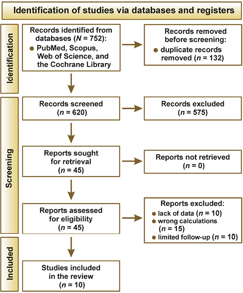
The systematic search yielded a total of 752 articles. After the removal of 132 duplicates, 620 titles and abstracts were screened. Of these, 45 full-text articles were assessed for eligibility, and 10 studies met the inclusion criteria for qualitative and quantitative analysis.1, 2, 3, 4, 5, 6, 7, 8, 9, 10 The study selection process is illustrated in the PRISMA 2020 flowchart (Figure 1). As this study was a systematic review and meta-analysis of previously published studies, no power calculation was required, and no new clinical data were collected.
Characteristics of the included studies
The 10 included studies involved a total of 412 patients and 684 implants, with follow-up periods ranging from 12 to 60 months. Eight studies were prospective in design, while 2 were retrospective cohort studies. Marginal bone loss was assessed using standardized periapical radiographs or cone-beam computed tomography CBCT in all cases. The type of abutment used (Ti-base vs. multi-unit) and implant location (anterior vs. posterior; maxilla vs. mandible) varied across studies. The mean patient age ranged from 42 to 68 years. Table 1 summarizes the main characteristics of the included studies, including study design, sample size, abutment type, follow-up duration, and MBL values.
Quantitative analysis of marginal bone loss
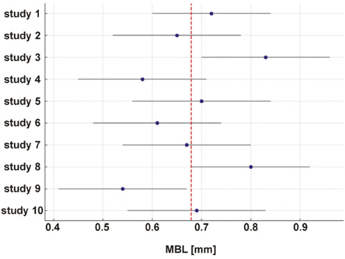
The pooled mean MBL across all studies was 0.68 mm (95% CI: 0.54–0.81 mm), as calculated using a random-effects model. The level of heterogeneity was moderate (I2 = 47%; Q = 16.8; p = 0.040). The forest plot of the meta-analysis is presented in Figure 2.
Subgroup analysis: Ti-base vs. multi-unit abutments
Subgroup analysis was conducted to compare MBL between the restorations supported by Ti-base abutments and those supported by MUAs. Although both groups demonstrated MBL values within the clinically acceptable range (<1.5 mm), no statistically significant difference was found between the 2 abutment types (p > 0.05). These findings suggest that both prosthetic solutions are comparable in terms of peri-implant bone preservation.
Risk-of-bias assessment
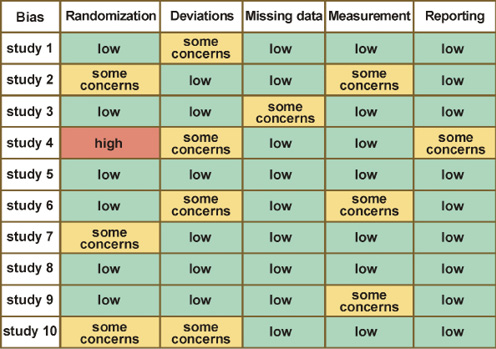
The Cochrane Risk of Bias 2.0 tool (RoB 2.0) was used to assess the methodological quality of the included studies across 5 domains: the randomization process; deviations from the intended interventions; missing data; the measurement of the outcome; and selective reporting. Two independent reviewers conducted the assessments, and any disagreement was resolved through discussion or consultation with a third reviewer. A visual summary of the domain-level risk-of-bias assessments is presented in Figure 3.
Discussion
The present systematic review and meta-analysis aimed to assess the extent of MBL in screw-retained zirconia prostheses supported by either Ti-base or multi-unit abutments. Based on 10 included clinical studies with varying study designs and follow-up periods, the pooled mean MBL was 0.68 mm, which was well below the clinically accepted threshold of 1.5 mm for implant success proposed by Albrektsson et al.11
Comparison of abutment types and outcomes
Across the analyzed studies, both abutment types demonstrated favorable bone stability over time. For instance, Vatenas et al., in an RCT, reported one of the lowest MBL values (0.54 mm) in patients rehabilitated with multi-unit abutments over a 12-month follow-up period.1 Similarly, Lind et al., in a case series, observed modest MBL (0.60 mm) with MUAs, and suggested that the vertical repositioning of the prosthetic interface may help mitigate early peri-implant bone remodeling.2
Conversely, Ti-base abutments also yielded acceptable outcomes, although slightly higher MBL values were observed in some cases. Bouhy et al.3 and Enkling et al.4 reported mean MBL values of 0.72 mm and 0.83 mm, respectively, in Ti-base-supported prostheses. Both studies hypothesized that the proximity of the restoration–implant interface to the bone crest in direct-to-implant configurations may result in greater bone remodeling, particularly under immediate or early loading protocols.5
These findings are consistent with the frameworks proposed by Al-Rimawi et al.5 and Yanagi et al.,6 which suggest that abutment height and its positioning relative to the mucosal seal may play an important role in preserving the crestal bone.
Design and methodological heterogeneity
A notable finding of this review is the substantial heterogeneity in study design, ranging from RCTs to retrospective case series. This variability may affect the strength and comparability of the evidence, and may partly account for the moderate heterogeneity observed in the meta-analysis (I2 = 47%).
For example, De Freitas et al., with a relatively long follow-up period of 18 months, reported a mean MBL of 0.61 mm in a Ti-base cohort; however, the study lacked randomization and blinding, increasing the risk of bias.7 In contrast, Goldstein et al., in a multicenter RCT, demonstrated lower MBL values with MUAs, and applied strict inclusion criteria and standardized measurement protocols.8
Such methodological differences – particularly in radiographic calibration, baseline measurement timing and abutment height standardization – limit the ability to draw definitive conclusions regarding the superiority of one abutment type over the other.8 Beyond prosthetic and mechanical factors, peri-implant inflammation is a key contributor to MBL and long-term implant failure. The early detection of inflammatory changes before radiographically detectable bone loss occurs is therefore essential. Recent studies have proposed non-invasive diagnostic approaches, including crevicular fluid analysis and implant sulcus thermography, as effective tools for the early identification of peri-implant mucositis and subclinical inflammation. It has been demonstrated that these techniques can detect inflammatory changes around implants even in asymptomatic patients, suggesting their potential role in preventive implant maintenance protocols.
These findings emphasize the importance of integrating biological monitoring with prosthetic planning to preserve peri-implant bone stability over time
Biomechanical and clinical implications
From a biomechanical standpoint, MUAs may provide stress-distributing benefits due to their supracrestal positioning, particularly in full-arch cases. This is supported by Romanos et al., who reported stable MBL with multi-unit configurations in edentulous patients undergoing immediate loading.9 However, other researchers cautioned that even minor mismatches at the abutment–implant interface may induce micromovements and bacterial infiltration,10, 12, 13, 14, 15 particularly when cementation is involved in Ti-base systems.16, 17, 18, 19, 20, 21, 22, 23, 24
Although Ti-base abutments offer a simplified and more direct connection between the implant and prosthesis, their clinical use is not without limitations. In particular, Ti-bases with reduced bonding height have been associated with an increased risk of decementation over time, especially when inadequate surface treatment or minimal bonding protocols are employed. These abutments rely heavily on the integrity of the adhesive interface, and the loss of cement retention may lead to prosthetic instability, complications, or even failure. Several studies have suggested that increased bonding height and optimized surface conditioning may improve the long-term retention and success of Ti-base restorations.11, 23, 24
Despite these differences, the overall MBL values remained within acceptable clinical limits across all included studies, supporting the use of both approaches according to individual anatomical, functional and esthetic considerations.
Limitations of the current evidence
This review is not without limitations. First, the number of high-quality RCTs remains limited, with only 4 included studies using randomization and blinding. Second, there is a lack of long-term (≥5 years) data on multi-unit-supported zirconia restorations. Furthermore, most studies lacked patient-reported outcome measures (PROMs) and did not report implant- or prosthesis-level complications in a standardized manner.
Future directions
Well-designed multicenter randomized trials with standardized radiographic protocols, longer follow-up durations and broader clinical endpoints are needed. Future studies should also investigate the effects of prosthetic screw access location, abutment height and surface characteristics on MBL.
Conclusions
This systematic review and meta-analysis demonstrated that screw-retained zirconia FDP supported by either Ti-base or multi-unit abutments are associated with minimal MBL, well within clinically acceptable limits. The pooled MBL across the 10 included clinical studies was 0.68 mm, with no statistically significant difference observed between the 2 abutment types.
While MUAs appear to offer slight biomechanical advantages, particularly in full-arch or immediate-loading scenarios, Ti-base abutments also demonstrated stable long-term outcomes when used in carefully selected cases. These findings suggest that both prosthetic platforms can be considered reliable in terms of peri-implant bone stability.
However, due to moderate heterogeneity among the available studies and the lack of long-term prospective data, clinical decision making should remain case-specific. Future high-quality RCTs with standardized radiographic protocols and comprehensive outcome reporting are essential to strengthen the evidence base.
Ethics approval and consent to participate
Not applicable.
Data availability
The datasets supporting the findings of the current study are available from the corresponding author on reasonable request.
Consent for publication
Not applicable.
Use of AI and AI-assisted technologies
Not applicable.