Abstract
Cyst enucleation is a common procedure performed in oral and maxillofacial surgery to remove cystic lesions. Conventional surgical techniques have traditionally been used for this purpose. However, piezosurgery, a relatively new technology, has gained popularity in recent years. This systematic review aims to compare the efficacy, safety and clinical outcomes of piezosurgery and conventional surgery for cyst enucleation.
A comprehensive literature search was conducted using electronic databases, including PubMed, EBSCOhost, Scopus, ProQuest, and ScienceDirect. Studies published from inception up to May 2023 were included. Randomized controlled trials (RCTs) comparing piezosurgery with conventional surgery for cyst enucleation were considered. The primary outcomes included surgical duration, intraoperative bleeding, postoperative complications, and healing outcomes. Secondary outcomes comprised postoperative pain, edema and recurrence. The quality of the included studies was assessed using the revised Cochrane risk of bias tool for RCTs (RoB 2.0).
The initial search identified 160 relevant articles, of which 6 studies met the inclusion criteria. Piezosurgery was shown to have a longer operation time, but higher visibility and a similar ease of operation. Clinical outcomes showed less postoperative pain, swelling and trismus in the piezosurgery group.
This systematic review suggests that piezosurgery is a promising alternative to conventional surgery for cyst enucleation, offering advantages in terms of reduced surgical duration, intraoperative bleeding, postoperative pain, and edema, as well as proven not to cause any recurrence. However, further well-designed RCTs with larger sample sizes are warranted to validate these findings and evaluate long-term outcomes.
Keywords: piezosurgery, enucleation, odontogenic cyst, conventional surgery
Introduction
Odontogenic cysts are common benign lesions that can arise from the epithelial remnants of tooth development or the inflammation of the dental pulp. They occur in 2.5% of the general population. These cysts can grow in size and cause bony expansion, the destruction of the surrounding bone, and the displacement of the adjacent teeth. They are usually asymptomatic and detected incidentally on routine radiographs. However, some cysts may cause pain, swelling or other complications.1
The treatment of odontogenic cysts varies depending on several factors; however, surgical intervention remains the mainstay of management. In recent years, more conservative surgical approaches have gained increasing support, as they are considered effective in treating odontogenic cysts while preserving the surrounding bone and teeth. Consequently, surgical treatment is increasingly designed to be as conservative as possible.2, 3, 4
Apart from the operative approach, more conservative techniques have also been developed for the treatment of odontogenic cysts. Piezosurgery is a specialized technique that uses non-heat-generating ultrasonic vibrations to cut and shape bone instead of conventional rotary instruments, which are more invasive and generate heat. It is thought to provide greater control and precision, and is associated with reduced trauma to the surgical site. Its use in oral and maxillofacial surgery has been reported to result in better clinical outcomes and fewer complications as compared to conventional techniques. However, little is known about its use in the surgical management of odontogenic cysts.2, 3, 4
The purpose of this systematic review was to compare the clinical outcomes of odontogenic cyst removal with piezosurgery and conventional surgery.
Methods
Protocol and registration
This literature review was conducted in accordance with the Cochrane Handbook for Systematic Reviews of Interventions, and reported in accordance with the Preferred Reporting Items for Systematic Reviews and Meta-Analyses (PRISMA) statement.
Eligibility criteria
Studies were screened based on the inclusion criteria of the population, intervention, comparison, outcome, and study design (PICOS) strategy. Articles focusing on other outcomes unrelated to piezosurgery for cyst removal, editorials, reviews, magazines, letters, and foreign articles were excluded.
Patients of any age, with good general health or controlled systemic disease, diagnosed with odontogenic cysts and indicated for jawbone cyst removal were included as the study population (P). The intervention investigated was piezosurgery (I), which was compared with conventional enucleation, the gold standard conservative treatment for odontogenic cysts (C). The primary outcomes (O) included the incidence of complications (e.g., hemorrhage, soft tissue injury, pain, swelling, and trismus), the duration of surgery, reduction in bone density, the ease of operation, and surgical visibility. Randomized clinical trials (RCTs), with or without blinding, as well as case series and case reports involving more than 5 cases, were eligible for inclusion (S).
Information sources
A comprehensive literature search was conducted in 5 electronic databases: PubMed; EBSCOhost; Scopus; ProQuest; and ScienceDirect. No date restrictions were applied, and all relevant studies published in English from database inception up to May 2023 were considered. In addition, the reference lists of the included studies were hand-searched to identify potentially eligible articles not captured by the electronic search.
Search strategy
The search strategy combined the keywords “enucleation” OR “cyst removal”, “piezosurgery” OR “piezoelectric”, and “odontogenic cyst” OR “jaw cyst”, together with relevant synonyms. The search terms and their combinations were adapted to the advanced search requirements of each database.
Data extraction
Data were extracted after the full texts of the included studies had been reviewed by the authors. The extracted data included: (1) first author and publication year; (2) study design; (3) sample size; (4) population characteristics; (5) cyst location; (6) treatment outcomes; (7) follow-up period; and (8) recurrence.
Quality assessment
The risk of bias was assessed using the revised Cochrane risk of bias tool for RCTs (RoB 2.0). The domains assessed included selection bias, performance bias, attrition bias, detection bias, and reporting bias. Three reviewers (UPG, LAL and SP) independently assessed the included studies. Each study was rated as having a low risk of bias, some concerns, or a high risk of bias according to the RoB 2.0 guidance. Studies in which all domains were rated as low risk were assigned an overall judgment of low risk of bias. Studies with at least one domain rated as having some concerns, but no domains rated as high risk, were judged to have some concerns. Studies with at least one domain rated as high risk were judged to have an overall high risk of bias.
Results
Study selection
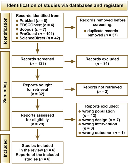
The search of 5 electronic databases yielded 160 records, of which 37 were duplicates. After duplicate removal, 123 records remained for title and abstract screening, and 91 were excluded. The remaining 32 reports were sought for full-text retrieval. Following full-text assessment, 29 articles were evaluated against the inclusion and exclusion criteria, and six studies were included in the review. The study selection process is presented in Figure 1.
Risk of bias
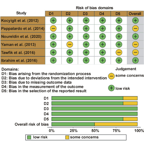
The risk of bias assessment showed that 3 studies had a low risk of bias, while the remaining 3 showed some concerns. A summary of the risk of bias assessment is presented in Figure 2.
Characteristics of the included studies
The characteristics of the 6 RCTs included in the review1, 2, 3, 4, 5, 6 are presented in Table 1. The dataset consists of the first author, year of publication, study design, population characteristics, cyst location, follow-up period, and clinical outcomes from both the intervention and comparison groups.
Outcomes
Intraoperative outcomes
Several intraoperative outcomes were identified from the included studies, including manipulation complexity, the ease of operation, visibility, and operation duration.
Manipulation complexity was reported in 2 studies – by Kocyigit et al.1 and Ibrahim et al.6 In the study by Kocyigit et al., manipulation complexity was observed in 5 patients in the conventional group and none in the piezosurgery group.1 In contrast, Ibrahim et al. reported no cases of manipulation complexity in either group.6
The ease of operation, visibility and operation duration were reported in 4 studies included in the analysis.1, 3, 4, 5 Kocyigit et al. reported that the piezosurgery group exceeded the expected operation time on average.1 In comparison with conventional surgery, enucleation using piezosurgery required a longer operation time, as reported by Noureldin and Melek (68.00 ±12.45 min vs. 44.43 ±12.74 min),3 Yaman and Suer (51.0 ±27.9 min vs. 36.0 ± 15.3 min)4 and Tawfik et al. (104.10 ±13.43 min vs. 89.10 ±10.92 min)5; statistical significance was reported in all 3 studies. The subjective ease of operation was similar between groups in the study by Noureldin and Melek, with no statistically significant difference observed.3 In contrast, visibility was significantly better with piezosurgery as compared to the conventional group, as reported by Yaman and Suer (89.4 ±5.2 vs. 79.8 ±9.4).5
Postoperative outcomes
Sequelae and complications were reported in all 6 included RCTs. The reported complications comprised hemorrhage, soft tissue damage, major perforation area, recurrence, neurosensory deficits, tissue dehiscence, and infection. The reported sequelae included pain, swelling and trismus, while some studies also evaluated changes in bone density.
Bone density changes were reported in 3 included studies – by Noureldin and Melek,3 Tawfik et al.5 and Ibrahim et al.6 In the study by Noureldin and Melek, bone defect reduction was significantly greater in the piezosurgery group as compared to the conventional group (46.58% vs. 33.04%).3 Similarly, higher bone density was observed in the piezosurgery group than in the control group in the studies by Tawfik et al. (84.00 ±7.74 vs. 75.50 ±6.39)5 and Ibrahim et al. (1,091.34 ±193.96 vs. 666.10 ±193.88).6
Pain, swelling and trismus were reported in 4 studies.2, 3, 5, 6 Pain was consistently lower in the piezosurgery group as compared to the control group, as reported by Pappalardo and Guarnieri (1.65 ±0.42 vs. 5.06 ±1.37),2 Noureldin and Melek (5.95 vs. 8.01; 3.81 vs. 6.71; 1.28 vs. 1.62)3 and Ibrahim et al. (3.43 ±0.98 vs. 7.71 ±0.95).6 Tawfik et al. also reported milder pain in the piezosurgery group than in the control group.5 Swelling was also reported to be lower in the piezosurgery group by Pappalardo and Guarnieri (15.00 ±5.92 vs. 40.50 ±6.50)2 and Tawfik et al. (32.65 ±1.55 vs. 35.08 ±1.06),5 with statistically significant differences. In addition, trismus was significantly lower in the piezosurgery group as compared to the control group (3.80 ±0.18 vs. 4.05 ± 0.13, as reported by Tawfik et al.5
Some complications were reported as having no occurrence in either group in some studies. Ibrahim et al. reported no cases of soft tissue damage or infection in either group,6 while Kocyigit et al. also reported no occurrence of soft tissue damage in either group.1
Discussion
Radicular cysts, the most common odontogenic cysts of the oral cavity, can be managed using various treatment modalities, including enucleation, marsupialization, decompression, and curettage.5, 7 Enucleation followed by the reconstruction of the bony defect is generally the preferred approach, although the spontaneous healing of the enucleated cyst cavity without reconstruction has also demonstrated favorable outcomes.8 In inflammatory odontogenic cysts, the incomplete removal of the cystic epithelium may increase the risk of recurrence. In addition, adjunctive procedures, such as apicoectomy or tooth extraction, performed during enucleation may adversely affect the prognosis.7, 8
Surgical enucleation aims to achieve the complete removal of the cystic epithelium, leaving no remnants around the interdental septa or the adjacent teeth. In recent years, piezosurgery, a technique that utilizes piezoelectric technology in oral and maxillofacial surgery, has gained popularity as an alternative to conventional surgical instruments.9 Introduced by Aro et al. approximately a century after Pierre Curie described the piezoelectric effect in 1880, this technology addresses several limitations of conventional instruments, and its application in bone surgery has become increasingly widespread over the past decade.10
The use of traditional hand instruments and surgical burs during cyst enucleation may have several drawbacks, potentially leading to intraoperative complications and an increased risk of recurrence.11 Rotary instruments, which are commonly used for this procedure, are considered more traumatic than piezosurgical devices. The high- and low-speed rotational movements used during bone removal may contribute to intraoperative and postoperative hemorrhage, which can obstruct the surgical field and make the complete removal of the cystic epithelium and residual tissue more challenging. In contrast, piezosurgery offers the advantage of reducing hemorrhage through the cavitation effect generated by ultrasonic micro-vibrations. This effect helps maintain a clear surgical field, facilitating the identification and removal of cystic remnants during the procedure.11, 12
Unlike conventional micro-saws, in which blood accumulation within the cutting area can impair visibility, piezosurgery maintains a nearly blood-free operative field during bone cutting procedures. This is attributed to the cavitation effect produced by the irrigation fluid and the ultrasonic vibrations generated by the instrument. The cavitation effect continuously clears blood from the surgical site, thereby improving visibility. Consequently, bleeding from the surrounding soft tissues is reduced, resulting in lower overall blood loss.13, 14 Furthermore, unlike conventional bone drilling or sawing, piezosurgery enables precise bone cutting while minimizing the risk of injury to the adjacent soft tissues, as it removes mineralized tissue through ultrasonic micro-vibrations rather than rotational or torsional forces.13
Another advantage of piezosurgery is its ability to provide continuous irrigation, thereby preventing bone overheating through the integrated saline coolant spray. This is achieved by applying light pressure to the handpiece while maintaining a steady flow of the cooling solution. The combination of continuous irrigation and effective temperature control not only improves the visibility of the surgical field, but also helps protect the bone from thermal injury.14
Compared with conventional rotary instruments or drills, piezosurgery requires only minimal pressure from the surgeon, which results in more precise bone cutting. Excessive pressure may restrict the movement of the instrument tip and generate excessive heat. When excessive pressure causes the tip to stop vibrating, indicating heat generation, the device emits a warning tone to alert the surgeon to discontinue cutting and prevent thermal injury to the bone. Piezosurgery is primarily designed for bone cutting while preserving the adjacent soft tissues, although modified settings have also been used for the excision of soft tissue lesions.15
Another advantage of piezosurgery is the availability of specialized tips, including straight and angulated designs, which enable precise manipulation, particularly in interdental areas. These tips facilitate access to the surgical site and may reduce the risk of major perforation of the cystic epithelium during enucleation. In contrast, conventional rotary instruments carry a greater risk of injury to the adjacent anatomical structures, such as nerves, the maxillary sinus and the nasal mucosa, particularly when used in anatomically challenging areas. Piezosurgery enhances surgical safety by selectively cutting mineralized tissue while minimizing injury to the surrounding soft tissues.11
For example, one study reported successful enucleation of a cyst located adjacent to the mental nerve using piezosurgery, with no temporary or permanent paresthesia observed.16 This finding highlights the potential safety advantages of piezosurgery when operating in close proximity to critical anatomical structures.
The impact of mechanical instruments on bone structure and cell viability is an important consideration in surgery, as even brief exposure to elevated temperatures may result in cellular damage and tissue necrosis. Previous studies have shown that bone harvested using conventional instruments, such as round burs, spiral implant burs or bone scrapers, may be unsuitable for grafting because of the absence of viable osteocytes and the predominance of nonviable bone tissue. In contrast, studies evaluating piezoelectric surgery have demonstrated improved bone cell viability, supporting earlier findings on the favorable effects of piezoelectric devices on bone chip morphology and cell survival during bone harvesting.14
Notably, Stübinger et al. demonstrated that autologous bone harvested from the zygomaticomaxillary region using a piezoelectric device could be successfully used for bone augmentation and the esthetic placement of oral implants following a 5-month healing period.7 Similarly, Happe reported that bone grafts harvested from the mandibular ramus using piezosurgery integrated successfully without significant complications, providing sufficient bone volume for implant placement.17 These clinical findings are further supported by in vitro studies demonstrating the presence of viable osteocytes and bone morphogenetic proteins in bone grafts harvested using piezosurgery.18
Piezosurgery offers a significant advantage in cystectomy, particularly when bone cutting is required near vital soft tissues, such as nerves and blood vessels, where the risk of mechanical or thermal injury must be minimized. One study reported that no nerve lesions occurred with piezosurgery, whereas surgery using rotary instruments resulted in reduced sensation lasting at least 1 week in 8% of patients.19 These findings are consistent with the study by Schaeren et al., which demonstrated that, even under worst-case conditions, the direct exposure of a nerve to piezosurgery did not transect the nerve, but might result in limited structural or functional alterations.9 In most cases, nerve regeneration can occur when the protective perineural sheath remains intact, unlike with conventional drills or oscillating saws. It was also observed that increased mechanical force applied to the nerve by the piezosurgery device resulted in greater tissue damage; however, this effect was not observed with ultrasonic vibration alone.16
The results of this study highlight a key advantage of piezoelectric instruments in the excision of cystic lesions, namely, the ability to perform precise ostectomy near delicate anatomical structures, such as the inferior alveolar nerve and the mental nerve, without causing damage. This advantage is attributed to the selective cutting mechanism of the device, which differentiates tissue hardness and acts primarily on mineralized structures. As the device automatically ceases cutting upon encountering non-mineralized tissues, it minimizes the risk of injury to soft tissues, including mucous membranes, nerves and blood vessels.20
The visual analog scale (VAS) scores for visibility indicate that ultrasonic surgery provides a significantly clearer surgical field as compared to conventional approaches, consistent with previous studies investigating ultrasonic surgery.21 Unlike conventional micro-saws, in which blood accumulation within the cutting area may impair visibility, ultrasonic surgery maintains a nearly blood-free operative field during bone cutting procedures. This is achieved through the cavitation effect generated by the distribution of the cooling fluid and the specific oscillation pattern of the instrument, which effectively clears blood from the surgical site and ensures optimal visibility throughout the procedure. Additionally, continuous saline irrigation prevents bone overheating, while intermittent micro-cavitation bursts contribute to hemostasis, resulting in a significant reduction in overall blood loss.22
The VAS scores for the ease of operation showed no significant differences between the piezosurgery and conventional surgery groups. Therefore, piezosurgery does not appear to provide a significant advantage in terms of improving operative ease as compared to conventional techniques. However, the frequent replacement of piezosurgery inserts and adjustments to machine settings, including power, oscillation and irrigation parameters according to the selected insert, may increase procedural complexity and operative time. Ongoing advances in piezosurgery technology are expected to address these limitations by introducing more user-friendly devices.23
The literature contains limited reports on the efficacy of piezosurgery in procedures involving soft tissues, such as cyst enucleation. However, several case reports and clinical studies have demonstrated favorable outcomes with the use of piezosurgery in the removal of jaw cysts,24 cystic ameloblastoma,13 and hyoid bone osteotomy during thyroglossal duct cyst surgery.16 These findings suggest that piezosurgery may be an effective approach for managing these procedures.
Regarding surgical outcomes, ultrasonic surgery was found to be superior to conventional surgery in terms of intraoperative bleeding, epithelial perforation, postoperative complications, and recurrence. However, it should be noted that ultrasonic surgery was associated with a longer overall surgical duration. These findings are consistent with previous studies, which also reported increased operative time with the use of ultrasonic surgery.16, 23
Although the effectiveness of the ultrasonic surgical approach in bone procedures has been consistently demonstrated, manual instruments remain necessary for certain soft tissue procedures and anatomically sensitive situations. In particular, manual instruments are preferred when operating around unerupted teeth in pediatric patients to prevent the potential displacement of tooth buds caused by ultrasonic oscillations from the surgical device. They are also favored when working near delicate anatomical structures, such as the orbital floor, during the the removal of large cystic lesions, as they allow surgeons to assess bone integrity through tactile feedback. Consequently, the development of new insert designs with different curves is needed to better accommodate these clinical requirements.23
Another drawback encountered during the use of ultrasonic surgery for cyst enucleation was insert breakage, with 2 inserts fracturing at their tips during procedures.23 Several factors may contribute to insert failure, including sterilization processes, material fatigue due to repeated use, manufacturing defects, or the inappropriate selection of machine parameters during operation.23
The effectiveness of ultrasonic surgery depends on the precise manufacturing and design of the ultrasonic surgical device, including its piezoelectric crystals, electronic control circuits, handpiece materials, and working tips (inserts). Although piezoelectric bone surgery is relatively recent advancement in oral and maxillofacial surgery for bone-related procedures, substantial technological progress has been made in both device design and surgical techniques over the past decade.24 Currently, several piezoelectric devices are available on the market, with newer versions continuing to be developed. It is important to recognize that similar piezoelectric units manufactured by different companies may demonstrate variations in performance. Furthermore, differences in insert design and performance may also influence clinical outcomes.14
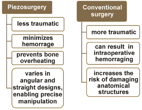
The schematic comparison of piezosurgery and conventional surgery is presented in Figure 3.
Conclusions
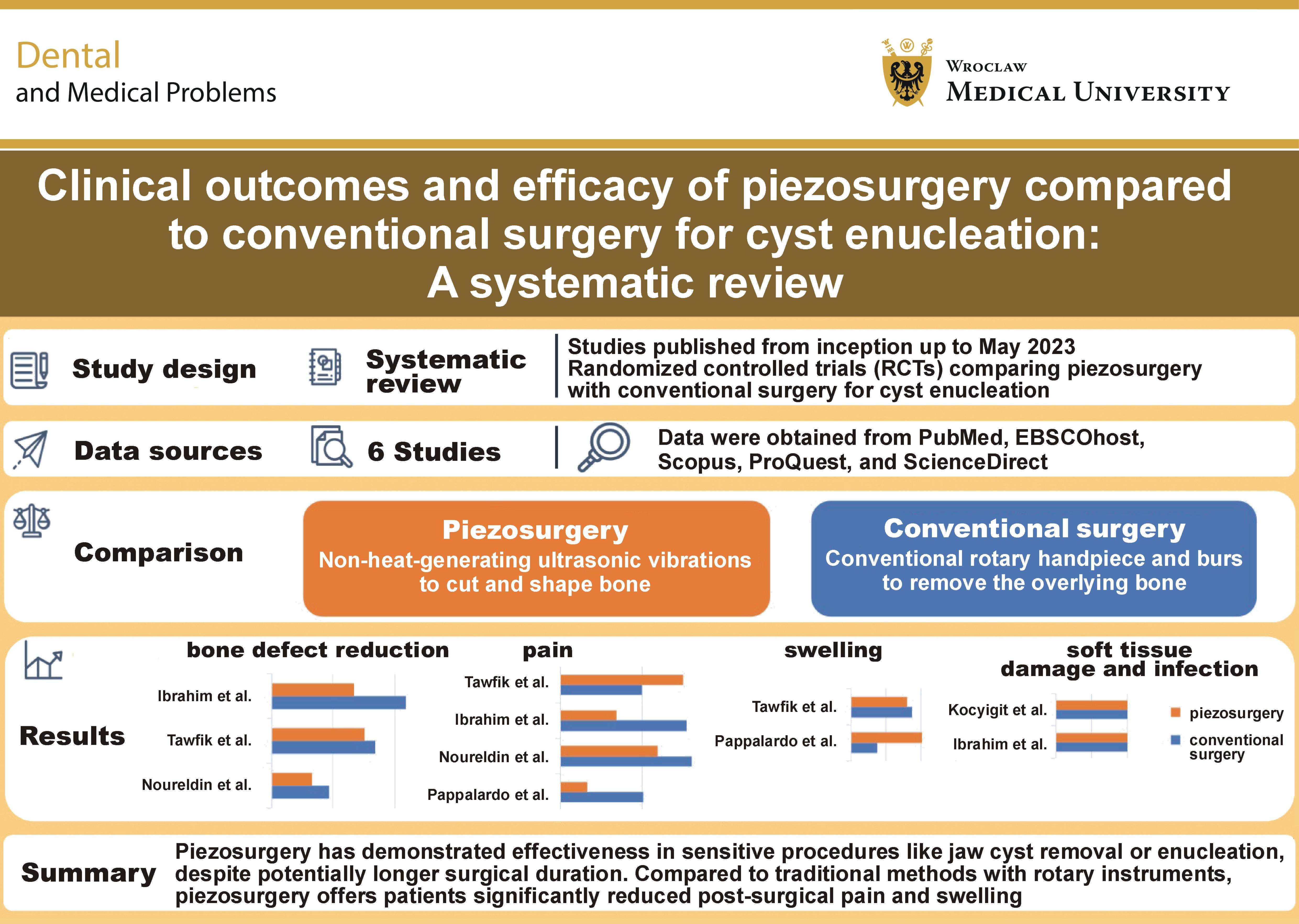
Piezosurgery has demonstrated effectiveness in sensitive procedures, such as jaw cyst removal and enucleation, despite potentially requiring longer operative times. Compared with conventional methods using rotary instruments, piezosurgery provides significant reductions in postoperative pain and swelling. It may be a more favorable approach for cyst enucleation due to its ability to preserve important anatomical structures and provide improved surgical visibility. Additionally, piezosurgery may promote better early healing of the bone defect following enucleation. Although ultrasonic surgery for jaw cyst enucleation increases operative time, it significantly improves visibility, making it advantageous for delicate surgical manipulation and reducing the risk of injury to vital structures, including neurovascular tissues.
Ethics approval and consent to participate
Not applicable.
Data availability
Not applicable.
Consent for publication
Not applicable.
Use of AI and AI-assisted technologies
Not applicable.