
One journal. Two disciplines. Shared excellence.

Dental and Medical Problems

Title abbreviation: Dent Med Probl
Journal Impact Factor (JIF 2025) – 4.3
Journal Citation Indicator (JCI 2025) - 1.38
Scopus CiteScore (2025) – 5.8
Index Copernicus Value (ICV 2024) – 178.25
MNiSW – 70 pts
ISSN 1644-387X (print)
ISSN 2300-9020 (online)
Periodicity – bimonthly


Download original text (EN)

Dental and Medical Problems

2026, vol. 63, nr 3, May-June, p. 793–804

doi: 10.17219/dmp/193767

Publication type: review

Language: English

License: Creative Commons Attribution 3.0 Unported (CC BY 3.0)

Download citation:

  • BIBTEX (JabRef, Mendeley)
  • RIS (Papers, Reference Manager, RefWorks, Zotero)

Cite as:


Alotaibi DH. Efficacy, safety, and health-related quality of life data of hyaluronic acid for the treatment of interdental papilla deficiency: A systematic review of randomized controlled trials. Dent Med Probl. 2026;63(3):793–804. doi:10.17219/dmp/193767

Efficacy, safety, and health-related quality of life data of hyaluronic acid for the treatment of interdental papilla deficiency: A systematic review of randomized controlled trials

Dalal H. Alotaibi1,A,B,C,D,E,F

1 Department of Periodontics and Community Dentistry, College of Dentistry, King Saud University, Riyadh, Saudi Arabia

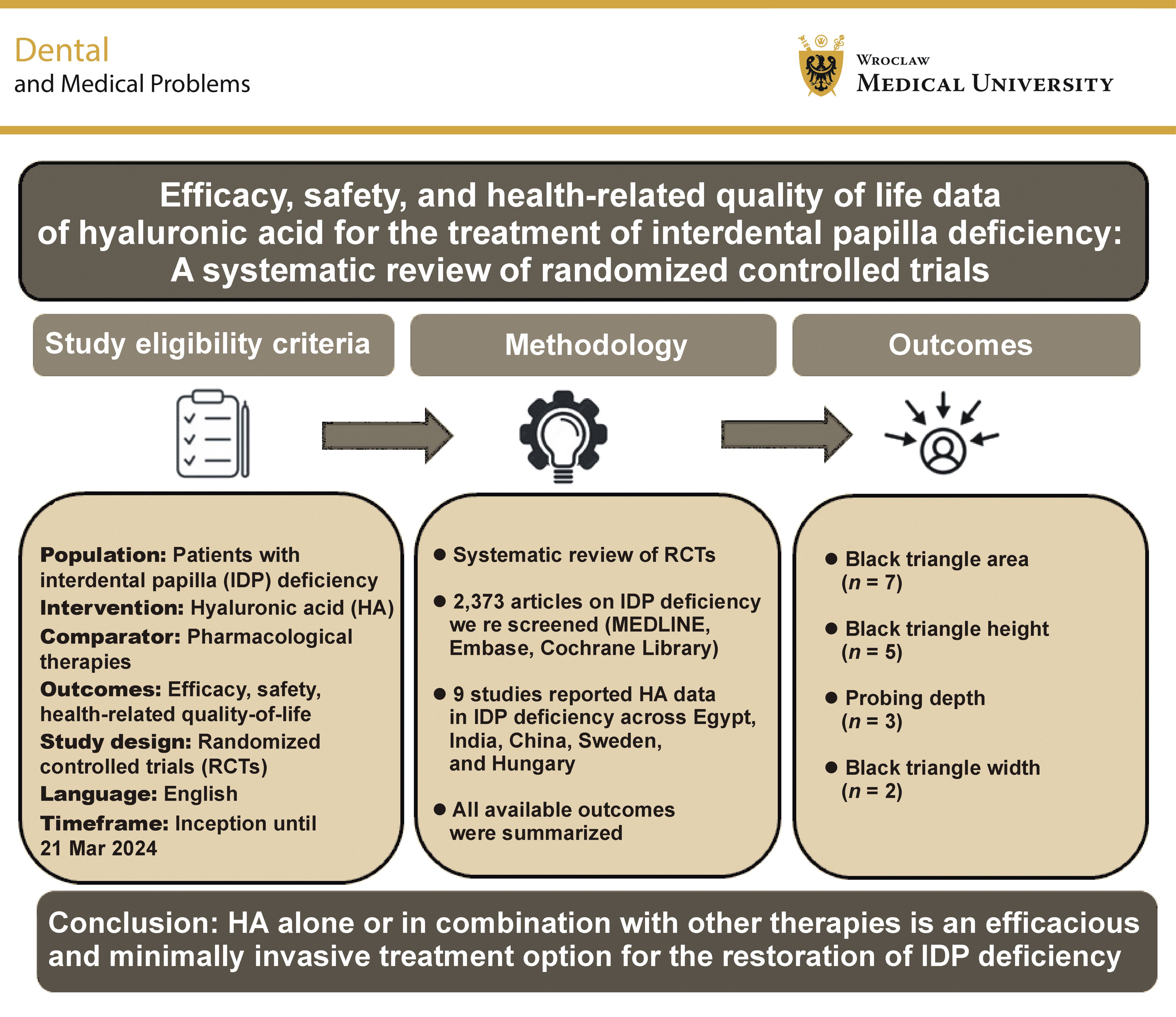
Graphical abstract


Graphical abstracts

Highlights


  • Hyaluronic acid (HA) appears to be an effective and minimally invasive treatment option for restoring interdental papilla (IDP) deficiency.
  • Most included RCTs reported improvement in black triangle dimensions following HA treatment.
  • Combination therapies may provide additional benefits compared with HA alone.
  • Evidence regarding HA safety remains limited.
  • None of the included studies evaluated health-related quality-of-life outcomes.

Abstract

The efficacy of hyaluronic acid (HA) in the treatment of interdental papilla (IDP) deficiency is well documented. This systematic review sought to evaluate clinical studies reporting efficacy, safety, and health-related quality of life (HRQoL) data for HA used in the treatment of IDP deficiency. This review was conducted and reported in accordance with the guidelines of the Preferred Reporting Items for Systematic Reviews and Meta-Analyses (PRISMA) methodology. A comprehensive search of electronic databases was conducted for published randomized controlled trials (RCTs) from their inception until March 21, 2024. A total of 9 studies met the inclusion criteria. The sample sizes in the included RCTs ranged from 10 to 31 participants, with the mean patient age between 26.7 and 46.1 years, predominantly females. Of the 9 eligible studies, 6 assessed the use of HA alone, while 3 investigated the effects of HA in combination with other therapies. The most common outcome measures assessed black triangle dimensions (i.e., area, height or width) followed by probing depth (PD). Based on the limited published studies, this review suggests that HA treatment (whether alone or in combination) may offer therapeutic benefits for patients with IDP defects. However, none of the included studies assessed the safety of HA and its effect on HRQoL. There is a need for further research on the long-term efficacy and safety of HA treatment, and its influence on HRQoL.

Keywords: periodontal disease, hyaluronic acid, efficacy, health-related quality of life, interdental papilla deficiency

Introduction

The interdental papilla (IDP) represents the visible anatomical part of the gingival tissue that extends from the interproximal alveolar crest to the contact point.1 Although the papilla is a small anatomical structure, it has a significant functional and esthetic value.1, 2 The deficiency or loss of IDP leads to an open gingival embrasure space, termed a “black triangle”, which adversely affects esthetics, increases the retention of food debris and contributes to phonetic problems.2 Therefore, the preservation and restoration of a deficient or lost IDP are essential. Nevertheless, the reconstruction of a lost IDP presents a significant challenge, often yielding unpredictable long-term outcomes, and remains a topic of debate and further investigation in the periodontal field.3

Several surgical methods, including pedicle flaps, semilunar coronally repositioned flaps, and regenerative procedures with connective tissue and bone grafts, have been proposed and used either alone or in combination to treat IDP loss. However, the invasiveness and uncertain effectiveness of surgical procedures, together with the anatomical location of IDP and its poor blood supply, still pose a major challenge.4, 5 Considering the potential limitations associated with surgical procedures, several non-surgical options (i.e., hemolaser therapy, orthodontic treatment, restorative treatment, and the injection of fillers and gingival tissue volumizers) have been reported to offer better therapeutic benefits; however, their use in routine dental practice remains limited.3, 6

Hyaluronic acid (HA), a naturally occurring polysaccharide, is present within the extracellular matrix (ECM) of various body tissues, including periodontal tissues.7 Hyaluronic acid has been demonstrated to play a significant role in regulating various biological processes, including anti-inflammatory responses, wound healing and tissue regeneration.8 Several controlled trials have reported the clinical efficacy of HA in the treatment of IDP deficiency.1, 5, 8, 9, 10, 11, 12, 13, 14 Moreover, evidence from the published systematic reviews indicates that HA treatment is efficacious in patients with IDP deficiency.3, 6, 7, 15, 16 However, none of the published reviews have reported on the health-related quality of life (HRQoL) of IDP-deficient patients treated with HA. Therefore, the aim of the current systematic review was to evaluate the efficacy and safety of HA use, and its effect on HRQoL in patients with IDP deficiency.

Methods

Study design

This systematic review was conducted and reported in accordance with the Preferred Reporting Items for Systematic Reviews and Meta-Analyses (PRISMA) statement.17 A protocol for the systematic review was developed and registered in the International Prospective Register of Systematic Reviews (PROSPERO) database (CRD42024547932).

Literature search

Electronic databases (MEDLINE, MEDLINE In-Process, Embase, and the Cochrane Library) were comprehensively searched from inception to March 21, 2024, via the Ovid platform. The search strategy used to retrieve relevant studies is presented in the supplementary material, openly available in https://figshare.com at doi:10.6084/m9.figshare.26863660. A gray literature search was performed, and the bibliographies of the published systematic reviews were screened. Additionally, a manual search of unpublished work, including theses and dissertations, was performed to identify any relevant information.

Study selection and data extraction

All retrieved citations were collated after duplicates were removed. The collated citations were screened by 2 independent reviewers (DA and SA). Initially, each citation was screened based on its title and abstract, using predefined eligibility criteria (Table 1).

Subsequently, the full-texts of potentially eligible studies were retrieved and further subjected to full-text screening, using a similar process. Any uncertainty regarding study inclusion at either stage was resolved through mutual discussion. Relevant data from the included studies were extracted by a single reviewer using a predefined MS Excel-based data extraction grid. The extracted data were thoroughly checked for transcription errors by a second reviewer.

The data extracted from the eligible studies included study information (i.e., primary author name, year of publication, country, number of deficiency sites, number of patients, age, sex, study design, intervention group, deficiency region, and treatment duration), and outcomes of interest (i.e., black triangle area, height and width, probing depth (PD), gingival index (GI), deficient volume, papillary deficiency height, satisfaction score, plaque index (PI), papilla presence index (PPI), modified papilla index score (MPIS), distance between the papilla tip and the contact point, gingival volume, clinical attachment level/loss (CAL), bleeding on probing (BoP), radiographic alveolar bone height, esthetic appearance, pain, signs of black triangle, and papillary marginal gingival index).

Quality assessment of the included studies

The Cochrane risk-of-bias assessment tool (RoB) was used to evaluate the methodological quality of the included RCTs.18 Two independent reviewers assessed the risk of bias for individual bias domains (i.e., selection, performance, detection, attrition, reporting, and any other biases not covered by these categories). The risk was reported as “low”, “high” or “unclear”. Any discrepancies between the reviewers were resolved through mutual discussion.

Results

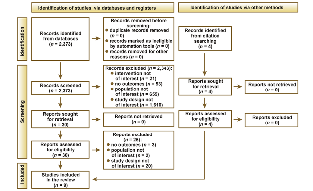
A total of 2,373 unique citations were obtained from the electronic database search. Following primary screening, 2,343 citations were excluded, as they did not meet the inclusion criteria, while 30 citations were subjected to full-text review. Of these, 5 studies were included in the systematic review. Additionally, 4 studies were identified through the bibliographic search, resulting in a total of 9 unique studies included in the final synthesis (Figure 1).

Study characteristics

The characteristics of the included studies are summarized in Table 2. All studies assessed patients with IDP deficiency. Four studies were conducted in Egypt, 2 in India, and 1 each in China, Sweden and Hungary. Of all included studies, 3 were double-blind RCTs, one was a single-blind RCT (i.e., investigator-blinded), and the remaining 5 studies did not report blinding information.

Of the 9 studies included in this review, only 2 studies explicitly specified outcomes assessed as primary and secondary outcomes. In the RCT conducted by Ni et al., the primary outcome measure was the height of each gingival papilla, while the area of each black triangle was assessed as the secondary outcome.1 In the randomized, comparative clinical study by Fakher et al., patient satisfaction was assessed as the primary outcome measure.5 Additionally, the evaluated secondary outcomes included changes in the surface area and height of the black triangle, clinical parameters (PD, PI and GI at the deficient papilla site), and pain levels 4 h after injection.

Three studies assessed outcomes for HA in combination with other interventions as compared to HA alone, while the remaining studies assessed outcomes for HA with different formulations (i.e., Flex Barrier, Revident) or with different comparators (i.e., physiological saline, untreated controls, placebo, or calcium hydroxylapatite). The treatment duration among the included studies ranged from a single administration to 6 months. The study population in the included RCTs ranged from 10 to 31 patients. The mean age of the patients included in the RCTs ranged from 26.7 to 46.1 years, and the participants were predominantly female. Most of the included RCTs used the Nordland and Tarnow classification19 to stratify IDP deficiency among patients (n = 6) (Table 2).

Outcome measures

Overall, most studies reported clinical outcomes, while only 3 studies reported patient-reported satisfaction following HA treatment. Among the studies that assessed clinical outcomes, black triangle area was the most frequently reported outcome (n = 7) followed by black triangle height (n = 5), PD (n = 3), black triangle width (n = 2), GI (n = 2), and CAL (n = 2). Other clinical outcomes reported included deficient volume, PI, IDP height, pain intensity, gingival volume, BoP, MPIS, radiographic alveolar bone height, esthetic appearance, clinical signs of black triangle, papillary marginal gingival index, and PPI (Table 3).

Most of the included RCTs (n = 8) reported that HA provided therapeutic benefits to patients with IDP deficiency. Hyaluronic acid was reported to be more effective in the treatment of IDP deficiency when used in combination with other interventions than HA alone. Specifically, HA in combination with plasma-rich growth factors (PRGF) was reported to provide significant improvement in papillary deficiency height, area and volume. Similarly, when HA was used in combination with injectable carboxymethyl chitosan hydrogel, improvement was reported in multiple clinical parameters (i.e., PI, GI, PD, and the surface area and height of the black triangle), while HA in combination with injectable platelet-rich fibrin (iPRF) was reported to improve black triangle height. Two studies reported beneficial effects of HA; however, the effect was not superior to that of the comparators (i.e., calcium hydroxylapatite and physiological saline).

Another study assessed the efficacy of single injections of 2 different HA preparations (i.e., Revident and Flex Barrier). Both demonstrated clinical applicability, with Revident providing longer-lasting improvement than Flex Barrier. Three RCTs compared the efficacy of HA with that of physiological saline. While 2 studies indicated a significant reduction in black triangle dimensions following the use of HA gel as compared to saline solution, the study by Ni et al. found no significant differences between HA gel and saline solution in terms of black triangle area and gingival papilla height.1 However, the results of in vitro analyses revealed a significantly increased rate of proliferation and migration of human gingival fibroblasts (HGF) following HA treatment, with the effect being more pronounced at higher concentrations.1

In the three studies that reported patient-reported satisfaction, a visual analog scale (VAS) was used to assess dental esthetics before and after HA treatment. All 3 studies reported significantly higher satisfaction with esthetic appearance following HA treatment.

Quality assessment of the included studies

One study had a low risk of bias across all the 7 domains, and 2 studies reported a low risk in 6 domains and an unclear risk of bias in 1 domain. Of the remaining studies (n = 6), 5 reported unclear risk of bias in 3 of the 7 domains and 1 study reported a high risk of bias in 1 domain (i.e., blinding of participants and personnel) (Table 4).

Overall, this review provides a descriptive summary of the included studies. The considerable heterogeneity across the included studies precluded the possibility of performing a meta-analysis.

Discussion

This systematic review provides the most contemporary evidence on the efficacy, safety and HRQoL outcomes of HA treatment in patients with IDP deficiency. Evidence from this review indicates that HA, whether used alone or in combination with other therapies (i.e., PRGF, injectable carboxymethyl chitosan hydrogel or iPRF), represents an effective and minimally invasive option for the treatment of IDP deficiency. It is important to highlight that none of the included RCTs provided data on the safety of HA treatment or its effect on HRQoL in this population.

The use of HA has garnered considerable interest for its potential role in restoring unesthetic defects in the papilla. Data from RCTs have shown favorable therapeutic benefits of HA in treating IDP deficiency.1, 3, 5, 8, 10, 11, 12, 13, 14 This review specifically aimed to synthesize the existing evidence on the efficacy, safety and HRQoL outcomes of HA treatment, and to provide meaningful insights into the use of HA treatment for patients with IDP defects. Across the 9 RCTs included in this literature review, HA was used either alone (n = 6)1, 9, 10, 11, 12, 13 or in combination with other therapies (n = 3) (i.e., PRGF, injectable carboxymethyl chitosan hydrogel or iPRF).5, 8, 14 Although there was considerable heterogeneity across the included studies in terms of population, sample size and treatment duration, HA was found to confer positive benefits as compared to HA alone or control arms. Consistent with previously published reviews,3, 6, 7, 15, 16 HA was reported to exert a positive effect on an array of clinical outcomes, including black triangle dimensions (i.e., area, height and width).1, 5, 8, 11, 12, 13, 14 Additionally, patients were found to be adequately satisfied with their esthetic appearance after HA treatment.5, 8, 11 This finding is of significant importance, as satisfaction with treatment is important for promoting acceptance of any intervention and improving health outcomes among patients with IDP deficiency.

The results of this review are consistent with those of previously published studies3, 6, 7, 15, 16 and closely resemble the findings of a recent systematic review,6 which identified HA injections as an effective and minimally invasive method for reconstructing a lost IDP. The key difference is that our review extends beyond clinical outcomes alone. We emphasize that, despite numerous clinical trials, the safety of HA and its effects on HRQoL in patients with IDP deficiency remain poorly investigated.

This review has some inherent limitations. The small sample sizes and limited number of RCTs included in this review are the major limitations. This highlights the need for robust, well-designed trials in this patient population. Another limitation of the review is the heterogeneity observed with regard to study design, treatment protocols and outcome measures, which prevents us from drawing robust conclusions on the efficacy and safety of HA in the treatment of IDP deficiency. As this review included results from RCTs only, its findings cannot be generalized to non-RCT interventions or real-world dental practice. Patients in the included studies had only a short-term exposure to HA, indicating a need for further research with longer treatment durations to better understand the long-term benefits of HA treatment. Most of the included RCTs in this review were judged to have an unclear risk of bias in some domains, which might have affected the study findings. Only studies published in English were included; thus, there is a high risk of missing important studies published in other languages. Finally, few studies reported data on safety outcomes, and no HRQoL data were reported across the included studies.

Despite these limitations, several strengths of the current systematic review merit consideration. First, this review was conducted in accordance with the PRISMA guidelines, which ensure the use of structured and optimal methodology to synthesize the evidence. The employed search methodology was extensive and rigorous, incorporating data from both published and unpublished studies, as well as manual and bibliographic search to ensure comprehensive coverage. This approach aimed to include studies from diverse populations and geographic regions, minimizing the risk of excluding any demographic groups. Finally, the studies included in this review were critically appraised using the Cochrane risk-of-bias tool, thereby adding further rigor to the review.

Conclusions

Based on the limited published evidence, this systematic review suggests that HA, either alone or in combination with other therapies, may be an effective and minimally invasive treatment option for the restoration of IDP deficiency. However, data on the safety of HA and its impact on HRQoL for IDP-deficient patients are lacking, highlighting the need for future clinical studies to address these aspects.

Ethics approval and consent to participate

Not applicable.

Data availability

The data presented within this manuscript have been extracted from 9 included studies. The data were readily available within the cited articles. The search strategy used for retrieving the studies for the current systematic review are openly available in https://figshare.com at doi:10.6084/m9.figshare.26863660.

Consent for publication

Not applicable.

Use of AI and AI-assisted technologies

Not applicable.
Tables


Table 1. Study eligibility criteria

Category

Inclusion criteria

Exclusion criteria

Population

Patients with IDP deficiency

• Healthy volunteers
• Patients without IDP deficiency

Intervention

Hyaluronic acid

Interventions other than hyaluronic acid

Comparator

Any pharmacological therapy

None

Outcomes

• Efficacy
• Safety
• Quality of life

Outcomes other than those specified in the inclusion criteria

Study design

• RCTs
• Systematic reviews and meta-analyses of RCTs/non-RCTsa

• Non-RCTs including comparative and non-comparative observational studies
• Preclinical studies
• Comments, letters, and editorials
• Case reports and case series
• Pharmacokinetic and economic studies

Language

English

Studies published in languages other than English

Time frame

From inception to March 21, 2024

None

IDP – interdental papilla; RCTs – randomized controlled trials.
Table 2. Characteristics of the included studies (N = 9)

Author,
year

Country

Study design

Number of patients

Number of sites

Mean age (SD)

Female, n (%)

Intervention groups

Classification/region

Tx duration

Ni,
20211

China

Double-blind RCT

Total: 24

HA: 34
Saline: 34

Total: 41.3 (7.73) years

Total: 19 (90.47)

• Saline
16 mg/mL HA gel

6 weeks

Fakher,
20235

Egypt

RCT

HA gel: 6
iPRF: 6
HA + iPRF: 6

Total: 20–50 years

• HA gel: 0.2 mL

Class I or II deficiency based on Nordland and Tarnow classification

6 weeks

Abdelkader,
20228

Egypt

RCT

HA + carboxymethyl chitosan hydrogel: 21
HA alone: 21

Total: 20–40 years

• HA: 0.2 mL of 50% HA gel
• HA + carboxymethyl chitosan hydrogel: 0.2 mL of 50% HA gel + 50% of chitosan gel

Class I or II deficiency in interbicuspidregion interdental space based on Nordland and Tarnow classification

Bertl,
20169

Sweden

Double-blind

HA: 11
Saline: 11

HA: 26.7 (5.2) years
Saline: 33.1 (6.0) years

HA: 5 (45.45)
Saline: 7 (70)

• HA: 1 mL contains 16 mg of cross-linked sodium hyaluronate and 2 mg Na-hyaluronate

Deficiency in the anterior maxilla

4 weeks

Shawky,
201710

Egypt

RCT

Total: 30

HA gel: 20
Radiesse: 20

Total: 25–35 years

Total: 30 (100)

• HA gel: 0.2 mL
• Radiesse: 0.2 mL

6 months

Abdelraouf,
201911

Egypt

Double-blind

HA: 5
Saline: 5

HA: 16
Saline: 14

Total: 32.55 (9.3) years

HA: 4 (80)
Saline: 4 (80)

• HA: 20 mg/mL

Class I or II deficiency based on Nordland and Tarnow classification

6 weeks

Mandel,
202012

Hungary

RCT

Revident: 16
Flex Barrier: 15

Revident (HA) Test: 48
Revident (HA) Untreated control: 32
Flex Barrier (HA) Test: 50
Untreated control: 30

Revident: 46.1 (12.3) years
Flex Barrier: 41.8 (13.8) years

Revident: 11 (68.75)
Flex Barrier: 12 (80)

• Flex Barrier: 0.1 mL per site
• Revident: Tx applied using gel

Class I or II
IDP loss in anterior region between canine teeth based on Nordland and Tarnow classification

Single administration

Kapoor,
202213

India

RCT

Total: 15

HA: 30
Saline: 30

HA: 37.13 (8.22) years
Placebo: 33.4 (7.89) years

• HA: 0.2 mL of 2% HA gel

Class I or II deficiency based on Nordland and Tarnow classification

6 weeks

Bal,
202314

India

Single-blind RCT

HA: 10
HA + PRGF: 11

HA: 18
HA + PRGF: 18

HA: 34.63 (5.22) years
HA + PRGF: 38.71 (8.4) years

• 0.8% of 0.2 mL HA gel
• 0.8% of 0.2 mL HA gel + 0.2 mL PRGF

Class I or Class II
IDP loss in the maxillary region based on Nordland and Tarnow classification

6 weeks

IDP – interdental papilla; iPRF – injectable platelet-rich fibrin; HA – hyaluronic acid; mg – milligram; mL –milliliter; PRGF – plasma rich in growth factors; RCT – randomized controlled trial; SD – standard deviation; Tx – treatment.
Table 3. Summary of the results from the included studies (N = 9)

Author, year

Outcome

Key takeaway

Ni,
20211

Mean (SD) papillary deficient height (mm)
Physiological saline
• Baseline: 2.99 (1.42)
• 6 months: 3.12 (1.36)
• 12 months: 3.26 (1.39)
HA gel
• Baseline: 3.25 (1.30)
• 6 months: 3.45 (1.27)
• 12 months: 3.53 (1.25)
Mean (SD) change from the baseline
Physiological saline
• 6 months: 0.135 (0.39) (p = 0.199)
• 12 months: 0.278 (0.45) (p = 0.006)
HA gel
• 6 months: 0.198 (0.34) (p = 0.011)
• 12 months: 0.280 (0.38) (p = 0.001)
Mean (SD) black triangle area (mm2)
Physiological saline
• Baseline: 1.78 (1.31)
• 6 months: 1.63 (1.26)
• 12 months: 1.46 (1.26)
HA gel
• Baseline: 1.90 (1.37)
• 6 months: 1.65 (1.32)
• 12 months: 1.45 (1.16)
Mean (SD) change from the baseline
Physiological saline
• 6 months: –0.150 (0.37) (p = 0.098)
• 12 months: –0.320 (0.50) (p = 0.004)
HA gel
• 6 months: –0.260 (0.42) (p = 0.007)
• 12 months: –0.450 (0.54) (p < 0.001)

This study suggest that the height of gingival papillae could be enhanced by injecting HA to address gingival papilla defects. Nevertheless, this effect is not better than that of a physiological saline

Fakher,
20235

Mean satisfaction score
HA gel
• Baseline: 3.8
• 6 months: 9.1
iPRF
• Baseline: 3.7
• 6 months: 7.8
HA gel + iPRF
• Baseline: 3.5
• 6 months: 8.7
Mean black triangle height (mm)
HA gel
• Baseline: 1.73
• 3 weeks: 1.4
• 6 weeks: 1.17
• 3 months: 0.72
6 months: 0.13
iPRF
• Baseline: 1.98
• 3 weeks: 1.68
• 6 weeks: 1.38
• 3 months: 1.2
• 6 months: 0.92
HA gel + iPRF
• Baseline: 2.5
• 3 weeks: 2.2
• 6 weeks: 1.93
• 3 months: 1.63
• 6 months: 1.33
Mean (SD) % of change of black triangle height
HA gel
• 3 weeks: –17.65
• 6 weeks: –31.18
• 3 months: –57.65
• 6 months: –92.35
iPRF
• 3 weeks: –15.15
• 6 weeks: –30.30
• 3 months: –39.39
• 6 months: –53.54

For gingival black triangles, iPRF and HA injections may be non-invasive alternatives

HA gel + iPRF
• 3 weeks: –12.00 (p=0.321)
• 6 weeks: –22.80 (p = 0.067)
• 3 months: –34.80 (p = 0.000)
• 6 months: –46.80 (p = 0.000)

Abdelkader,
20228

Mean (SD) plaque index
HA + carboxymethyl chitosan hydrogel
• Baseline: 1.43 (1.27)
• 3 months: 0.29 (0.49)
• 6 months: 0.0 (0.0)
HA alone
• Baseline: 1.43 (1.27)
• 3 months: 0.57 (0.79)
• 6 months: 0.14 (0.38)
Mean (SD) gingival index
HA + carboxymethyl chitosan hydrogel
• Baseline: 1.14 (1.07)
• 3 months: 0.0 (0.0)
• 6 months: 0.0 (0.0)
HA alone
• Baseline: 1.14 (1.21)
• 3 months: 0.29 (0.49)
• 6 months: 0.0 (0.0)
Mean (SD) probing depth (units not reported)
HA + carboxymethyl chitosan hydrogel
• Baseline: 3.79 (0.76)
• 3 months: 3.07 (0.19)
• 6 months: 2.79 (0.27)
HA alone
• Baseline: 3.86 (0.90)
• 3 months: 3.29 (0.49)
• 6 months: 3.14 (0.48)
Mean (SD) black triangle height (mm)
HA + carboxymethyl chitosan hydrogel
• Baseline: 1.98 (0.45)
• 3 months: 1.71 (0.45)
• 6 months: 1.33 (0.47)
HA alone
• Baseline: 2.23 (0.40)
• 3 months: 2.03 (0.40)
• 6 months: 1.79 (0.34)
Mean (SD) black triangle area (mm2)
HA + carboxymethyl chitosan hydrogel
• Baseline: 0.66 (0.20)
• 3 months: 0.34 (0.11)
• 6 months: 0.19 (0.07)
HA alone
• Baseline: 0.68 (0.15)
• 3 months: 0.42 (0.08)
• 6 months: 0.24 (0.05)
Mean (SD) satisfaction score
HA + carboxymethyl chitosan hydrogel
• Baseline: 3.29 (1.11)
• 6 months: 8.86 (0.69)
HA alone
• Baseline: 4.29 (1.11)
• 6 months: 9.14 (0.90)

Injectable carboxymethyl chitosan hydrogel and HA gel Tx of the black triangle of the IDP result in greater improvements in clinical results. After six months, there was no statistically significant difference in the number of black triangles between the two groups

Bertl,
20169

Percentage of patients with a MPISa of 2
HA
• Baseline: 11 (100.0)
• 3 months: 11 (100.0)
• 6 months: 11 (100.0)
Saline
• Baseline: 10 (100.0)
• 3 months: 10 (100.0)
• 6 months: 10 (100.0)
Mean (SD) distance between PT-CPb (mm)
HA
• Baseline: 2 (1.1)
• 3 months: 1.8 (0.7)
• 6 months: 1.9 (0.8)

Injecting HA near the implant-supported crowns in the front of the upper jaw did not lead to any noticeable increase in volume of the lacking papillae

Saline
• Baseline: 2.3 (1.2)
• 3 months: 2.2 (1.2)
• 6 months: 2.2 (1.2)
Mean (SD) change from the baseline
HA
• 3 months: –0.23 (0.68) (p = 0.296)
• 6 months: –0.09 (0.63) (p = 0.640)
Saline
• 3 months: –0.10 (0.39) (p = 0.443)
• 6 months: –0.15 (0.41) (p = 0.279)
Mean (SD) black triangle area (mm2)
HA gel
• Baseline: 0.51 (0.31)
• 3 months: 0.47 (0.20)
• 6 months: 0.52 (0.26)
Saline
• Baseline: 0.51 (0.25)
• 3 months: 0.49 (0.23)
• 6 months: 0.54 (0.27)
Mean (SD) change from the baseline
HA gel
• 3 months: –0.04 (0.15) (p = 0.413)
• 6 months: –0.01 (0.10) (p = 0.734)
Saline
• 3 months: –0.02 (0.07) (p = 0.424)
• 6 months: 0.03 (0.10) (p = 0.382)
Mean (SD) change from the base in gingival volume (mm2)
HA gel
• 3 months: 0.10 (0.23)
Saline
• 3 months: –0.02 (0.23)
Mean (SD) probing depth (mm)
HA gel
• Baseline: 2.0 (0.5)
• 3 months: 2.0 (0.4)
• 6 months: 2.1 (0.4)
Saline
• Baseline: 2.0 (0.6)
• 3 months: 2.0 (0.6)
• 6 months: 2.1 (0.4)
Mean (SD) change from the baseline
HA gel
• 3 months: –0.05 (0.35) (p = 0.680)
• 6 months: 0.10 (0.47) (p = 0.484)
Saline
• 3 months: 0.03 (0.63) (p = 0.902)
• 6 months: 0.06 (0.53) (p = 0.718)
Mean (SD) CAL (mm)
HA gel
• Baseline: 2.1 (0.5)
• 3 months: 2.0 (0.4)
• 6 months: 2.2 (0.4)
Saline
• Baseline: 2.2 (0.7)
• 3 months: 2.2 (0.6)
• 6 months: 2.2 (0.4)
Mean (SD) change from the baseline
HA gel
• 3 months: –0.10 (0.34) (p = 0.341)
• 6 months: 0.05 (0.39) (p = 0.709)
Saline
• 3 months: 0.01 (0.62) (p = 0.950)
• 6 months: 0.06 (0.52) (p = 0.941)

Mean (SD) percent of BoP
HA gel
Baseline: 9.1 (12.6)
• 3 months: 25.0 (25.0)
• 6 months: 25.0 (25.0)
Saline
• Baseline: 32.5 (16.9)
• 3 months: 45.0 (30.1)
• 6 months: 27.5 (18.4)

Mean (SD) change from the baseline
HA gel
• 3 months: 15.9 (16.9) (p = 0.011)
• 6 months: 15.9 (25.7) (p = 0.067)
Saline
• 3 months: 12.5 (24.3) (p = 0.138)
• 6 months: –5.0 (25.8) (p  = 0.555)
Mean (SD) percent of plaque
HA gel
• Baseline: 31.8 (37.2)
• 3 months: 38.6 (34.2)
• 6 months: 40.9 (35.8)
Saline
• Baseline: 32.5 (29.0)
• 3 months: 42.5 (29.0)
• 6 months: 27.5 (24.9)
Mean (SD) change from the baseline
HA gel
• 3 months: 6.8 (38.9) (p = 0.574)
• 6 months: 9.1 (49.1) (p = 0.553)
Saline
• 3 months: 10.0 (35.8) (p = 0.399)
• 6 months: –5.0 (36.9) (p = 0.678)
Mean (SD) radiographic ABH (mm)
HA gel
• Baseline: 3.6 (0.7)
• 6 months: 3.6 (0.7)
Saline
• Baseline: 3.3 (1.3)
• 6 months: 3.1 (1.3)
Mean (SD) change from the baseline
HA gel
• 6 months: 0.06 (0.23) (p = 0.444)
Saline
• 6 months: 0.18 (0.26) (p=0.080)
Mean (SD) esthetic appearance measured using VASc – Patient
HA gel
Baseline: 21.8 (23.8)
• 3 months: 11.2 (9.1)
• 6 months: 13.7 (11.7)
Saline
• Baseline: 30.2 (26.6)
• 3 months: 17.9 (15.6)
• 6 months: 18.1 (13.0)
Mean (SD) change from the baseline
HA gel
• 3 months: –10.6 (22.2) (p = 0.143)
• 6 months: –8.1 (23.0) (p = 0.269)
Saline
• 3 months: –12.3 (20.8) (p = 0.094)
• 6 months: –12.1 (20.0) (p = 0.088)
Mean (SD) esthetic appearance measured using VASc – Examiner
HA gel
• Baseline: 23.6 (8.4)
• 3 months: 20.8 (5.0)
• 6 months: 19.9 (6.2)
Saline
• Baseline: 27.7 (14.9)
• 3 months: 25.4 (9.5)
• 6 months: 24.8 (10.5)

Mean (SD) change from the baseline
HA gel
• 3 months: –2.7 (6.9) (p = 0.222)
• 6 months: –3.6 (7.7) (p = 0.146)
Saline
3 months: –2.3 (6.7) (p = 0.304)
6 Months: –2.9 (5.6) (p = 0.135)

Mean (SD) pain intensity measured using VAS
HA gel
• During injection
– 1st injection: 57.1 (23.6) (p = 0.388 vs saline)
– 2nd injection: 56.2 (19.4) (p = 0.507 vs saline)
• Within 1 week after injection
–1st injection: 27.6 (28.6) (p = 0.034 vs saline)
–2nd injection: 25.8 (38.3) (p = 0.064 vs saline)
Saline
• During injection
– 1st injection: 49.2 (16.3)
– 2nd injection: 61.9 (19.3)
• Within 1 week after injection
– 1st injection: 5.0 (13.8)
– 2nd injection: 1.7 (5.4)

Shawky,
201710

Percentage of sites with PMGId
HA gel
• Baseline
– PMGI score 1: 20.0 (100.0)
– PMGI score 2: 0.0 (0.0)
– PMGI score 3: 0.0 (0.0)
• 3 weeks
– PMGI score 1: 20.0 (100.0)
– PMGI score 2: 0.0 (0.0)
– PMGI score 3: 0.0 (0.0)
• 3 months
– PMGI score 1: 20.0 (100.0)
– PMGI score 2: 0.0 (0.0)
– PMGI score 3: 0.0 (0.0)
• 6 Months
– PMGI score 1: 18.0 (90.0)
– PMGI score 2: 2.0 (10.0)
– PMGI score 3: 0.0 (0.0)
Radiesse gel
• Baseline
– PMGI score 1: 20.0 (100.0)
– PMGI score 2: 0.0 (0.0)
– PMGI score 3: 0.0 (0.0)
• 3 weeks
– PMGI score 1: 17.0 (85.0)
– PMGI score 2: 1.0 (5.0)
– PMGI score 3: 2.0 (10.0)
• 3 months
– PMGI score 1: 18.0 (90.0)
– PMGI score 2: 2.0 (10.0)
– PMGI score 3: 0.0 (0.0)
• 6 Months
– PMGI score 1: 18 (90.0)
– PMGI score 2: 2.0 (10.0)
– PMGI score 3: 0.0 (0.0)
Percentage of sites with PPIe
HA gel
• Baseline
– PPI score 1: 0.0 (0.0)
– PPI score 2: 17.0 (85.0)
– PPI score 3: 3.0 (15.0)
• 3 weeks
– PPI score 1: 15.0 (75.0)
– PPI score 2: 5.0 (25.0)
– PPI score 3: 0.0 (0.0)
• 3 months
– PPI score 1: 11.0 (55.0)
– PPI score 2: 9.0 (45.0)
– PPI score 3: 0.0 (0.0)

It was concluded that both fillers were biocompatible and safe for Tx of IDP deficiency. Though, Radiesse gel was more effective and long-lasting compared with HA.

• 6 Months
– PPI score 1: 10 (50.0)
– PPI score 2: 9.0 (45.0)
– PPI score 3: 1.0 (5.0)
Radiesse gel
• Baseline
– PPI score 1: 0.0 (0.0)
– PPI score 2: 16.0 (80.0)
– PPI score 3: 4.0 (20.0)

• 3 weeks
– PPI score 1: 18.0 (90.0)
– PPI score 2: 2.0 (10.0)
– PPI score 3: 0.0 (0.0)
• 3 months
– PPI score 1: 18 (90.0)
– PPI score 2: 2.0 (10.0)
– PPI score 3: 0.0 (0.0)
• 6 months
– PPI score 1: 17.0 (85.0)
– PPI score 2: 2.0 (15.0)
– PPI score 3: 0.0 (0.0)
Probing depth (median, min–max)
HA gel
• Baseline: 1.0 (1.0–1.3)
• 3 weeks: 1.0 (1.0–1.5)
• 3 months: 1.0 (1.0–1.5)
• 6 months: 1.0 (1.0–1.5)
Radiesse gel:
• Baseline: 1.0 (1.0–2.0)
• 3 weeks: 1.0 (1.0–2.0)
• 3 months: 1.0 (1.0–2.0)
• 6 months: 1.0 (1.0–2.0)
Mean (SD) probing depth
HA gel
• Baseline: 1.06 (0.12)
• 3 weeks: 1.10 (0.21)
• 3 months: 1.10 (0.21)
• 6 months: 1.10 (0.21)
Radiesse gel
• Baseline: 1.05 (0.22) (p = 0.317)
• 3 weeks: 1.08 (0.24) (p = 0.432)
• 3 months: 1.10 (0.31) (p = 0.485)
• 6 months: 1.10 (0.31) (p =0.485)
Median (min–max) CAL
HA gel
• Baseline: 0.0 (0.0–2.0)
• 3 weeks: 0.0 (0.0–1.5)
• 3 months: 0.0 (0.0–1.5)
• 6 months: 0.0 (0.0–2.0)
Radiesse gel
Baseline: 0.0 (0.0–2.0)
• 3 weeks: 0.0 (0.0–1.0)
• 3 months: 0.0 (0.0–1.0)
• 6 months: 0.0 (0.0–1.0)
Mean (SD) CAL
HA gel
• Baseline: 0.20 (0.52)
• 3 weeks: 0.13 (0.39)
• 3 months: 0.13 (0.39)
• 6 months: 0.15 (0.49)

Radiesse gel
• Baseline: 0.20 (0.52) (p = 1.000)
• 3 weeks: 0.05 (0.22) (p = 0.534)
• 3 months: 0.05 (0.22) (p = 0.534)
• 6 months: 0.05 (0.22) (p = 0.534)
Median (min–max) PPIe
HA gel
• Baseline: 2.0 (2.0–3.0)
• 3 weeks: 1.0 (1.0–2.0)
• 3 months: 1.0 (1.0–2.0)
• 6 months: 1.5 (1.0–3.0)

Radiesse gel
• Baseline: 2.0 (2.0–3.0)
• 3 weeks: 1.0 (1.0–0.0)
• 3 months: 1.0 (1.0–2.0)
• 6 months: 1.0 (1.0–2.0)
Mean (SD) PPIe
HA gel
• Baseline: 2.15 (0.37)
• 3 weeks: 1.25 (0.44)
• 3 months: 1.45 (0.51)
• 6 months: 1.55 (0.60) Radiesse gel
• Baseline: 2.20 (0.41) (p = 0.681)
• 3 weeks: 1.10 (0.31) (p = 0.218)
• 3 months: 1.10 (0.31) (p = 0.014)
• 6 months: 1.15 (0.37) (p = 0.018)
Mean (SD) height of the IDP
HA gel
• Baseline: 3.81 (0.49)
• 3 weeks: 4.31 (0.67)
• 3 months: 4.28 (0.64)
• 6 months: 4.10 (0.59)
Radiesse gel
• Baseline: 3.81 (0.49) (p = 0.897)
• 3 weeks: 5.12 (0.40) (p < 0.001)
• 3 months: 5.09 (0.43) (p < 0.001)
• 6 months: 5.10 (0.43) (p < 0.001)
Median (min–max) % of change of IDP height
HA gel
• 3 weeks: 8.6 (0.0–36.5)
• 3 months: 4.5 (0.0–38.1)
• 6 months: 2.2 (–0.7–37.8)
Radiesse gel
• 3 weeks: 30.3 (0.2–82.6)
• 3 months: 28.9 (0.2–83.0)
• 6 months: 28.9 (0.2–83.0)
Mean (SD) % of change of IDP height
HA gel
• 3 weeks: 13.3 (13.4)
• 3 months: 12.9 (13.8)
• 6 months: 7.9 (12.5)
Radiesse gel
• 3 weeks: 35.3 (18.6) (p = 0.001)
• 3 months: 34.5 (18.6) (p = 0.001)
• 6 months: 34.8 (18.6) (p = 0.001)

Abdelraouf,
201911

Mean (SD) change from the baseline in black triangle height (mm)
HA gel
• 0–3 months: –0.31 (0.25) (p = 0.025)
• 3–6 months: –0.06 (0.17) (p = 0.822)
• 0–6 months: –0.25 (0.26) (p = 0.047)
Saline
• 0–3 months: –0.07 (0.18)
• 3–6 months: 0.04 (0.13)
• 0–6 months: –0.03 (0.13)
Mean (SD) % change from baseline in black triangle area
HA gel
• 0–3 months: –36.5 (24.4) (p < 0.001)
• 3–6 months: –11.8 (30.3) (p = 0.224)
• 0–6 months: –45.0 (28.5) (p < 0.001)
Saline
• 0–3 months: –0.9 (10.6)
• 3–6 months: 0.9 (8.9)
• 0–6 months: –2.0 (11.4)
Mean (SD) satisfaction score
HA gel
• 6 months: 45.0 (12.65) (p = 0.002)
Saline
• 6 months: 27.86 (12.65)

HA gel was successfully used to reconstruct interdental papillary deficiencies, and patient satisfaction levels were encouraging.

Mandel,
202012

Mean (SD) black triangle area (%) (pixel count)
Revident (test)
• Baseline: 100.0 (0.0)
• Immediately after Tx: 81.0 (16.38)
• 1 week: 84.8 (18.9)
• 1 month: 86.1 (22.6)
Revident (untreated control)
• Baseline: 100.0 (0.0)
• Immediately after Tx: 100 (0.0)
• 1 week: 99.3 (3.6)
• 1 month: 100.4 (4.6)
Flex barrier (test)
• Baseline: 100 (0.0)
• Immediately after Tx: 83.8 (17.1)
• 1 week: 91.2 (13.24)
• 1 month: 96.0 (8.8)
Flex barrier (untreated control)
• Baseline: 100.0 (0.0)
• Immediately after Tx: 100 (0)
• 1 week: 100.0 (1.1)
• 1 month: 100.1 (0.9)

This study showed that both Flex Barrier and Revident have clinical applicability. However, Revident showed longer-lasting improvements compared with Flex Barrier. Additional trials are necessary to enhance the effectiveness of multiple-application protocols for the treatment of gingival black triangles.

Kapoor,
202213

Mean (SD) black triangle area (units not reported)
HA gel
• Baseline: 1.58 (0.46)
• 3 weeks: 1.20 (0.37)
• 3 months: 0.98 (0.15)
• 6 months: 0.42 (0.26)
Placebo
• Baseline: 1.54 (0.54)
• 3 weeks: 1.86 (0.62)
• 3 months: 1.93 (0.55)
• 6 months: 1.85 (0.52)
Mean (SD) black triangle width (units not reported)
HA gel
• Baseline: 2.81 (1.14)
• 3 weeks: 2.10 (1.06)
• 3 months: 1.21 (0.64)
• 6 months: 0.55 (0.46)
Placebo
• Baseline: 2.96 (1.00)
• 3 weeks: 3.17 (0.97)
• 3 months: 2.96 (0.99)
• 6 months: 2.93 (0.97)
Mean (SD) black triangle height (units not reported)
HA gel
• Baseline: 3.86 (2.01)
• 3 weeks: 2.80 (1.59)
• 3 months: 1.32 (0.76)
• 6 months: 0.58 (0.40)
Placebo
• Baseline: 5.02 (1.84)
• 3 weeks: 5.31 (1.44)
• 3 months: 4.97 (1.45)
• 6 months: 4.95 (1.34)
Percentage of sites with no clinical signs of black triangle
• HA gel: 63.3%
• Placebo: 0.0%
Percentage of sites with clinically observable black triangle (%)
• HA gel: 36.7%
• Placebo: 13.3%
Percentage of sites with no change (%)
• HA gel: 0.0%
• Placebo: 90%

HA is an alternative noninvasive volumizing therapy for interdental papilla deficiency. Because it does not result in an unfavorable response, it is biocompatible. Gingival fibroblasts' proliferative and migrating capacities are significantly impacted by HA gel.

Bal,
202314

Mean (SD) black triangle width (mm)
HA
• Baseline (n = 18): 0.94 (0.35)
• 3 weeks (n = 18): 0.81 (0.29)
• 6 weeks (n = 18): 0.68 (0.32)
• 12 weeks (n = 17): 0.60 (0.36)
HA + PRGF
• Baseline (n = 18): 0.77 (0.24)
• 3 weeks (n = 18): 0.69 (0.21)
• 6 weeks (n = 18): 0.64 (0.21)
• 12 weeks (n = 17): 0.54 (0.19)
Mean (SD) change from the baseline
HA
• 3 weeks (n = 18): 0.14 (0.22) (p = 0.019)
• 6 weeks (n = 18): 0.26 (0.30) (p = 0.002)
• 12 weeks (n = 17): 0.34 (0.36) (p = 0.001)
HA + PRGF
• 3 weeks (n = 18): 0.08 (0.1) (p = 0.003)
• 6 weeks (n = 18): 0.13 (0.12) (p < 0.001)
• 12 weeks (n = 17): 0.23 (0.17) (p < 0.001)
Mean (SE) Tx difference (HA vs. HA + PRGF)
• Baseline: 0.18 (0.1) (p = 0.08)
• 3 weeks: 0.12 (0.08) (p = 0.16)
• 6 weeks: 0.04 (0.09) (p = 0.63)
• 12 weeks: 0.05 (0.1) (p = 0.59)
Mean (SD) black triangle height (mm)
HA
• Baseline (n = 18): 1.28 (0.69)
• 3 weeks (n = 18): 1.18 (0.68)
• 6 weeks (n = 18): 1.04 (0.53)
• 12 weeks (n = 17): 0.89 (0.61)
HA + PRGF
• Baseline (n = 18): 1.12 (0.32)
• 3 Weeks (n = 18): 0.90 (0.11)
• 6 Weeks (n = 18): 0.56 (0.20)
• 12 Weeks (n = 17): 0.33 (0.21)
Mean (SD) change from the baseline
HA
• 3 weeks (n  = 18): 0.10 (0.06) (p < 0.001)
• 6 weeks (n = 18): 0.24 (0.17) (p < 0.001)
• 12 weeks (n = 17): 0.41 (0.23) (p < 0.001)
HA + PRGF
• 3 weeks (n = 18): 0.22 (0.3) (p < 0.007)
• 6 weeks (n = 18): 0.56 (0.3) (p < 0.001)
• 12 weeks (n = 17): 0.80 (0.3) (p < 0.001)
Mean (SE) Tx difference (HA vs HA + PRGF)
• Baseline: 0.16 (0.18) (p = 0.37)
• 3 weeks: 0.28 (0.16) (p = 0.09)
• 6 weeks: 0.49 (0.13) (p = 0.001)
• 12 weeks: 0.56 (0.16) (p = 0.001)
Mean (SD) black triangle area (mm2)
HA
• Baseline (n = 18): 0.61 (0.45)
• 3 weeks (n = 18): 0.54 (0.49)
• 6 weeks (n = 18): 0.42 (0.41)
• 12 weeks (n = 17): 0.36 (0.44)
HA + PRGF
• Baseline (n = 18): 0.42 (0.16)
• 3 weeks (n = 18): 0.31 (0.11)
• 6 weeks (n = 18): 0.17 (0.06)
• 12 weeks (n = 17): 0.09 (0.05)

While HA gel has been shown to be a promising injectable treatment for interdental papillary deficiency, PRGF may also be utilized in conjunction with HA to provide a notable adjuvant effect. More clinical research with longer follow-up times, larger sample sizes, and tooth shape standardization is needed to fully understand the adjuvant effect of PRGF in combination with HA.

Mean (SD) change from the baseline
HA
• 3 weeks (n = 18): 0.07 (0.13) (p = 0.028)
• 6 weeks (n = 18): 0.19 (0.15) (p < 0.001)
• 12 weeks (n = 17): 0.25 (0.19) (p < 0.001)
HA + PRGF
• 3 weeks (n = 18): 0.11 (0.12) (p = 0.001)
• 6 weeks (n = 18): 0.25 (0.14) (p < 0.001)
• 12 weeks (n = 17): 0.34 (0.16) (p < 0.001)

Mean (SD) percent change from baseline
HA
• 3 weeks (n = 18): 12.07 (8.96)
• 6 weeks (n = 18): 20.24 (10.8)
• 12 weeks (n = 17): 57.62 (21.78)

HA + PRGF
• 3 weeks (n = 18): 15.94 (14.41) (p = 0.340)
• 6 weeks (n = 18): 49.30 (18.01) (p = 0.000)
• 12 weeks (n = 17): 77.42 (16.7) (p = 0.006)
Mean (SE) Tx difference (HA vs. HA + PRGF)
• Baseline: 0.19 (0.11) (p = 0.11)
• 3 weeks: 0.22 (0.12) (p = 0.07)
• 6 weeks: 0.24 (0.10) (p = 0.019)
• 12 weeks: 0.27 (0.11) (p = 0.017)
Mean (SD) deficient volume (mm3)
HA
• Baseline (n = 18): 1.79 (1.32)
• 3 weeks (n = 18): 1.20 (1.15)
• 6 weeks (n = 18): 0.71 (0.67)
• 12 weeks (n = 17): 0.46 (0.61)
HA + PRGF
• Baseline (n = 18): 1.42 (0.62)
• 3 weeks (n = 18): 0.80 (0.31)
• 6 weeks (n=18): 0.35 (0.15)
• 12 weeks (n = 17): 0.12 (0.07)
Mean (SD) change from the baseline
HA
• 3 weeks (n = 18): 0.59 (0.49) (p<0.001)
• 6 weeks (n = 18): 1.08 (0.74) (p<0.001)
• 12 weeks (n = 17): 1.33 (0.86) (p<0.001)
HA + PRGF
• 3 weeks (n = 18): 0.63 (0.39) (p < 0.001)
• 6 weeks (n = 18): 1.07 (0.55) (p = 0.000)
• 12 weeks (n = 17): 1.33 (0.60) (p = 0.000)
Mean (SD) percent change from baseline
HA
• 3 weeks (n = 18): 23.60 (15.87)
• 6 weeks (n = 18): 56.03 (20.55)
• 12 weeks (n = 17): 81.42 (17.20)
HA + PRGF
• 3 weeks (n = 18): 42.28 (11.91) (p = 0.000)
• 6 weeks (n = 18): 73.14 (13.83) (p = 0.006)
• 12 weeks (n = 17): 91.19 (5.70) (p = 0.033)
Mean (SE) Tx difference (HA vs. HA + PRGF)
• Baseline: 0.37 (0.34) (p = 0.29)
• 3 weeks: 0.41 (0.28) (p = 0.16)
• 6 weeks: 0.36 (0.16) (p = 0.032)
• 12 weeks: 0.33 (0.15) (p = 0.032)

ABH – alveolar bone height; BoP – bleeding on probing; CAL – clinical attachment level; HA – hyaluronic acid; IDP – interdental papilla; iPRF – injectable platelet-rich fibrin; MPIS – modified papilla index score; mm – millimeter; PMGI – papillary marginal gingival index; PPI – papilla presence index; PT–CP – distance between the papilla tip and the contact point; SD – standard deviation; SE – standard error; Tx – treatment.
Table 4. Quality assessment of the included randomized controlled trials (RCTs) (N = 9), using the Cochrane risk-of-bias assessment tool (RoB)18

Author, year

Random sequence generation

Allocation concealment

Blinding of participants and personnel

Blinding of outcome assessment

Incomplete outcome data

Selective reporting

Other bias, ideally prespecified

Ni, 20211

Low risk

Unclear risk

Low risk

Low risk

Low risk

Low risk

Low risk

Fakher, 20235

Low risk

Unclear risk

Unclear risk

Unclear risk

Low risk

Low risk

Low risk

Abdelkader, 20228

Low risk

Unclear risk

Unclear risk

Unclear risk

Low risk

Low risk

Low risk

Bertl, 20169

Low risk

Unclear risk

Low risk

Low risk

Low risk

Low risk

Low risk

Shawky, 201710

Low risk

Unclear risk

Unclear risk

Unclear risk

Low risk

Low risk

Low risk

Abdelraouf, 201911

Low risk

Low risk

Low risk

Low risk

Low risk

Low risk

Low risk

Mandel, 202012

Low risk

Unclear risk

Unclear risk

Unclear risk

Low risk

Low risk

Low risk

Kapoor, 202213

Low risk

Unclear risk

Unclear risk

Unclear risk

Low risk

Low risk

Low risk

Bal, 202314

Low risk

Low risk

High risk

Low risk

Low risk

Low risk

Low risk

Figures


Fig. 1. PRISMA (Preferred Reporting Items for Systematic Reviews and Meta-Analyses) flowchart for study selection

References (19)

  1. Ni J, Zhong Z, Wu Y, Shu R, Wu Y, Li C. Hyaluronic acid vs. physiological saline for enlarging deficient gingival papillae: A randomized controlled clinical trial and an in vitro study. Ann Transl Med. 2021;9(9):759. doi:10.21037/atm-20-7599
  2. Zhang Y, Hong G, Zhang Y, Sasaki K, Wu H. Minimally invasive procedures for deficient interdental papillae: A review. J Esthet Restor Dent. 2020;32(5):463–471. doi:10.1111/jerd.12608
  3. Faé DS, de Aquino SN, Bello Correa FO, Rabelo CC, Farias Pontes AE, Araujo Lemos GA. Hyaluronic acid gel as a nonsurgical approach for the interdental papillary defects: A systematic review. Dent Rev. 2023;3(1):100066. doi:10.1016/j.dentre.2023.100066
  4. Firkova E. Minimally invasive reconstruction of deficient papillae with hyaluronic acid – treatment protocol and 6-months results. J IMAB. 2020;26(4):3408–3415. doi:10.5272/jimab.2020264.3408
  5. Fakher I, Hazzaa H, Abdelgawad N. Use of injectable hyaluronic acid gel and injectable platelet-rich fibrin in the treatment of gingival black triangles: A randomized clinical trial. Al-Azhar J Dent. 2023;10(2):471–477. doi:10.58675/2974-4164.1510
  6. Alsharif SB, Aljahdali B. The use of hyaluronic acid injection for treatment of black triangle and reconstruction of lost interdental papilla in anterior teeth: A systematic review. Acta Odontol Scand. 2024;83:371–391. doi:10.2340/aos.v83.40864
  7. Makdisi J, Akbari S, Zayeri F, AslRoosta H, Yaghobee S. Application of hyaluronic acid for treatment of interdental papillary deficiency: A systematic review and meta-analysis. Front Dent. 2023;20:19. doi:10.18502/fid.v20i19.12867
  8. Abdelkader EM, Ebrahem MA. Management of black triangle of interdental papilla using injectable carboxymethyl chitosan hydrogel with and without hyaluronic acid gel: Comparative study. Minia J Med Res. 2022;33(2):111–120. doi:10.21608/mjmr.2022.251003
  9. Bertl K, Gotfredsen K, Jensen SS, Bruckmann C, Stavropoulos A. Can hyaluronan injections augment deficient papillae at implant-supported crowns in the anterior maxilla? A randomized controlled clinical trial with 6 months follow-up. Clin Oral Implants Res. 2017;28(9):1054–1061. doi:10.1111/clr.12917
  10. Shawky H, Darwish MM. Clinical application of Radiesse and hyaluronic acid gel for treatment of papillae deficiencies in the esthetic zone. Egypt Dent J. 2017;63:533–545. doi:10.21608/edj.2017.75003
  11. Abdelraouf SA, Dahab OA, Elbarbary A, El-Din AM, Mostafa B. Assessment of hyaluronic acid gel injection in the reconstruction of interdental papilla: A randomized clinical trial. Open Access Maced J Med Sci. 2019;7(11):1834–1840. doi:10.3889/oamjms.2019.478
  12. Mandel I, Farkasdi S, Varga G, Nagy ÁK. Comparative evaluation of two hyaluronic acid gel products for the treatment of interdental papillary defects. Acta Stomatol Croat. 2020;54(3):227–237. doi:10.15644/asc54/3/1
  13. Kapoor S, Bhardwaj A. Reconstruction of interdental papilla in esthetic zone using hyaluronic acid gel: A clinical prospective study. J Pharm Res Int. 2022;34(10B):43–58. doi:10.9734/jpri/2022/v34i10B35525
  14. Bal A, Panda S, Mohanty R, et al. Effectiveness of hyaluronic acid gel injection with and without PRGF for management of interdental papillary loss: A randomized clinical trial. J Funct Biomater. 2023;14(2):114. doi:10.3390/jfb14020114
  15. Sanchez-Perez A, Vela-García TR, Mateos-Moreno B, Jornet-García A, Navarro-Cuellar C. Systematic review and meta-analysis of the use of hyaluronic acid injections to restore interproximal papillae. Appl Sci. 2021;11(22):10572. doi:10.3390/app112210572
  16. Patel M, Guni A, Nibali L, Garcia-Sanchez R. Interdental papilla reconstruction: A systematic review. Clin Oral Investig. 2024;28(1):101. doi:10.1007/s00784-023-05409-0
  17. Page MJ, McKenzie JE, Bossuyt PM, et al. The PRISMA 2020 statement: An updated guideline for reporting systematic reviews. BMJ. 2021;372:n71. doi:10.1136/bmj.n71
  18. Higgins JP, Altman DG, Gøtzsche PC, et al.; Cochrane Statistical Methods Group. The Cochrane Collaboration’s tool for assessing risk of bias in randomised trials. BMJ. 2011;343:d5928. doi:10.1136/bmj.d5928
  19. Nordland WP, Tarnow DP. A classification system for loss of papillary height. J Periodontol. 1998;69(10):1124–1126. doi:10.1902/jop.1998.69.10.1124