Abstract
Control of the physical parameters of Er:YAG and Er,Cr:YSGG laser settings may optimize and improve their effectiveness for cavity preparation in deciduous teeth while ensuring patient comfort and safety during treatment.
This systematic review aimed to identify the optimal ranges of laser parameters (frequency, power, applicator type and diameter, pulse duration, and water spray settings) for Er:YAG and Er,Cr:YSGG lasers used in the treatment of carious lesions involving the enamel and dentin of deciduous teeth in children.
Scientific databases were systematically searched, and the included articles were assessed for risk of bias. The Cochrane risk-of-bias tool was modified according to the requirements of this systematic review.
Ten randomized studies met the inclusion criteria. Four were in vitro studies conducted on extracted deciduous teeth, and 6 were clinical trials involving children. Er:YAG lasers were used in 6 studies, whereas Er,Cr:YSGG lasers were used in 4 publications.
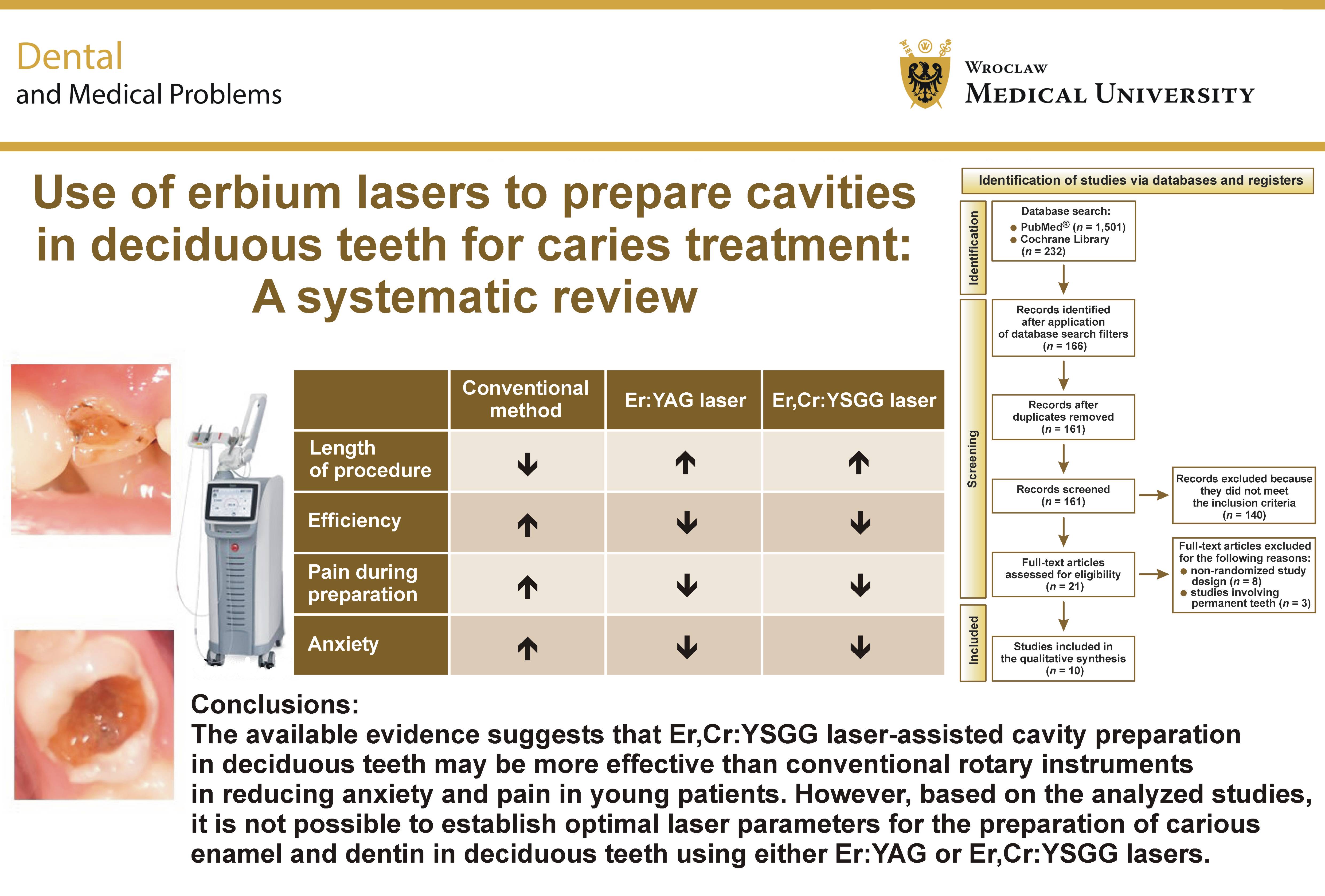
The available evidence suggests that Er,Cr:YSGG laser-assisted cavity preparation in deciduous teeth may be more effective than conventional rotary instrumentation in reducing anxiety and pain in young patients. However, based on the analyzed studies, it is not possible to establish optimal laser parameters for the preparation of carious enamel and dentin in deciduous teeth using either Er:YAG or Er,Cr:YSGG lasers. Furthermore, the included studies provide limited information regarding the laser parameters used.
Keywords: pain, dental caries, primary teeth, lasers, efficacy
Introduction
The treatment of carious disease in deciduous teeth consists of the limited removal of diseased, cariously altered tooth tissues and replacing them with a filling material. It is aimed at inhibiting the caries process, restoring the tooth function, preserving the vital pulp, and preventing the formation of secondary caries. Therefore, the currently used methods of managing dental caries follow the principles of minimally invasive dentistry (MID).1, 2, 3
The methods of minimally invasive treatment of carious cavities include pit and fissure sealing, caries infiltration, preventive resin restoration (PRR-1 and PRR-2), atraumatic restorative treatment (ART), chemomechanical caries removal (CMCR), air abrasion, kinetic cavity preparation (KCP), laser cavity preparation, sonic cavity preparation, ultrasonic cavity preparation, and steel crowns.
One method of treating carious cavities in deciduous teeth is the use of erbium lasers (Er:YAG and Er,Cr:YSGG) for the preparation of hard dental tissues. This is possible due to the presence of water in hard tissues, which is the main chromophore that efficiently absorbs the energy of the electromagnetic wave generated by these lasers (2,940 nm for Er:YAG, close to the maximum absorption of water; 2,780 nm for Er,Cr:YSGG). It is also worth mentioning the second chromophore, hydroxyapatite, which also absorbs these wavelengths well and is abundant in the hard tissues of teeth. The Er:YAG and Er,Cr:YSGG lasers are the most used and safest lasers for the preparation of dental hard tissues. They are characterized by the highest absorption in water and hydroxyapatite compared with the Nd:YAG laser (1,064 nm) and the CO2 laser (10,600 nm).4, 5, 6, 7
The first mechanism of action of the high-power laser occurs through the obliteration of tubules by the partial fusion of dentin substrates. The low-power laser decreases the chronic inflammatory process present in the cells by activating sodium and potassium pumps and obliterating dentinal tubules through the formation of tertiary dentin. It can also promote analgesic, anti-inflammatory and biomodulatory effects.8 The second mechanism of action of the high-power laser is based on the ablation phenomenon. The tissue is exposed to laser radiation, causing the water contained within it to be rapidly transformed into steam, and the tissue is fragmented into fine particles. Ablation makes the procedure very precise. If the laser pulses are of high power (high energy and short duration), the rate of ablation is greater than the rate at which heat penetrates the tissues. The penetration depth of the Er:YAG laser in enamel is 7 μm, which is 3 times smaller than that of the Er,Cr:YSGG laser. In dentin, the penetration depth of the Er:YAG laser is 5 μm and is also 3 times shallower than that of the Er,Cr:YSGG laser. The ablation threshold is the minimum amount of laser radiation pulse energy required to initiate the ablation process. The ablation threshold for enamel when using Er:YAG and Er,Cr:YSGG lasers is 12–20 J/cm2, and for dentin, 8–14 J/cm2.9 The most important factor is to deliver adequate energy in the shortest possible time to achieve effective ablation without damaging the tissues. Laser treatment is well tolerated by patients and is considered safe, quick and painless.8
The main chromophore in enamel and dentin for both the Er:YAG and Er,Cr:YSGG lasers is the water present between the hydroxyapatite crystals. Hydroxyapatite itself is also a chromophore. It is very important to consider the differences in water content between enamel and dentin. It is also necessary to compare the structural differences between primary and permanent teeth, as well as between immature or newly erupted teeth and aged teeth.
Healthy enamel is a highly mineralized tissue composed of 85% hydroxyapatite, 12% water and 3% organic tissue. Healthy dentin is less mineralized than enamel. It contains approx. 45–47% mineral components, 30–33% organic tissue and 20–24% water. The water content of carious tissue ranges from approx. 27% to 54%, which is higher than that of healthy tissues and depends on the stage of the carious lesion. Differences in the content of the main chromophore within the tissue determine the selection of the appropriate laser parameters according to the tissue being prepared. As a rule, the more hydrated the tissue, the lower the laser parameters should be. This is the main reason why the laser energy level must be decreased during dentin ablation, because both healthy and carious dentin have a higher water content than enamel.
Primary teeth have a higher water content in enamel and dentin. Additionally, the enamel layer is much thinner than that of permanent teeth, which is why ablation occurs much more rapidly than in the enamel of permanent teeth.
The use of the Er:YAG laser or the Er,Cr:YSGG laser to prepare carious cavities in primary teeth must be performed using appropriate parameters. The energy level and frequency should be lower than those used for the treatment of permanent teeth. Other settings are also very important, including the water spray, air cooling, and the type and diameter of the laser applicator.
Primary teeth have several ultrastructural differences compared with permanent teeth (Table 1).10, 11
Advantages of the use of erbium lasers include:
• the laser beam can operate in contact or non-contact mode;
• the laser tip does not have to touch the tooth; cutting efficiency is greatest when the tip is positioned approx. 1 mm from the target tissue;
• the ideal impact angle for laser ablation of enamel is perpendicular to the orientation of the enamel prisms;
• reduction of bacteria: a study by Hibst et al.12 showed that bacteria beneath the prepared surface were eliminated during laser cavity preparation to a depth of 300–400 μm;
• reduction of bacteria in combination with the use of antiseptics and lasers was reported by Golob Deep et al.13 They also demonstrated a reduction in dentin hypersensitivity using high-power lasers. For this purpose, high-power lasers were applied for an average of 43 s, delivering 1–20 pulses during a single visit, as reported by Pion et al.8;
• minimal heat increase in the pulp chamber;
• no smear layer after cavity preparation with laser use;
• no vibration.
Due to the differences in wave absorption, although the described lasers belong to the same family, there are important differences between them. Table 2 presents a comparison of the properties of the Er:YAG and Er,Cr:YSGG lasers. 9, 14, 15, 16, 17
Our study aims to critically evaluate, through a systematic review of the literature, randomized clinical trials with respect to the setting parameters of erbium lasers (Er:YAG and Er,Cr:YSGG) in the conservative treatment of deciduous teeth in children.
Material and methods
Focused question
Is it possible to precisely determine the optimal ranges of Er:YAG and Er,Cr:YSGG laser settings (energy, power, frequency, applicator size and diameter, and amount of air-water spray) for the preparation of cariously affected enamel and dentin in deciduous teeth?
Protocol, information sources, search strategy, and study selection
The review was prepared in accordance with the PRISMA (Preferred Reporting Items for Systematic reviews and Meta-Analyses) guidelines.18 Details of the study protocol are presented in Table 3.
Electronic searches were conducted from 2001 to May 29, 2024. The PubMed® and Cochrane Library databases were searched using the following Medical Subject Headings (MeSH) terms, keywords, and their combinations: (“Erbium YAG laser” OR “Er:YAG laser” OR “Er-YAG laser” OR “Er,Cr:YSGG” OR “Erbium Chromium YSGG” OR “2940 nm” OR “2780 nm”) AND (“primary teeth” OR “deciduous teeth” OR “caries” OR “enamel” OR “dentin”).
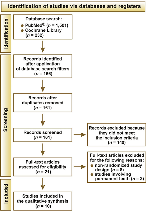
After applying the exclusion criteria (publications from the last 23 years (2001–2024), English language, and randomized trials only), the initial 1,501 articles retrieved from PubMed® were reduced to 138, and the initial 232 articles retrieved from the Cochrane Library were reduced to 28. During the initial screening phase, the titles and abstracts of these articles were independently screened and assessed by 2 reviewers according to the same criteria. Any disagreements were satisfactorily resolved through discussion.
A duplicate filter was then applied, resulting in the removal of 5 publications and leaving 161 articles for further evaluation.
After narrowing the selection criteria, 140 articles on permanent teeth and adhesion were excluded.
Subsequently, the full texts of the 21 articles were examined, and a final exclusion of 11 studies was made. Ultimately, 10 publications were included in the review (Figure 1).
Eligibility criteria
The following inclusion criteria were considered in the review:
• randomized trials only;
• use of the Er:YAG or Er,Cr:YSGG laser for the preparation of carious cavities in the deciduous teeth of children;
• presence of a negative control group;
• at least 10 patients (10 teeth) included in the study group;
• reporting of the laser operating parameters (pulse duration, frequency, power, type of laser applicator, and amount of air-water spray);
• a minimum follow-up period of 3 months;
• in vivo and in vitro studies.
Studies were excluded if they used laser other than Er:YAG and Er,Cr:YSGG, were duplicate publications, reported data from studies with the same clinical approval number, or investigated the use of the Er:YAG or Er,Cr:YSGG laser in general medicine, cosmetology, aesthetic medicine, or the conservative treatment of permanent teeth.
Quality assessment and risk of bias across studies
Following data extraction, the included articles were further evaluated by assessing their risk of bias. The Cochrane risk-of-bias tool17 was modified according to the requirements of this systematic review (Table 4). The studies were classified according to the total number of “YES = 1” responses to the posed questions.19 For the current study, the risk of bias was assessed according to the following score ranges:
• high risk: 0–3;
• moderate risk: 4–6;
• low risk: 7–9.
After analyzing the quality of the publications, 1 study received a score of 7, corresponding to a low risk of bias.20 Two studies received a score of 6, corresponding to a moderate risk of bias,21, 22 and the remaining 6 publications received 5 points, which also corresponded to a moderate risk of bias.22, 23, 24, 25, 26, 27
Any disagreements were resolved through discussion until consensus was reached.
Results
Study selection
A total of 185 studies were initially identified for analysis. Screening of the abstracts and titles excluded 99 studies. Twenty-one studies underwent full-text analysis.14, 17, 20, 21, 22, 23, 24, 25, 26, 27, 28, 29, 30, 31, 32, 33, 34, 35, 36, 37, 38 Of these, 11 were excluded because they did not meet the predefined inclusion criteria (Table 5).17, 28, 29, 30, 31, 32, 33, 35, 36, 37, 38 The main reasons for exclusion were non-randomized study design (8 studies28, 29, 30, 31, 32, 34, 35, 37) and studies involving permanent teeth (3 studies17, 33, 38).
General characteristics of the included studies
Ten studies were included.14, 20, 21, 22, 23, 24, 25, 26, 27, 36 Table 6 provides an overview of the articles that met the inclusion criteria. Four studies were in vitro laboratory investigations14, 23, 24, 27 and 6 were in vivo clinical studies.20, 21, 22, 25, 26, 36 No studies have comprehensively translated the results of laboratory investigations into clinical applications.
All 10 eligible studies were randomized. Four were conducted on extracted deciduous teeth,14, 23, 24, 27 whereas 6 were clinical trials involving children.20, 21, 22, 25, 26, 36 In 6 studies, the Er:YAG laser was used,20, 21, 22, 24, 26, 27 while 4 studies used the Er,Cr:YSGG laser.14, 23, 25, 36 None of the publications directly compared the 2 laser systems (Er:YAG and Er,Cr:YSGG). All studies included a control group. Among the in vitro studies, the smallest sample consisted of 48 teeth24 and the largest of 100 extracted teeth.23 The remaining in vitro studies included 50 teeth14 and 80 teeth.27 Among the in vivo studies, the smallest sample consisted of 50 teeth,25 while the largest included 84 teeth.20 The remaining studies included 80 teeth,21, 26 60 teeth36 and 70 teeth.22
All included studies compared cavity preparation in deciduous teeth using Er:YAG or Er,Cr:YSGG lasers with conventional rotary instrumentation. The studies evaluated the effectiveness and efficiency of the use of the Er:YAG laser in the removal of caries in deciduous teeth,20, 21, 22, 25, 26, 36 microleakage after cavity preparation with the Er,Cr:YSGG laser,23 sealing of the lacquer after the use of the Er,Cr:YSGG laser,14 and the acceptance of laser cavity preparation by pediatric patients.21, 22, 26, 36 The reported results indicate that the use of the Er:YAG laser for the preparation of deciduous teeth was less effective than, or comparable to, the conventional method for deep cavity preparation, whereas caries removal from the side walls of the cavity was more effectively achieved using the conventional method.20 Analysis of pain perception showed that most children experienced no pain during laser cavity preparation8, 21, 22, 25, 26, 36 and children remained calmer during treatment, despite procedures taking more than twice as long as conventional rotary treatment.21 Laser-assisted cavity preparation was reported to be more comfortable for children.22, 25, 36 The use of erbium lasers for cavity preparation in deciduous teeth does not eliminate the need for enamel etching with 37% orthophosphoric acid.14, 24
Characteristics of individual studies in terms of laser settings
Table 7 summarizes the laser settings used in the included studies. Information extracted from the articles included laser wavelength and the operating parameters, namely power, frequency, fluence, pulse duration, and the amount of air-water spray.
In 7 publications, the fluence values were not reported.14, 22, 24, 25, 26, 27, 36 In 2 studies, fluence values ranged from 39 J/cm2 to 55.5 J/cm2, although separate values for enamel and dentin were not provided. In only 1 study were fluence values reported separately for enamel and dentin, ranging from 17.8 J/cm2 to 26.8 J/cm2 for dentin and from 44.6 J/cm2 to 53.6 J/cm2 for enamel.23 The laser power was not reported in 8 studies.20, 21, 22, 23, 24, 26, 27, 36 In the remaining studies, the reported power ranged from 2.5 W to 6 W.14, 25 Pulse energy was reported in 5 studies20, 21, 22, 24, 27 and ranged from 50 mJ to 700 mJ. Frequency was provided in all studies and ranged from 2 Hz,24 through 4 Hz,20 10 Hz,21 and 20 Hz,14, 23 up to 25 Hz.25 Pulse duration was not reported in 5 studies.21, 22, 24, 26, 36 Two publications noted pulse durations ranging from 140 μs to 200 μs,14, 23 one study reported a pulse duration of 100 μs,20 and another reported 140 μs.34, 39 The study by Wang et al. described the use of the microsecond pulsing laser (MSP) mode without providing its numerical value.27 Information regarding air-water spray was reported in all studies; however, different units were used. Water spray was expressed either as percentages, ranging from 60% to 85%,14, 23, 25, 27 or as flow rates, ranging from 5 mL/min to 6 mL/min.20, 24 Air cooling was determined in only 4 studies,14, 23, 25, 27 with values of 40%, 55% and 90%. The remaining 6 publications did not report air cooling settings.20, 21, 22, 24, 26, 36
Characteristics of the laser applicators, types of primary teeth, and treated surfaces
The characteristics of the laser applicators, types of primary teeth treated, and treated surfaces are presented in Table 8.
The type of applicator used was not specified in 6 out of 10 studies; neither its shape nor its length was given.20, 21, 22, 25, 26, 36 The remaining 4 publications specified the applicator used: a G6tip sapphire tip23; a 2051 handpiece24; a 6-mm long sapphire tip14; and optical fiber with an R14 handle.27 In 4 cases,22, 24, 26, 36 the diameter of the applicator was not reported. The remaining 6 tests were carried out with the use of applicators with a diameter of 600 μm,14, 23, 25, 27 800 μm21 or 900 μm.20 Only 3 publications reported the angle of the laser beam during operation.21, 25, 27 None of the included studies described the operator’s hand movements during laser cavity preparation. One publication did not specify which primary teeth were treated with the laser.25 The remaining 9 studies identified the treated teeth as deciduous molars,14, 20, 22, 26, 27, 36 canines23, 24 and incisors.21 Four studies20, 21, 23, 25 specified the treated tooth surfaces: occlusal surfaces20, 25; Black’s Class V cavities23; and Black’s Class III and IV cavities.21 Six studies did not report the treated areas.14, 22, 24, 26, 27, 36 In 7 cases, the laser was operated in a non-contact mode.14, 20, 21, 23, 24, 25, 27 However, the distance between the applicator and the target tissue varied. Four studies reported a working distance of 1 mm,14, 21, 23, 27 1 study did not report the value,6 one noted a distance of 17 mm,24 and another – of 7 mm.20
Discussion
The presented results demonstrate the small number of published articles investigating the use of Er:YAG and Er,Cr:YSGG lasers in laboratory and clinical studies for the preparation of hard tissues in deciduous teeth in children, as well as the complete lack of similar systematic reviews on this topic. In addition, most of the published articles do not provide complete data on the laser operating parameters, including energy, power, frequency, applicator type and diameter, and the amount of air-water spray. Furthermore, they do not distinguish between the parameters used for individual hard tissues, namely enamel and dentin.
With regard to frequency, considerable variation was observed, ranging from 2 Hz4 to 25 Hz.6 A frequency of 20 Hz14, 23 was used in 2 studies, whereas 25 Hz was reported in 1 publication.25 None of the researchers differentiated the frequency according to whether the laser was used on enamel or dentin. In the studies included in this review, the frequency values used with the Er,Cr:YSGG laser ranged from 20 Hz to 25 Hz,14, 23, 25 whereas those used with the Er:YAG laser ranged from 2 Hz to 10 Hz.20, 21, 24 None of the analyzed studies explained why the frequencies used with the Er,Cr:YSGG laser were considerably higher than those used with the Er:YAG laser. Likewise, no such relationship has been described in other published studies.6, 7, 17, 33 When preparing hard dental tissues, frequency appears to be an important parameter. The speed of the Er:YAG and Er,Cr:YSGG35, 40, 41, 42, 43, 44, 45, 46, 47, 48, 49 lasers depends not only on the laser parameters and the operator’s experience but also on the chemical composition of the hard dental tissues (enamel and dentin). Enamel consists of 95% of hydroxyapatite, 4% water and 1% collagen fibers, whereas dentin contains approx. 70% hydroxyapatite, 20% collagen fibers and 10% water.44 Carious hard tissues contain more water than healthy tissues, which means that laser processing of dentin is faster than that of enamel, and diseased tissue is removed more easily than healthy tissue. Therefore, careful control of the laser parameters during cavity preparation is essential to comply with the principles of MID. During enamel preparation, higher frequency settings than during dentin preparation may be appropriate, particularly near the intraventricular wall.31, 46
Similarly, fluence was not reported in 2 studies.24, 25 When each study is analyzed individually, it is not possible to determine which laser parameters were used for specific tooth surfaces or which parameters were applied to enamel and dentin during caries removal. One publication reported the fluence, applicator type and diameter, use of the non-contact mode at a distance of 1 mm from the target area, the type of tooth treated (canines), and the cavity type (Class V).23 It did not describe the treated surface or the operator’s movements during the procedure.23 The study by Rossi et al. appears to be one of the most comprehensive among the included studies and provides the most useful information for determining appropriate Er,Cr:YSGG laser settings for enamel and dentin preparation in primary teeth with Class V cavities.23
After comparing the studies on deciduous teeth included in this review with studies on permanent teeth that were excluded from the analysis because they were outside the scope of this review, a considerable lack of consistency in reporting laser operating parameters was observed. Furthermore, the available studies do not provide tissue-specific parameters for enamel and dentin. Apart from reporting the laser wavelength, the published studies generally fail to provide essential information, including the type of handpiece, applicator type, applicator diameter, operating mode (contact or non-contact), working distance, angle of laser application, operator movement, type of tissue treated (enamel or dentin), type of teeth treated (primary, immature permanent, or mature permanent), treated tooth surface, and laser operating parameters, including pulse duration, frequency, pulse energy, power, fluence, water spray, and air cooling. Among the analyzed studies, wavelength, frequency and water spray were the only parameters consistently reported. However, water spray was expressed either as mL/min or as percentages, making direct comparison impossible because no validated method exists for converting percentage values into water flow rates.
Two of the reviewed papers20, 25 investigated cavity preparation on the occlusal surfaces of deciduous molars. The first paper evaluated the Er:YAG laser,20 whereas the second investigated the Er,Cr:YSGG laser.25 The reported frequencies differed substantially: 4 Hz20 vs. 25 Hz.25 In the first study, the fluence was 39 J/cm2, and in the second study, no fluence value was provided. The first paper also reported pulse energy and pulse duration, making it possible to establish baseline data for Er:YAG laser preparation of occlusal surfaces in primary teeth. However, the authors did not specify whether these parameters were used for enamel or dentin preparation, which is an important limitation. The provided information is insufficient to establish basic Er,Cr:YSGG laser parameters for enamel and dentin preparation on occlusal surface of deciduous teeth.25 Valério et al.20 used a 900-μm applicator in a non-contact mode with a focal distance of 7 mm, whereas Johar et al.25 used a 600-μm applicator in a non-contact mode but did not specify either the working distance or the applicator type. Neither study indicated whether the reported parameters referred to enamel or dentin preparation.
Following the analysis of the reported laser settings, although 7 publications reported the applicator diameter,8, 14, 20, 21, 23, 24, 25 only 4 specified the applicator type.14, 23, 24, 27 None reported the angle between the applicator and the treated tissue, and none described the operator’s hand movements. All studies used the non-contact technique, reporting working distances of 1 mm,14, 21, 23 7 mm,20 17 mm,24 or not specifying the distance from the treated surface.25 However, the rationale for selecting these distances was never explained.
The included studies also provide only general information regarding applicator type and diameter. In contrast, several studies involving permanent teeth, although excluded from this review, described more detailed treatment protocols. Among these, Kato et al. described laser cavity preparation in both permanent and deciduous teeth using the Er:YAG laser.34 They reported pulse duration, applicator type and diameter, as well as separate laser parameters for enamel and dentin. However, they did not describe the operator’s movements or distinguish between laser settings used for deciduous and permanent teeth. Consequently, their findings cannot be used to establish standardized laser parameters for either dentition.34
Another excluded study by Eren et al. assessed pain during Er,Cr:YSGG laser treatment of permanent teeth in children.33 The authors reported different air-water spray settings for enamel (85% air and 85% water) and dentin (65% air and 55% water). They also described the head and the applicator used and were the only authors to report the operator’s movements: “During preparation, the sapphire tip (600 μm) was moved carefully back and forth and up and down to remove caries.” The authors compared treatment duration with conventional method in first permanent molars of children aged 7–10 years. Laser preparation required an average of 7.4 min, whereas mechanical preparation required approx. 3.7 min.33 Liu et al. likewise compared treatment times in primary teeth and found that Er:YAG laser preparation required approx. 2.35 times longer than conventional methods.21
Overall, laser preparation requires nearly twice as much time as conventional rotary instrumentation. However, pain is one of the most important factors contributing to uncooperative behavior in pediatric patients. The reviewed studies indicate that approx. 70% of children preferred laser preparation, compared with 30% who preferred conventional treatment. Although laser preparation is slower, children cooperate more readily, remain calmer during treatment, and frequently choose laser treatment again during subsequent visits.21, 25, 33 These advantages make laser-assisted treatment less traumatic and more acceptable for pediatric patients.
The only conclusion resulting from the presented analysis is that studies investigating Er:YAG and Er,Cr:YSGG lasers have been published with similar frequency.
The main common finding to all published studies is that erbium lasers provide advantages because they operate without direct contact with the tissues of the prepared tooth and without vibration.21, 24, 33, 34, 39, 40 Approximately 90% of children accept laser dental treatment,33 while patients aged 10 years or older generally show no preference between laser and conventional treatment.34, 36 The reduction in turbine noise appears to be particularly important for younger children, taking into account comfort during work.21, 24, 33, 34, 39, 40, 41 In most of the presented cases, local anesthesia was not required during laser treatment.14, 20, 21, 23, 24, 25, 33, 34, 39, 40 Laser cavity preparation enables minimally invasive and selective removal of infected carious tissues.34, 39, 42, 43 The use of laser techniques in hard tissues produces little or no thermal effect on the dental pulp.34, 39, 44, 45 Dentin prepared with the Er:YAG laser is free of a smear layer, and the dentinal tubules remain open and visible.34, 43, 45, 47, 48, 49
Removal of carious tissue from the side walls of the cavities is more difficult with laser preparation than with conventional methods because the laser applicator does not function in the same manner as a drill. In contrast, preparation of infected dentin on the intraventricular wall is comparable with conventional mechanical preparation.20
The use of laser technology represents a new philosophy of treatment for both dentists and patients. Laser-assisted preparation enables young patients to undergo treatment in a more comfortable and less stressful environment.
The limited amount of published information regarding laser treatment of primary teeth justifies further research in this area and highlights the need to establish standardized treatment protocols for laser preparation of hard tissues in primary teeth.
Based on the available evidence, there is a clear need for further laboratory and clinical studies. Future publications should consistently report the following information: laser wavelength; head type; applicator type and diameter; operating mode (contact or non-contact); working distance; angle of laser application; operator movement; tissue treated (enamel or dentin); tooth type (primary, immature permanent, or mature permanent); treated tooth surface; pulse duration; frequency; pulse energy; power; fluence; water spray; and air cooling.
The use of the Er,Cr:YSGG laser for cavity preparation in deciduous teeth may be more effective than conventional rotary instruments in reducing anxiety and pain in young patients. Therefore, the Er,Cr:YSGG laser may represent an alternative to conservative treatment in children.36 The use of the Er:YAG laser for cavity preparation is also associated with minimal pain and discomfort, thereby reducing the need for local anesthesia and providing a more comfortable treatment option for children than the conventional method.22 Additionally, erbium laser technology may enable more precise cavity preparation than traditional methods, resulting in improved adaptation and bonding of the filling material. This may contribute to greater restoration longevity and a lower risk of recurrent caries in deciduous teeth.26 Following Er:YAG laser irradiation of dentin, dentinal tubules may undergo morphological changes. Laser energy can induce partial sealing or closure of the dentinal tubules, thereby reducing their diameter. This sealing effect can help reduce dentin hypersensitivity and improve the bond strength of restorative materials to the treated surface.27, 37
When Er:YAG lasers are used for cavity preparation in primary teeth, specific laser parameters influence treatment effectiveness. Commonly recommended parameters include:
• Pulse duration: Short pulse durations (approx. 50–100 μs) are preferred for precise ablation with minimal thermal damage to the surrounding tissues;
• Energy density: Fluence should be optimized (approx. 20–100 mJ/cm2) to ensure efficient ablation while preventing overheating of the tooth structure;
• Repetition rate: The repetition rate may vary (2–20 Hz), depending on the desired ablation efficiency and thermal effects. Higher repetition rates may accelerate cavity preparation but require careful monitoring to prevent tissue damage;
• Water spray or air-water cooling: The use of water spray or air-water cooling helps dissipate heat generated during laser ablation and reduces the risk of thermal injury to the dental pulp.
Although laser-assisted cavity preparation in deciduous teeth shows considerable promise, additional studies are required to establish the optimal irradiation parameters for clinical use.38
The development of erbium family lasers in pediatric dentistry, particularly for the treatment of deciduous teeth, is likely to progress in several directions:
1. Improved treatment efficiency: Continued advances in laser technology and optimization of operating parameters may improve precision, shorten treatment time and enhance clinical outcomes during caries removal, cavity preparation and soft tissue procedures in deciduous teeth;
2. Minimally invasive treatment: Erbium lasers facilitate minimally invasive procedures that preserve healthy tooth structure, reduce the need for anesthesia, and improve patient comfort during dental treatment, making them particularly advantageous in pediatric dentistry;
3. Preventive applications: Further research may explore the use of erbium lasers for preventive procedures, such as enamel conditioning, fissure sealant application and surface modification to enhance the resistance of deciduous teeth to caries;
4. Treatment of patients with special needs: Erbium lasers may provide a gentle and efficient alternative for managing dental conditions in children who have difficulty tolerating conventional dental treatments.
5. Combination therapies: Combining erbium laser treatment with other dental modalities, such as fluoride therapy, sealants or antimicrobial agents, may further improve the effectiveness of caries prevention and management in deciduous teeth.
The future application of erbium family lasers in pediatric dentistry appears promising, offering innovative approaches to the treatment of deciduous teeth with improved precision, minimal discomfort and reduced anxiety for young patients. By addressing important challenges in pediatric dental care, including fear of dental procedures, pain management and patient cooperation, erbium lasers have the potential to revolutionize the treatment experience and improve oral health outcomes in children.
Overall, the ongoing development and clinical implementation of erbium laser technology in pediatric dentistry represent a promising direction toward safer, more effective and more patient-friendly dental care for children.
Limitations
One limitation of this systematic review is the limited number of high-quality studies available on the use of erbium family lasers for cavity preparation in deciduous teeth. Consequently, it is difficult to draw definitive conclusions because of the limited amount of robust scientific evidence. The available studies are generally small, employ different methodologies, and often report inconsistent outcomes, making comprehensive evaluation of the effectiveness and safety of erbium lasers challenging.
Several additional limitations should be considered when interpreting studies on laser treatment of carious lesions in deciduous teeth:
• Sample size and diversity: The limited availability of pediatric patients with carious lesions may restrict both sample size and population diversity, affecting the statistical power and limiting the generalizability of the findings;
• Ethical considerations: Clinical studies involving pediatric populations require strict ethical standards, including informed parental consent, child assent when appropriate, and careful attention to patient comfort and safety throughout treatment;
• Long-term follow-up: Assessment of long-term clinical outcomes is challenging due to the transient nature of primary dentition and the potential loss of follow-up over time;
• Variability in tooth morphology: Deciduous teeth exhibit diverse anatomical features, making it difficult to standardize laser treatment protocols across different tooth types and stages of dental development.
Research involving deciduous teeth is challenging because of the limited availability of suitable teeth and their eventual replacement by permanent dentition. Consequently, studies involving primary teeth often include smaller sample sizes than studies conducted on permanent teeth. Researchers may face difficulties in recruiting sufficiently large study populations because of ethical considerations, patient age and the natural shedding process of these teeth.
Despite the potential limitations in sample size, studies involving deciduous teeth remain essential for improving our understanding of pediatric oral health and for developing appropriate treatment strategies tailored to this age group. These limitations should therefore be considered when designing and interpreting research involving deciduous teeth to make informed decisions regarding pediatric dental care.
Conclusions
Based on the available evidence, it is currently not possible to establish optimal Er:YAG and Er,Cr:YSGG laser parameters for the preparation of carious enamel and dentin in deciduous teeth. Analysis of the included studies demonstrates that only limited information is available regarding the laser operating parameters used during treatment. Individual data values are missing to determine the final laser settings. The information reported in the available studies is incomplete and insufficient to allow reliable assessment of the physical laser parameters.
Ethics approval and consent to participate
Not applicable.
Data availability
The datasets generated and/or analyzed during the current study are available from the corresponding author on reasonable request.
Consent for publication
Not applicable.
Use of AI and AI-assisted technologies
Not applicable.