One journal. Two disciplines. Shared excellence.

Dental and Medical Problems

Title abbreviation: Dent Med Probl
Journal Impact Factor (JIF 2025) – 4.3
Journal Citation Indicator (JCI 2025) - 1.38
Scopus CiteScore (2025) – 5.8
Index Copernicus Value (ICV 2024) – 178.25
MNiSW – 70 pts
ISSN 1644-387X (print)
ISSN 2300-9020 (online)
Periodicity – bimonthly


Download original text (EN)

Dental and Medical Problems

2026, vol. 63, nr 3, May-June, p. 707–733

doi: 10.17219/dmp/215136

Publication type: review

Language: English

License: Creative Commons Attribution 3.0 Unported (CC BY 3.0)

Download citation:

  • BIBTEX (JabRef, Mendeley)
  • RIS (Papers, Reference Manager, RefWorks, Zotero)

Cite as:


de Oliveira Souza AIS, Raposo J, da Silva JB, et al. Physical therapy interventions for awake and sleep bruxism: What has been the approach over the last decades? A systematic review of the evidence and research gaps. Dent Med Probl. 2026;63(3):707–733. doi:10.17219/dmp/215136

Physical therapy interventions for awake and sleep bruxism: What has been the approach over the last decades? A systematic review of the evidence and research gaps

Ana Izabela Sobral de Oliveira Souza1,A,B,C,D,E,F, Jakelline Raposo2,3,B,C,D, Jordana Barbosa da Silva1,B,C,D, Maryllian Albuquerque Vieira4,B,C, Gesa Hahn1,A,B,C, Harry von Pierkartz1,A,C,E,F, Susan Armijo-Olivo1,5,6,A,C,D,E,F

1 Faculty of Business and Social Sciences, Osnabrück University of Applied Sciences, Germany

2 University of Pernambuco, Recife, Brazil

3 Federal Institute of Education, Science and Technology of Alagoas, Brazil

4 Physiotherapy Department, Federal University of Pernambuco, Recife, Brazil

5 Faculty of Rehabilitation Medicine, University of Alberta, Edmonton, Canada

6 Faculty of Medicine and Dentistry, University of Alberta, Edmonton, Canada

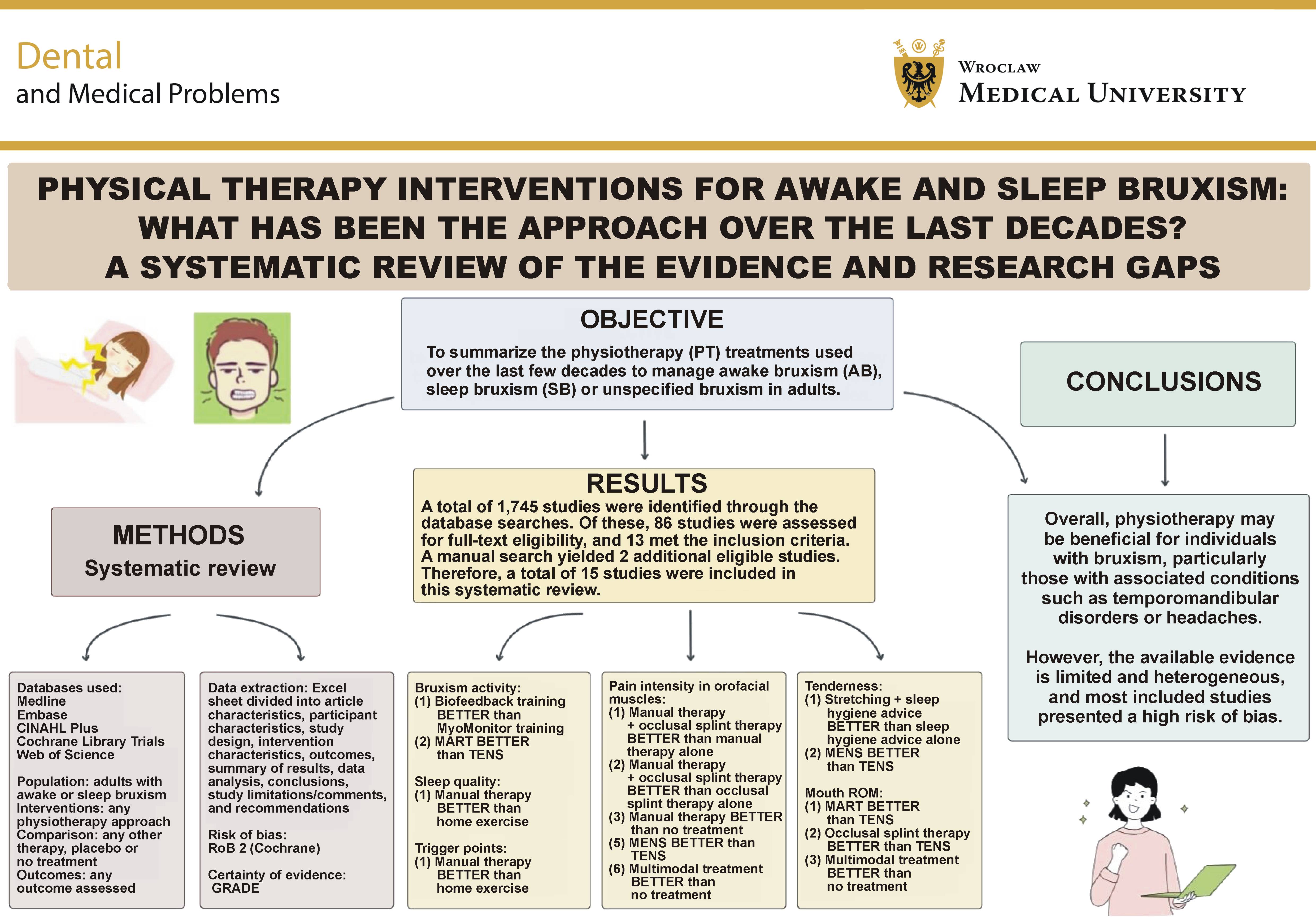
Graphical abstract


Graphical abstracts

Highlights


  • Physiotherapy has been increasingly used to manage the adverse consequences of bruxism, although supporting evidence remains limited and of low quality.
  • Muscle stretching, massage therapy and adjunctive interventions, including occlusal splints, show potential for alleviating the adverse consequences of bruxism.
  • Future research should adopt the latest bruxism sub-classification.
  • A biopsychosocial approach integrating patient education, motor control exercises and behavioral interventions may improve the therapeutic and rehabilitative management of bruxism.

Abstract

The objective of this systematic review was to summarize the physiotherapy (PT) treatments used over the last few decades to manage awake bruxism (AB), sleep bruxism (SB) or unspecified bruxism in adults. Searches were conducted in 5 databases, and all stages of the review were performed independently by 2 reviewers. A total of 1,745 records were identified through the database searches. Of these, 86 studies underwent full-text assessment, and 13 met the inclusion criteria. An additional manual search identified 2 further eligible studies, resulting in a total of 15 studies included in this systematic review. A variety of PT modalities were reported in the literature. Manual therapy (MT) combined with occlusal splint therapy or kinesiotaping demonstrated positive effects on clinical outcomes, including orofacial pain, sleep quality, the number of trigger points, and jaw disability in individuals with bruxism. Overall, PT may be beneficial for individuals with bruxism, particularly those with associated conditions such as temporomandibular disorders (TMD) or headaches. However, the available evidence is limited and heterogeneous, and most included studies presented a high risk of bias.

Keywords: physical therapy modalities, bruxism, systematic review, evidence gaps

Introduction

Musculoskeletal physiotherapy focuses on dysfunctions involving muscles, joints, bones, nerves, tendons, and related structures, with the primary aims of alleviating pain and improving function. It encompasses techniques such as manual therapy (MT), exercise, electrotherapy, relaxation techniques, movement therapy, kinesiotherapy, and patient education to address pain, fatigue and mobility limitations according to individual clinical needs.1, 2, 3, 4, 5, 6, 7 One area of physiotherapy (PT) addresses dysfunctions affecting the head, face and neck, including the stomatognathic system,2, 3, 4, 7 with a particular emphasis on temporomandibular disorders (TMD).8 Within this field, bruxism, a masticatory muscle behavior that may or may not be associated with TMD,8, 9, 10, 11 has gained increasing attention from physiotherapists.2 International consensus statements defining the characteristics of this behavior were published in 2013 and 2018, and were subsequently updated in 2025.9, 12, 13, 14

According to the international consensus, bruxism is not classified as a disorder but may be considered a risk factor when associated with adverse health outcomes, such as pain, severe mechanical tooth wear, and tooth damage.12 Bruxism is categorized into awake bruxism (AB), characterized by repetitive or sustained masticatory muscle activity involving tooth contact and/or jaw bracing or thrusting during wakefulness, and sleep bruxism (SB), characterized by rhythmic or non-rhythmic masticatory muscle activity during sleep.12 Bruxism can be assessed using self-report, clinical examination, and instrumental methods such as electromyography (EMG) and polysomnography.15, 16 Each assessment method captures a different aspect of bruxism.12 Self-report reflects an individual’s awareness of the behavior, whereas clinical examination may identify potential signs associated with bruxism, such as tooth wear, rather than the behavior itself. Instrumental assessments provide objective measures of masticatory muscle activity.12, 15

The prevalence of bruxism in the general population is increasing. A review published in 2024 estimated a pooled prevalence of 22%, with rates of 23% for AB and 21% for SB. Both types were reported more frequently in women than in men.17

There is also evidence that psychosocial factors are associated with bruxism, particularly AB.18, 19, 20 Studies suggest that stress, anxiety, depression, and certain personality traits contribute to the development of AB. For example, Manfredini and Lobbezoo reported that AB is strongly associated with psychosocial factors and psychopathological symptoms, whereas SB does not demonstrate the same pattern of associations.18 Anxiety, stress sensitivity, depression, and specific personality traits have consistently been linked to AB, while the evidence relating psychosocial factors to SB remains less consistent.18, 21 However, the etiology of bruxism is multifactorial and may differ between SB and AB, with lifestyle factors playing an important role in both.22, 23, 24 Furthermore, bruxism and its adverse consequences have been shown to negatively affect quality of life, jaw function, motor activity, pain, and oral health.2, 24

When bruxism is associated with adverse outcomes, interventions are often required to manage the behavior and mitigate its consequences, such as pain and tooth wear.2 An important aspect of its clinical presentation is its possible association with TMD.10 Although this relationship remains controversial, several studies have investigated the association between bruxism and TMD.10, 11 Emodi-Perlman and Eli, for example, discussed the prevalence of AB and SB and suggested that excessive bruxing activity may contribute to dysfunction of the stomatognathic system, orofacial pain, and TMD.25 A recent meta-regression analysis by Zieliński et al. estimated the global co-occurrence of bruxism and TMD to be 17%, with marked geographical variation. The authors also found that each 1% increase in the proportion of female participants was associated with a 4.4% increase in the probability of co-occurring TMD and bruxism.11 These findings highlight the frequent coexistence of both conditions and the influence of geographical and demographic factors on this association.11

Occlusal splint therapy remains the most common intervention for individuals with bruxism, particularly SB, and is often combined with pharmacotherapy under the supervision of a dentist.4, 26 These approaches aim to protect the dentition, reduce masticatory muscle tension, improve sleep quality, and alleviate orofacial pain and headaches, when present.26 Physiotherapy has not yet been established as a standard approach for managing the adverse consequences of bruxism. However, several PT interventions may offer clinical benefits, particularly for individuals presenting with pain, jaw dysfunction and increased muscle tension.2, 27

Numerous systematic reviews2, 26, 27 have evaluated interventions for individuals with bruxism presenting with symptoms such as pain, jaw disability and muscle tension. One review investigated the effectiveness of PT for both AB and SB and examined 7 treatments, including electrotherapy and cognitive behavioral therapy, which were the most frequently studied and showed the most favorable results for reducing muscle activity and pain.2 Another systematic review focused solely on the management of SB and its associated symptoms through pharmacotherapy, oral appliances, biofeedback, and cognitive behavioral therapy.4 However, these reviews2, 4 were published more than 5 years ago. Since then, the concept of bruxism has evolved considerably, with important updates to its definition and assessment. Bruxism is now understood as a motor behavior of the masticatory muscles rather than a disorder, supporting an interdisciplinary management approach that includes professionals with expertise in human movement, such as physiotherapists.2, 3 Consequently, earlier reviews may not have captured important PT-related evidence. Furthermore, the review focused on SB restricted its literature search to studies published between 2007 and 2015, potentially limiting the identification of relevant evidence.4 Although a variety of PT strategies were reported, relatively few studies were included, possibly because of the limited search strategy.2

A review published by our group in 2023 examined biofeedback as a primary intervention for individuals with AB.27 The review demonstrated positive effects of auditory and visual biofeedback in reducing non-functional masticatory muscle activity and the frequency of bruxism-related muscle events. However, it also identified important limitations, including methodological heterogeneity and an exclusive focus on masticatory muscle activity without evaluating other clinically relevant outcomes that could be significant for those experiencing negative consequences of bruxism.27

As research in this field has expanded in recent years, several new randomized controlled trials (RCTs) and clinical trials (CTs) have investigated PT interventions for individuals with AB and SB.28, 29, 30, 31, 32 Consequently, existing systematic reviews are now outdated.2, 4, 26, 27 There is therefore a need to synthesize the most recent evidence regarding the effectiveness of PT interventions for managing bruxism and its associated adverse outcomes. In addition, it is important to determine whether these interventions are consistent with the current understanding of bruxism.

This systematic review aims to critically evaluate and synthesize the current evidence regarding PT interventions for managing bruxism and its adverse consequences, when present, including masticatory muscle activity, muscle tension, jaw pain, functional impairment, muscle tenderness, and compromised oral health-related quality of life. Furthermore, it aims to assess the methodological quality and risk of bias of the included studies and to formulate clear directions for future research in the field of bruxism and PT.

The following questions were addressed in this review: What PT interventions have been used to manage AB and/or SB, either alone or in combination with associated conditions, and how effective are these interventions compared with other treatment approaches? Specifically, how do these interventions affect muscle activity (measured by EMG or other tools), reduce the adverse consequences of bruxism, including pain, muscle tenderness and jaw disability, and improve the quality of life of individuals with bruxism?

Material and methods

This systematic review was conducted in accordance with the PRISMA (Preferred Reporting Items for Systematic reviews and Meta-Analyses) statement.33

The review was registered in PROSPERO under registration No. CRD42021251182. All stages of the review were conducted independently by at least 2 trained reviewers. All extracted data are available from the corresponding author upon reasonable request.

Search strategy

The search strategy was developed by 2 investigators (AISOS and SAO) using the Population/Intervention/Comparison/Outcome (PICO)34 framework under the supervision of an experienced Health Sciences Librarian. The final search strategy was based on a list of keywords derived from Medical Subject Headings (MeSH) and related to the concepts of bruxism and PT. The search was restricted to RCTs and CTs. No restrictions were applied regarding publication date, language or publication status. The electronic search was last updated in July 2023, and a manual search was conducted through November 2024 in the following databases: MEDLINE (Ovid MEDLINE® All), Embase (Ovid interface), CINAHL Plus (EBSCOhost interface), Cochrane Library Trials (Wiley interface), Web of Science (Science Citation Index Expanded (SCIE), Social Sciences Citation Index (SSCI), Arts & Humanities Citation Index (AHCI), Emerging Sources Citation Index (ESCI)). The details of the search strategy can be found in Appendix 1 (available on request from the corresponding author). Manual searches, including grey literature, were performed by screening the reference lists of the included papers using the Scopus database. In addition, members of the review team, who are experts in the field, were consulted to identify any ongoing trials on this topic.

Eligibility criteria

The inclusion and exclusion criteria were defined according to the PICO framework.

Population

This systematic review included studies involving adults (>18 years) with AB and/or SB. Bruxism could have been diagnosed by a dentist using standardized assessment methods, such as EMG, polysomnography, the oral health checklist, or other validated instruments, or identified through participants’ self-report (Appendix 2). The classification of bruxism followed the international consensus on the assessment of bruxism.10 Studies were excluded if they focused solely on children or adolescents, lacked clear information regarding bruxism classification, focused exclusively on dental conditions (e.g., missing teeth, toothache) unrelated to bruxism, or included participants with specific comorbidities, such as systemic diseases, rheumatoid conditions, arthralgia, cancer, neuropathic pain, or previous temporomandibular joint (TMJ) or neck surgery.

Studies were grouped according to the type of bruxism reported, as follows:

• AB and SB – studies that included participants with both AB and SB without reporting separate analyses;

• AB – studies including only participants with AB;

• SB – studies including only participants with SB;

• unspecified bruxism – studies that did not specify whether participants had AB or SB.

Whenever possible, information regarding whether bruxism was associated with other conditions was extracted (Appendix 2). When studies reported only the presence of bruxism without describing associated symptoms (e.g., pain), this limitations was highlighted.

Interventions of interest

Studies evaluating any form of PT that could potentially reduce the adverse consequences of bruxism, including pain, muscle tenderness and jaw disability, were eligible. Interventions of interest included MT, exercise therapy and PT techniques aimed at promoting muscle relaxation, such as stretching, massage, myofascial release, and progressive muscle relaxation. In addition, interventions commonly used within PT practice, including acupuncture, dry needling, massage therapy, electrotherapy (e.g., transcutaneous electrical nerve stimulation (TENS) and microcurrent electrical neuromuscular stimulation (MENS)), trigger point therapy, and general PT (i.e., combinations of different PT approaches), were considered eligible (Appendix 2).

Studies evaluating PT combined with other interventions were included only when the specific effects of the PT component could be identified and analyzed separately. Studies in which the effect of PT could not be isolated were excluded. Interventions based primarily on psychological approaches, such as behavioral or relaxation therapies, were also excluded.

Comparison

Eligible comparator interventions included placebo, no treatment, pharmacological therapy, behavioral interventions, occlusal splint therapy, and any other therapeutic approach.

Outcomes

All reported outcomes were considered (Appendix 2). The primary outcome was masticatory muscle activity (i.e., bruxism behavior), including clenching, grinding, jaw thrusting, and jaw bracing. Secondary outcomes included pain intensity, muscle tenderness, jaw function, jaw activity, mastication, quality of life, and patient-reported outcomes. Any additional outcomes evaluated in the included studies were also extracted.

Study design

This systematic review included RCTs and CTs, as these designs provide the highest level of evidence for evaluating treatment effectiveness. All other types of studies, such as cross-sectional studies, cohort studies, case reports, narrative reviews, systematic reviews, meta-analyses, commentaries, letters to the editor, conference papers, book chapters, protocol registrations, abstracts without full text, and animal studies, were excluded.

Time points

All assessment time points reported in the included studies were considered, including baseline, immediate post-intervention, short-term, medium-term, and long-term follow-up.

Data screening

Study selection

Search results were imported into EndNote (https://endnote.com) and subsequently uploaded to the Covidence platform (https://www.covidence.org), which was used to manage the screening process. The PRISMA flowchart was used to document the numbers of identified, duplicated, selected, and excluded studies.35

Two independent reviewers screened the titles and abstracts according to the predefined eligibility criteria. Full texts were obtained for all potentially eligible studies and for studies with insufficient information in the title or abstract. The reviewers then independently assessed the full-text articles while remaining blinded to each other’s decisions. Disagreements were resolved through consensus. If consensus could not be reached, the third reviewer made the final decision.

Data extraction

The data was extracted and organized using a pilot-tested Microsoft Excel form developed specifically for this review. The extraction form was created following a standardized process and was refined through iterative discussions among the reviewers. One reviewer independently verified all extracted information and organized the data into tables for further analysis. A second reviewer double-checked all extracted data. Any disagreements were resolved through discussion and consensus, with arbitration by a third reviewer when necessary.

Extracted information included, but was not limited to, article characteristics, participant characteristics, study design, intervention characteristics, outcomes, summary of results, data analysis, conclusions, study limitations/comments, and recommendations. When quantitative data was missing, the authors were contacted to request the unavailable information.

Risk of bias assessment

Assessment of the risk of bias (RoB) was conducted concurrently with data extraction. Two independent reviewers evaluated the quality of the included studies using a compiled set of items (CSoI) and the revised Cochrane risk-of-bias tool (RoB 2).36, 37 The RoB 2 evaluates 5 domains: the randomization process; deviations from the intended interventions; missing outcome data; measurement of the outcome; and selection of the reported results.36, 38 Each domain was rated as presenting low RoB, unclear (some concerns) RoB, or high RoB. An overall risk-of-bias judgment was assigned to each study according to standard guidelines.36, 39, 40, 41 Any disagreements were resolved by consensus.

Strategy for data synthesis

The data was summarized narratively, resulting in a descriptive analysis of the included studies. Evidence tables were prepared to summarize study characteristics, interventions, outcomes, and findings. Data synthesis was based on the PT intervention applied (e.g., therapeutic exercises, MT, and other modalities). Within each intervention category, studies were further grouped according to the type of bruxism (SB, AB, both SB and AB, or unspecified bruxism, with or without other conditions associated) and the outcomes evaluated (e.g., masticatory muscle activity, quality of life, self-reported pain, mandibular function, range of motion (ROM)). This approach facilitated the synthesis of findings despite the substantial methodological heterogeneity across studies.

Certainty of the evidence

The overall certainty of the evidence was assessed using the GRADE (Grading of Recommendations Assessment, Development and Evaluations) approach.42 According to Guyatt et al., the certainty of evidence is classified as high, moderate, low, or very low.42 The GRADE assessment considers the following domains: study design; risk of bias; inconsistency of results; indirectness of the evidence (not generalizable); imprecision (insufficient data); and other factors, such as reporting bias.42

Results

Search results

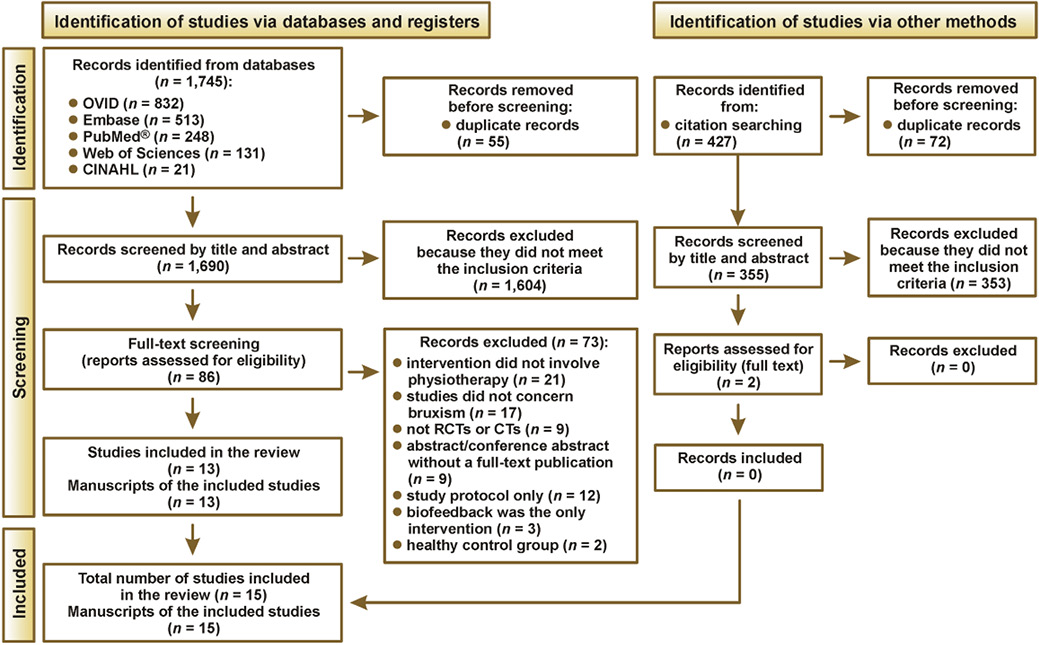
A total of 1,745 studies were identified through the database searches. Of these, 86 studies were assessed for full-text eligibility, and 13 met the inclusion criteria. The PRISMA flow diagram provides a detailed overview of the study selection process and the reasons for exclusion (Figure 1). Additional information on excluded studies is provided in supplementary materials (Appendix 6). A manual search based on the citations of the included studies was also performed. Of the 427 records identified, 72 were duplicated and 353 were excluded after screening because they did not meet the inclusion criteria. This search yielded 2 additional eligible studies. Therefore, a total of 15 studies were included in this systematic review.

Description of the included studies

The included studies investigated different classifications of bruxism. Six studies (40.0%) included participants with SB,28, 43, 44, 45, 46, 49 one (6.7%) included participants with both AB and SB,30 four (26.7%) did not specify the type of bruxism,29, 31, 47, 48 three (20.0%) included participants with unspecified bruxism associated with TMD,13, 50, 51 and 1 study (6.7%) included only participants with AB.32 Sample sizes ranged from 12 to 100 participants, and the mean age ranged from 18 to 60 years. Most studies included both male and female participants, whereas 2 studies enrolled only women.32, 43 Detailed study characteristics are presented in Table 1 and Table 2.

It is important to highlight that 60% of the included studies evaluated pain as an outcome. Most participants included in these studies presented bruxism associated with orofacial pain. Seven studies (46.7%) evaluated MT,29, 30, 31, 32, 43, 49, 51 including massage and conventional MT (i.e., combination of MT techniques). Three studies (20.0%) examined kinesiotaping,28, 29, 30 3 studies (20.0%) investigated TENS,46, 48, 50 and 5 (33.3%) evaluated exercise-based interventions,31, 32, 44, 45, 46, 47 including stretching and/or active relaxation techniques such as massed negative practice (MNP), Jacobson’s progressive muscle relaxation (PMR), and muscular awareness relaxation training (MART). Three studies (20.0%) used biofeed­back as a comparator intervention,13, 32, 45 1 study (6.7%) evaluated MENS,48 and 1 study (6.7%) investigated MyoMonitor training.13 Physiotherapy interventions were compared with several alternative treatments, including occlusal splint therapy (n = 5, 33.3%),28, 43, 45, 49, 50 sham treatment such as sham TENS (n = 1, 6.7%) or sham biofeedback (n = 1, 6.7%),46 and no treatment (n = 4, 26.7%).43, 47, 49, 51 Several outcomes were assessed using different tools. Detailed study characteristics are presented in Table 1 and Table 2.

The most frequently investigated outcomes were pain (n = 9, 60%), muscle activity measured by EMG (n = 5, 33.3%), tenderness assessed using the pressure pain threshold (PPT) (n = 4, 26.7%) quality of life (n = 4, 26.7%), and mouth range of motion (ROM) (n = 4, 26.7%), among others. Outcome assessment time points varied across studies. Most studies performed evaluations immediately after treatment,13, 28, 29, 30, 31, 32, 43, 44, 46, 47, 48, 49, 51 whereas others included follow-up assessments ranging from 1 to 6 months.45, 50

Because of the considerable heterogeneity in bruxism classification, interventions and outcome measures across the included studies, a meta-analysis was not feasible. Therefore, the effects of each PT intervention compared with other treatment approaches are described below.

Interpretation of the clinical results

Manual therapy treatment

Manual therapy vs. home exercise

Kadıoğlu et al. compared the effects of MT and home exercise on orofacial pain in individuals with SB associated with headaches and parafunctional habits.31 Pain intensity at rest, during activity and during sleep was assessed using a visual analogue scale (VAS). Both interventions reduced pain across all assessment conditions, with no significant differences between groups. However, the point estimates favored home exercise for reducing pain at rest (mean difference (MD) (95% confidence interval (CI)) = −1.84 (−3.84, 0.16)), pain during activity (MD (95% CI) = −1.45 (−3.80, 0.90)) and pain during sleep (MD (95% CI) = −1.70 (−4.32, 0.92)) (Appendix 4, Fig. A4.1, A4.2 and A4.3). According to the GRADE assessment, the certainty of the evidence for these outcomes was rated as very low (Appendix 3, Table A3).31

The same study also evaluated sleep quality using the Pittsburgh Sleep Quality Index (PSQI). Manual therapy resulted in significantly greater improvement than home exercise (MD (95% CI) = 3.28 points (0.67, 5.89); Appendix 4, Fig. A4.4).31 Quality of life, assessed using the short form health survey (SF-36), improved in both groups without significant between-group differences, although the point estimates favored MT (MD (95% CI) = −15.66 points (−32.17, 0.85); Appendix 4, Fig. A4.5). Similarly, stress levels assessed using the perceived stress scale (MD (95% CI) = 1.47 points (−1.97, 4.91); Appendix 4, Fig. A4.6) and jaw function assessed using the Fonseca Anamnestic Index (FAI) (MD (95% CI) = 6.34 points (−5.42, 18.10); Appendix 4, Fig. A4.7), as well as the number of trigger points improved in both groups, without any significant differences noted. In contrast, MT resulted in significantly fewer trigger points (MD (95% CI) = 2.07 points (0.06, 3.98); Appendix 4, Fig. A4.8). The certainty of the evidence for all outcomes was rated as very low according to the GRADE assessment (Appendix 3; Table A3).31

Manual therapy vs. manual therapy + kinesiotaping

Volkan-Yazici et al. compared MT alone to MT combined with kinesiotaping in individuals with both SB and AB associated with orofacial pain. Pain intensity was measured using a Likert scale.30 Both groups demonstrated improvements in pain intensity following treatment, and jaw pain decreased more in the KTMT group compared with the MT group (p < 0.05; (MD (95% CI) = −0.34 points (−0.90, 0.22); Appendix 4, Fig. A4.9). The certainty of the evidence was rated as very low according to GRADE (Appendix 3, Table A3).

One study reported in 2 publications29, 30 compared MT alone to MT combined with kinesiotaping in individuals with both SB and AB presenting tenderness of the orofacial and cervical muscles. Tenderness was assessed bilaterally in the masseter, temporalis and trapezius muscles. No significant differences between the groups were observed in tenderness reduction (Appendix A4, Fig. A4.10). The certainty of the evidence was rated as very low (Appendix 3, Table A3).

Manual therapy vs. occlusal splint therapy

De Paula Gomes et al. compared 4 treatment groups: massage therapy; occlusal splint therapy; massage combined with occlusal splint therapy; and no treatment in individuals with SB associated with TMD.43 Orofacial pain intensity was measured using the numeric rating scale (NRS) after 4 weeks of treatment. Combined therapy (massage plus occlusal splint therapy) produced significantly greater reductions in pain intensity than massage alone (MD (95% CI) = −2.51 points (−3.57, −1.45)), occlusal splint therapy alone (MD (95% CI) = 2.37 points (1.37, 3.37)), and no treatment (MD (95% CI) = 4.41 points (3.55, 5.27)). Massage therapy alone also resulted in greater pain reduction than no treatment (MD (95% CI) = 1.90 points (0.90, 2.90)). However, no significant differences were observed between massage therapy and occlusal splint therapy alone (MD (95% CI) = −0.14 points (−1.26, 0.98)) (Appendix A4; Fig. A4.11). The certainty of the evidence for all comparisons was rated as very low according to GRADE (Appendix 3, Table A3).43

The same research group evaluated bruxism activity by measuring maximal voluntary contraction (MVC) of the masseter and temporalis muscles using EMG.49 The authors reported greater reductions in bruxism activity following combined treatment (massage + occlusal splint therapy) than after either massage therapy or occlusal splint therapy alone. However, numerical data were not reported, so the data could not be represented in a forest plot. The certainty of the evidence was rated as very low (Appendix 3, Table A3).49

Manual therapy vs. no treatment

Capellini et al. compared massage therapy with no treatment in individuals with unspecified bruxism associated with TMD.51 Bruxism activity was evaluated by measuring the root mean square of the EMG signal at the mandibular rest position (RMS-MRP) of the masseter and temporalis muscles.51 No significant between-group differences were observed in RMS-MRP for any of the 4 muscles evaluated across the 4 EMG assessment sessions. The authors also reported reductions in orofacial pain following massage therapy compared with no treatment. However, numerical outcome data was not provided; therefore, quantitative synthesis was not possible. The certainty of the evidence was rated as very low according to GRADE (Appendix 3, Table A3).51

Stretching

Muscle active stretching vs. sleep hygiene advice (control group)

Gouw et al. compared active stretching combined with sleep hygiene advice with sleep hygiene advice alone in individuals with SB without any additional symptoms.44 Bruxism activity was divided into subcategories based on the frequency of bruxism episodes (episodes/hour of sleep) and bruxism bursts (bursts/hour of sleep). Mouth ROM was evaluated using 3 measures: active maximal mouth opening (MMO), passive MMO and pain-free MMO. Tenderness was assessed using PPT. Neither intervention produced significant reductions in the frequency of bruxism bursts or episodes, and no significant differences were observed between the groups following treatment (Appendix A4, Fig. A4.12). Similarly, no significant between-group differences were found for active MMO (MD (95% CI) = 1.30 mm (−6.79, 9.39)), pain-free MMO (MD (95% CI) = 3.20 mm (−5.73, 12.13)) and passive MMO (MD (95% CI) = −0.20 mm (−8.03, 7.63); Appendix A4, Fig. A4.13). In contrast, participants receiving active stretching combined with sleep hygiene advice demonstrated significantly greater improvements in tenderness than those receiving sleep hygiene advice alone (MD (95% CI) = 1.00 kg/cm2 (0.05, 1.95); Appendix A4, Fig. A4.14); According to the GRADE assessment, the certainty of the evidence for all outcomes was rated as very low (Appendix 3, Table A3).44

Exercise therapy

MyoMonitor training vs. biofeedback training

Wieselmann-Penkner et al. compared MyoMonitor training with biofeedback training in individuals with unspecified bruxism associated with tenderness of the masticatory muscles.13 Electromyographic activity of the right and left masseter and temporalis muscles was measured weekly throughout the whole treatment period and after each of the 3 treatment sessions. Overall, no significant between-group differences were observed after the first or third treatment sessions for any of the muscles evaluated (Appendix A4, Fig. A4.15,A4.17). However, following the second treatment session, participants in the MyoMonitor group demonstrated significantly greater reductions in bruxism activity for both the right masseter (MD (95% CI) = −2.33 (−4.53, −0.13)) and the left masseter (MD (95% CI) = −2.22 (−4.27, −0.17)) compared to those in the biofeedback group (Appendix A4, Figure A4.16). The certainty of the evidence for all comparisons was rated as very low according to the GRADE assessment (Appendix 3, Table A3).

Massed negative practice vs. occlusal splint therapy + strength exercise + biofeedback

Pierce and Gale compared MNP with occlusal splint therapy, strength exercise, nocturnal biofeedback, and diurnal biofeedback in individuals with SB without associated symptoms. Bruxism activity was assessed by the frequency of bruxism episodes per hour of sleep (EMG frequency) and the duration of bruxism activity per hour of sleep (EMG duration).45 No significant differences were observed among the treatment groups in either outcome at the end of the treatment or at the one-month follow-up. Numerical data was not reported; therefore, the results could not be represented in a forest plot. The certainty of the evidence was rated as very low according to the GRADE assessment (Appendix 3, Table A3).45

Massed negative practice vs. education therapy (Jacobson’s progressive muscle relaxation) and no treatment

Heller and Forgione compared MNP with education therapy based on Jacobson’s PMR and with no treatment in individuals with unspecified bruxism without associated symptoms.47 No significant differences in bruxism activity were observed among the groups at the end of the treatment or at the one-month follow-up (Appendix A4, Fig. A4.18,A4.19). The certainty of the evidence was rated as very low according to the GRADE assessment (Appendix 3, Table A3).47

Kinesiotaping

Kinesiotaping vs. occlusal splint therapy

Keskinruzgar et al. compared kinesiotaping with occlusal splint therapy in individuals with SB without associated symptoms. The outcomes evaluated were pain intensity, muscle tenderness and mouth ROM.28 No significant between-group differences were observed for pain intensity, although the point estimates slightly favored kinesiotaping (MD (95% CI) = 1.40 points (−0.34, 3.14)) (Appendix A4; Fig. A4.20). Similarly, no significant between-group differences were found for tenderness of either the masseter muscle (MD (95% CI) = −1.43 (−4.14, 1.28)) (Appendix A4; Fig. A4.21) or the temporalis muscle (MD (95% CI) = −2.13 (−5.75, 1.49)) (Appendix A4; Fig. A4.22), as well as for active MMO (MD (95% CI) = −0.88 mm (−6.47, 4.71)) (Appendix A4, Fig. A4.22). The certainty of the evidence for all outcomes was rated as very low according to the GRADE assessment (Appendix 3, Table A3).

Transcutaneous electrical nerve stimulation (TENS)

TENS vs. sham TENS vs. muscular awareness relaxation training

Treacy compared TENS, sham TENS and MART, an active relaxation technique, in individuals with SB associated with symptoms of craniomandibular disorders.46 Participants in the MART group demonstrated significantly greater improvements in mouth ROM than those receiving either active or sham TENS (< 0.05). No significant differences were observed between TENS and sham TENS groups (> 0.05). Bruxism burst was measured using EMG recordings of the frontalis and masseter muscles at rest [µV]. Participants receiving MART demonstrated significantly greater reductions in bruxism activity than those receiving TENS (p < 0.05), whereas no significant differences were observed between active and sham TENS (p > 0.05). Numerical outcome data was not reported; therefore, the results could not be presented in a forest plot. The certainty of the evidence was rated as very low according to the GRADE assessment (Appendix 3, Table A3).46

TENS vs. occlusal splint therapy

Doǧu et al. compared the effect of TENS with occlusal splint therapy in individuals with unspecified bruxism associated with TMD and myofascial pain.50 The outcomes evaluated included pain intensity, muscle tenderness, quality of life, and ROM. Both groups demonstrated reductions in pain intensity after treatment; however, no clinically or statistically significant differences were noted between the groups either immediately after treatment or at the one-month follow-up. Numerical outcome data was not reported; therefore, the results could not be presented in a forest plot.50

Muscle tenderness was assessed at 3 anatomical regions of the masseter muscle bilaterally after treatment and at the one-month follow-up. Immediately after treatment, occlusal splint therapy resulted in significantly greater improvements than TENS for the right masseter origin (MD (95% CI) = −0.90 kg/cm2 (−1.70, −0.10)), right masseter insertion (MD (95% CI) = −0.97 kg/cm2 (−1.71, −0.19)), left masseter origin (MD (95% CI) = −1.60 kg/cm2 (−2.64, −0.56)), left masseter body (MD (95% CI) = −1.05 kg/cm2 (−1.94, −0.16)), and left masseter insertion (MD (95% CI) = −1.80 kg/cm2 (−2.68, −0.92)) (Appendix A4, Fig. A4.23). At the one-month follow-up, no significant differences were noted between the groups (Appendix A4, Fig. A4.24).

Quality of life was evaluated using 4 domains of the SF-36 questionnaire. No significant differences were observed between the groups immediately after treatment or at the one-month follow-up (Appendix A4, Fig. A4.25,A4.26). According to the GRADE assessment, the certainty of the evidence for these outcomes was rated as low (Appendix 3, Table A3).

TENS vs. MENS

Rajpurohit et al. compared MENS with TENS in individuals with unspecified bruxism associated with masticatory muscle pain.48 Pain intensity was the primary outcome. The authors reported significantly greater reductions in pain intensity and muscle tenderness following MENS than TENS (p < 0.0495). However, numerical outcome data was not provided; thus, quantitative analysis was not possible. The certainty of the evidence was rated as very low according to the GRADE assessment (Appendix 3, Table A3).48

Multimodal treatment

Multimodal treatment vs. waiting list

Von Piekartz et al. compared a multimodal PT program consisting of MT, exercise, biofeedback, and education with a waiting-list control group in individuals with AB associated with TMD.32 No significant between-group differences were observed following treatment for mouth opening (MD (95% CI) = −4.18 mm (−11.28, 2.92)), mandibular protrusion (MD (95% CI) = 0.44 mm (−1.56, 2.44)), or mouth lateral deviation (MD (95% CI) = 0.20 mm (−1.51, 1.91)) (Appendix A4, Fig. A4.27,A4.28,A4.29). Similarly, no significant between-group differences were found for neck disability (MD (95% CI) = 6.94 points (−1.51, 15.39)) (Appendix A4, Fig. A4.30) or for the pain disability index (MD (95% CI) = 4.88 points (−3.52, 13.28)) (Appendix A4, Fig. A4.31). The certainty of the evidence for all outcomes was rated as very low according to the GRADE assessment (Appendix 3, Table A3).

A summary of the results for each comparison is presented in Table 3 (matrix of results).

Risk of bias assessment

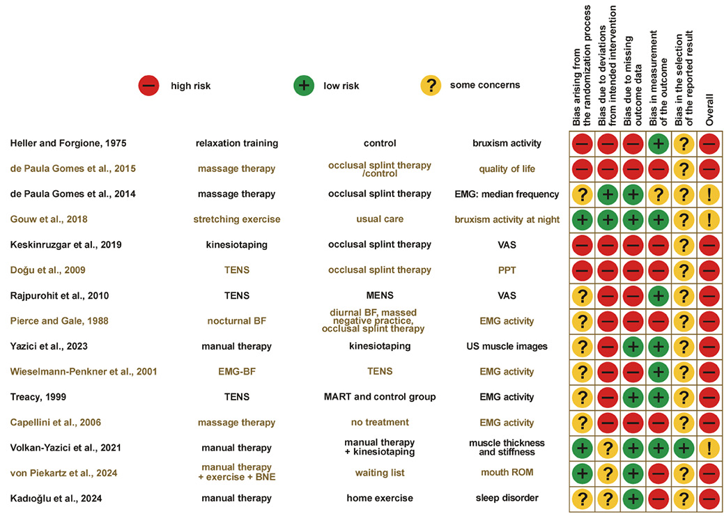
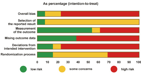
The majority of studies (n = 12, 80%) were considered to be at high RoB,13, 28, 29, 31, 32, 43, 45, 46, 47, 48, 50, 51 whereas three (20%) studies were judged to have some concerns.30, 44, 49 No study was classified as having a low RoB (Figure 2). Among studies rated as having a high RoB or some concerns, the most common methodological limitations were related to deviations from intended intervention domains, missing outcome data, and outcome measurement (Figure 3) (Appendix 5).

Certainty of the evidence

The certainty of the evidence for each outcome evaluated in this review is presented in Appendix 3. Overall, the certainty of the evidence was rated as very low, primarily because of small sample sizes, the high risk of bias and high heterogeneity of the studies. Considerable heterogeneity was observed regarding interventions, comparator treatments and outcome measures. Consequently, the data could not be pooled.

Discussion

This systematic review included 15 studies; however, synthesizing the findings was challenging due to substantial heterogeneity in the interventions evaluated, treatment dosage and intensity, comparator groups, and methods used to classify bruxism. Overall, the findings suggest that PT may reduce symptoms associated with bruxism, such as pain and muscle tenderness. Physiotherapy showed potential benefits compared with no treatment and demonstrated effects comparable to other conservative interventions, such as occlusal splint therapy. Combined approaches, particularly MT in conjunction with occlusal splints or kinesiotaping, showed the most promising results. However, the overall body of evidence was limited by methodological shortcomings and considerable clinical heterogeneity, including variations in interventions, diagnostic criteria and outcome measures. Consequently, firm conclusions regarding the effectiveness of PT cannot be drawn, underscoring the need for more standardized research in this field.

The 2025 International Consensus on Bruxism redefined the condition. However, most studies included in this review were conducted before publication of the updated consensus and therefore relied on outdated definitions. The findings of these studies were interpreted in light of the updated consensus. Five studies28, 43, 44, 45, 46 assessed SB using tools consistent with the diagnostic criteria of the American Academy of Sleep Medicine, combining clinical evaluation with self-reported measures. Only 2 studies incorporated instrumental assessments (EMG, PSG), despite PSG remaining the gold standard for assessing SB. Moreover, the PSG protocols and diagnostic thresholds differed between the studies. Awake bruxism was assessed only via self-reports29, 30, 32 often in combination with SB, whereas several older studies used poorly defined criteria.13, 31, 47, 48 These findings highlight the need for future studies to adopt standardized methods for assessing bruxism.

The findings of the present review are consistent with those of previous reviews,2, 4, 27 which concluded that PT may benefit individuals with bruxism who experience associated symptoms such as pain or muscle tenderness, regardless of whether AB or SB is present.2, 4, 27 However, the overall quality of the available evidence remains insufficient to support definitive clinical recommendations. Across the included studies, PT interventions were used to reduce pain, alleviate muscle tenderness, improve ROM, decrease excessive masticatory muscle activity, and ultimately improve quality of life. Although the available evidence suggests that these interventions may be beneficial, only a limited number of randomized and controlled clinical trials have been conducted, and most are characterized by a high risk of bias and substantial methodological limitations.

Physiotherapy may alleviate symptoms associated with bruxism through several complementary mechanisms. Manual therapy and myofascial release techniques target hyperactive masticatory muscles directly, reducing muscle tension, and trigger point sensitivity, thereby reducing pain and potentially decreasing excessive mechanical loading of the dentition. Stretching and guided exercises may improve muscle flexibility, coordination and jaw ROM, contributing to more efficient neuromuscular function.52 Neuromodulatory interventions, including TENS and low-level laser therapy, may inhibit nociceptive signaling, modulate trigeminal motor reflexes, and decrease hyperexcitability of the jaw-closing muscles.53 Furthermore, PT interventions incorporating relaxation training or biofeedback may reduce sympathetic nervous system activation, reducing stress-related parafunctional muscle activity, particularly in individuals with AB.45, 47 Collectively, these interventions target both peripheral musculoskeletal system and central neuromodulator mechanisms, supporting a multidimensional approach to managing bruxism and its adverse consequences.

With respect to bruxism activity, the available evidence suggests that improvements may be achieved with combined interventions, particularly MT combined with occlusal splint therapy and MART. Similarly, massage combined with occlusal splint therapy resulted in greater reductions in pain intensity than massage alone. The authors propose that combining these 2 approaches could enhance local blood flow and nutrient delivery, thereby improving muscle activity (i.e., normalizing muscle function) and reducing pain. Doǧu et al. reported that occlusal splint therapy was more effective than TENS in reducing tenderness of the masticatory muscles.50 This difference may reflect the distinct mechanisms of action of the 2 interventions. Transcutaneous electrical nerve stimulation primarily provides temporary analgesia through neuromodulatory mechanisms, acting predominantly on pain modulation without addressing the primary cause.53 In contrast, occlusal splints act mechanically by stabilizing the relationship between the arches and reducing excessive loading of masticatory muscles associated with bruxism.43 These results are consistent with previous concepts related to occlusal splint treatment, which emphasize its role in bruxism symptom relief. Occlusal splints are reversible and non-invasive and may provide sustained symptom relief when used consistently. However, their effectiveness depends largely on patient adherence to the prescribed treatment regimen.

Regarding jaw ROM, no significant differences between groups were observed in most comparisons. Neither stretching nor kinesiotaping demonstrated clinically meaningful improvements in active, pain-free, or passive ROM.44 It is important to note, however, that restricted jaw ROM is rarely reported in individuals with bruxism.

An additional consideration is the coexistence of bruxism with other clinical conditions. Three studies included participants with bruxism and associated TMD, making it difficult to determine whether the observed changes in outcomes were due to TMD symptoms, bruxism, or both. Symptoms such as pain and limited ROM are more prevalent in individuals with TMD than in those with bruxism alone. Consequently, findings from studies involving participants with both conditions cannot be directly compared with studies that included individuals with bruxism only.

Risk of bias and certainty of evidence

As previously mentioned, the RoB varied across the included studies. Most studies were classified as having a high risk of bias, several raised some concerns, and none were judged to have a low risk of bias. The most common methodological limitations were inadequate reporting of the randomization process and insufficient information regarding the blinding of participants, therapists, and outcome assessors. Blinding is particularly challenging in PT trials because interventions are typically delivered directly by therapists or performed by patients, increasing the potential for performance bias. Among the studies included in this review, only 2 described their blinding procedures,43, 49 1 study had no blinding,44 and the remaining 12 provided no information of blinding, indicating a substantial risk of bias in this domain.

Inadequate randomization and allocation concealment may substantially influence treatment effects. When participant allocation is non-random or predictable, selection bias may occur, whereby patients with characteristics such as greater motivation or less severe symptoms are preferentially allocated to the intervention group. Similarly, inadequate allocation concealment may allow therapists or investigators to influence group assignment, resulting in systematic differences between groups at baseline. These methodological flaws may exaggerate the apparent effectiveness of PT, particularly for subjective outcomes such as self-reported pain and muscle tenderness, which are susceptible to expectation and placebo effects. For instance, in studies with poorly implemented allocation procedures, improvements reported in the intervention group could partially reflect baseline differences rather than a true therapeutic effect. Additionally, poorly executed randomization and allocation increase the risk of selection, performance and attrition bias.39, 40, 41

Additional shortcomings further contribute to attrition bias. Most studies provided limited information regarding study limitations, participant adherence, or reasons for missing outcome data. Failure to account appropriately for missing data may lead to biased or overestimated treatment effects.39 Furthermore, the long-term effectiveness of PT remains unclear, as only 1 study included long-term follow-up, whereas the duration of treatment was generally short, with the exception of the study by Treacy,46 which lasted 4 months. Collectively, these limitations warrant cautious interpretation of the reported effectiveness of PT for individuals with bruxism.

The certainty of the evidence, assessed using the GRADE approach, was predominantly rated as very low due to methodological limitations and high heterogeneity among the studies. The majority of studies included a small number of participants, further limiting the strength of the evidence.

Clinical implications

This review provides a comprehensive overview of PT interventions that have been investigated for the management of bruxism with the aim of determining the most effective intervention. Although the available evidence does not allow definitive recommendations regarding the most effective intervention, several approaches appear promising for improving pain, muscle tenderness, and, when impaired, maximal mouth opening immediately after treatment. Overall, PT can be a good alternative strategy for improving pain, MMO, and tenderness (when present) immediately after treatment, especially when carried out in an interdisciplinary way. Manual therapy, active relaxation techniques and multimodal PT programs could be considered the first treatment option to manage the consequences of bruxism. Importantly, in the included studies, PT was used to manage the adverse consequences of bruxism, and not the bruxism behavior.

Research implications

The optimal PT approach for individuals with bruxism remains unclear due to methodological weaknesses, small sample sizes, short intervention periods, and inconsistent methods for diagnosing and classifying bruxism. Future trials should adopt standardized, consensus-based diag­nostic criteria and validated assessment tools for both AB and SB to improve the comparability of study findings. Methodological quality should be strengthened through robust randomization procedures, appropriate allocation concealment, comprehensive reporting of blinding, par­ticipant adherence, and study limitations. In addition, strategies for handling missing data should be clearly described and based on appropriate statistical methods to minimize the risk of bias.

Future research should also establish adequate treatment durations and include sufficiently long follow-up periods to exert measurable effects. This will ensure reporting of long-term outcomes to evaluate therapeutic benefits over a longer period. Larger, adequately powered clinical trials are needed, following recommendations by Zielinski et al.54 regarding minimum sample sizes for PT research. Reporting standardized effect sizes and measures of clinical importance would further improve the interpretation and clinical applicability of future findings. Additionally, comparative trials examining different types of bruxism within the same study design would further elucidate potential variations in intervention outcomes.

The evidence synthesized in this review largely reflects earlier concepts of bruxism, which emphasized its adverse musculoskeletal consequences, including pain and dysfunction. As knowledge of bruxism continues to evolve, specialized orofacial physiotherapists increasingly recognize its multifactorial nature and complexity. Therefore, future interventions may integrate musculoskeletal treatment with neuromuscular, behavioral and psychological strategies.55 These methods may combine bruxism neuroscience education (BNE), motor control training, and parafunctional behavior-specific exercises tailored to each patient.32 Such multidimensional approaches may influence future research outcomes. Finally, it should be acknowledged that most studies included in this review were published more than 5 years ago. Given the considerable advances in both bruxism research and PT practice since then, further high-quality studies based on current conceptual frameworks are needed.

Strengths and limitations

A major strength of this systematic review is its comprehensive search strategy, which was developed in collaboration with an experienced librarian and conducted without language or publication date restrictions. The broad PICO-based eligibility criteria enabled the inclusion of a wide range of PT approaches for bruxism. However, this broad scope also resulted in significant heterogeneity, limiting comparisons across studies and precluding quantitative synthesis. In addition, several studies inadequately reported numerical outcome data, preventing the calculation of effect sizes or independent verification of the reported findings. Although attempts were made to contact study authors to obtain missing information, this was not always possible, particularly for older publications, further impacting data verification and consistency.

Although broad inclusion criteria were used, only RCTs and CTs were included in this review. Although this decision reduced the number of eligible studies, these designs provide the highest level of evidence for evaluating treatment effectiveness. Additionally, most studies did not specify whether participants with bruxism were included or excluded, partly because of evolving definitions. Only one study focused exclusively on AB, whereas the remaining studies investigated SB, mixed populations, or unspecified bruxism. The present review adopted the most recent consensus definitions,9, 12 highlighting the importance of distinguishing between bruxism types. Future studies should clearly define and report bruxism subtypes, staying consistent to the updated guidelines and enhancing the consistency and comparability of research findings.

Finally, the considerable heterogeneity in intervention type, treatment intensity, duration, comparator groups, and outcome measures prevented the formulation of specific clinical recommendations. Accordingly, the findings of this review should be interpreted as a summary of the currently available evidence regarding PT interventions for managing the adverse consequences of bruxism rather than as definitive recommendations for clinical practice.

Conclusions

Physiotherapy for the management of bruxism remains an emerging area of research, with important gaps in the current evidence. Existing studies have primarily focused on reducing the adverse consequences of bruxism in symptomatic individuals. Methodological limitations, heterogeneous interventions, and evolving definitions of bruxism continue to limit the interpretation and generalizability of the available evidence. Recent advances in the understanding and assessment of bruxism have enabled a clearer distinction between the mechanisms underlying awake and sleep bruxism and have emphasized that currently available assessment tools measure different aspects of the behavior rather than the behavior itself. These developments provide an opportunity for future research to adopt more standardized, precise, and clinically meaningful methodologies. Given that bruxism is currently understood as a motor behavior, physiotherapists, with their expertise in the understanding and modulation of movement are well positioned to contribute to its management. When integrated within an interdisciplinary framework, PT interventions may help alleviate the musculoskeletal and neuromodulatory consequences of bruxism, offering a holistic approach to mitigating symptoms and improving patient outcomes. Future research should focus on rigorous, well-designed trials to establish evidence-based PT protocols that are both effective and sustainable.

Ethics approval and consent to participate

Not applicable.

Data availability

The datasets generated and/or analyzed during the current study, including the Appendix, are available from the corresponding author on reasonable request.

Consent for publication

Not applicable.

Use of AI and AI-assisted technologies

Not applicable.
Tables


Table 1. Characteristics of the included studies (N = 15)

Variable

n (%)

Country

Turkey

5 (33.3)

Brazil

3 (20.0)

USA

1 (6.7)

Austria

1 (6.7)

India

1 (6.7)

Netherlands

1 (6.7)

England

1 (6.7)

Sweden

1 (6.7)

Germany

1 (6.7)

Publication date

before 2000

3 (20.0)

2000–2010

4 (26.7)

after 2010

8 (53.3)

Study design

RCT

15 (100.0)

Trial registered

yes

4 (26.7)

no

2 (13.3)

not reported

9 (60.0)

Ethical approval

yes

9 (60.0)

not reported

6 (46.1)

Funding

not reported

10 (66.7)

no funding

3 (30.0)

government

2 (13.3)

Total sample size

≤50

11 (73.3)

>50 but <100

4 (26.7)

Number of interventions

4

2 (13.3)

3

2 (13.3)

2

11 (73.3)

Bruxism classification tool*

VAS (subject-based)

4 (26.7)

questionnaire based on the classification (subject-based)

2 (13.3)

Physiotherapy modalities*

manual therapy (massage therapy)

7 (46.7)

kinesiotaping

3 (20.0)

TENS

3 (20)

exercise

5 (33.3)

MENS

1 (6.7)

MyoMonitor training

1 (6.7)

BNE

1 (6.7)

Other treatment*

occlusal splint

5 (33.3)

occlusal adjustment

1 (6.7)

sleep hygiene advice

1 (6.7)

Sham/No treatment*

sham TENS

1 (6.7)

no treatment

4 (26.7)

Bruxism classification

AB and SB (together)

1 (6.7)

AB

1 (6.7)

SB

6 (40.0)

unspecified bruxism combined with TMD

3 (20.0)

unspecified bruxism

4 (26.7)

Outcomes*

pain

9 (60.0)

bruxism/EMG activity

6 (40.0)

quality of life

4 (26.7)

ROM

4 (26.7)

stress level

1 (6.7)

jaw disability

1 (6.7)

neck disability

1 (6.7)

EMG/PSG (device-based)

5 (33.3)

self-reported (subject-based)

3 (20.0)

RCT – randomized controlled trial; TENS – transcutaneous electrical neuromuscular stimulation; TMD – temporomandibular disorders; ROM – range of motion; EMG – electromyography; VAS – visual analog scale; MENS – microcurrent electrical neuromuscular stimulation; BNE – bruxism neuroscience education; AB – awake bruxism; SB – sleep bruxism; PSG – polysomnography; * numbers do not add up since several studies were included in these categories.
Table 2. Summary of the interventions, comparators, outcomes, and main findings of the included studies

Study details

Participants

Intervention group

Comparison group(s)

Outcome/measurements

Results

Author: Gouw et al.44
Year: 2018
Design: RCT
Objective: To determine the effectiveness of stretching of the masticatory muscles for SB
Funding: Independent grant from TMJ Solutions BV
Setting: Universities and University Medical Centres of Nijmegen and Utrecht; waiting rooms of several general practitioners, dentists and physical therapists

N = 24 (M = 9; F = 15)
Age: 34.6 (9.1) years (stretching: 34.1 (6.39) years; control: 35.1 (11.5) years)
Bruxism classification: SB
Evaluation tool:
Device-based assessment – PSG (>4 bruxism episodes/h of sleep or >25 bruxism bursts/h of sleep)
Additional symptoms: None

Muscle-stretching exercises + sleep hygiene advice (MS) (n = 12)
Description: Static masticatory muscle-stretching exercises with a stretching device
Duration of the session: Unclear
Duration of the treatment: Unclear
Number of sessions: 10
Times/week: 2
Time point/Follow-up: End of treatment

Control group (sleep hygiene advice) (CO) (n = 12)
Description: Patients received advice to control the symptoms before sleep. Practices and habits that can promote a good sleep quality, to rule out non-specific aspects.
Duration of the session: NA
Duration of the treatment: NA
Number of sessions: NA
Times/week: NA
Time point/Follow-up: End of treatment

(1) Bruxism episodes [h/sleep]:
Ambulant polysomnography
(2) Bruxism bursts [h/sleep]:
Ambulant polysomnography
(3) Sleep duration [min]:
Ambulant polysomnography
(4) Sleep quality:
15-item validated sleep quality scale
(5) MMO pain-free [mm]: ruler
(6) Maximal voluntary bite force:
Measured using a modified device developed by Radboudumc
(6) Tenderness [kg/cm
2]:
Masseter PPT measured with the Wagner algometer
(7) Mandibular function:
MFIQ
(8) Disability:
Graded Chronic Pain Scale (GCPS)

(1) Bruxism episodes: MS vs. CO
Increased in both groups compared to baseline, but more in the intervention group (within-group analysis); however, there was moderate evidence against the null hypothesis between groups, slightly favoring the control group, which presented fewer bruxism episodes (
p = 0.066).
(2) Bruxism bursts: MS vs. CO
Increased in both groups, but more in the intervention group; however, there was only moderate evidence against the null hypotheses between groups, slightly favoring the control group, which presented fewer bruxism bursts (
p = 0.049).
(3) Sleep duration: MS vs. CO
Increased in the intervention group (within-group analysis); weak evidence against the null hypothesis, with no difference between the groups (
p = 0.646).
(4) Sleep quality: MS vs. CO
Weak evidence against the null hypothesis, with no difference between the groups (
p = 0.923).
(5) Pain-free MMO: MS vs. CO
Statistically significant differences between the groups,
favoring the intervention group (
p = 0.020).
(6) Tenderness: MS vs. CO
Statistically significant differences between the groups,
favoring the MS group (
p = 0.036).
(7) MFIQ: MS vs. CO
Weak evidence against the null hypothesis, with no
difference between the groups (
p = 0.787).

Author: Capellini et al.51
Year: 2006
Design: RCT
Objective: To study the effect of massage on pain relief and/or EMG changes in individuals with bruxism
Funding: Fundacao de Amparo a Pesquisa do Estado de São Paulo (FAPESP - protocol: 0214144/0)
Setting: Faculty of Science and Technology, State University of São Paulo

N = 12 (M = 2; F = 10)
Age: 19–22 years
(MT: 20.3 (1.2) years; CG: 20.5 (1.4) years)
Bruxism classification: Unspecified bruxism combined with TMD
Other associated diagnosis: Myogenic TMD
Evaluation tool:
Clinically based assessment/subject-based assessment
Questionnaire: Clinical evaluation (only TMD)
Additional symptoms: Masticatory muscle pain

Massage therapy (MT) (n = 6)
Description: Massage on the masseter and anterior temporalis. The technique was based on classic massage: superficial and deep sliding, friction, compressive stationary and with distension kneading, rolling, and superficial sliding. The intermediate substance used was mineral oil.
Duration of the session: 30 min
Duration of the treatment: 3 weeks
Number of sessions: 15
Times/week: 5
Time point/Follow-up: The 1
st EMG signal assessment occurred before the treatment and the others in the 1st, 15th and 30th days after treatment. Pain was assessed before and after the 1st and 15th massage sessions.

Control group (CG) (n = 6)
Description: No treatment
Duration of the session: NA
Duration of the treatment: NA
Number of sessions: NA
Times/week: NA
Time point/Follow-up: The 1
st EMG signal assessment occurred before the treatment and the others on the 1st, 15th, and 30th days after treatment. Pain was assessed before and after the 1st and 15th massage sessions.

(1) Pain [points]: VAS
(2) Masseter and temporalis activity
a) Mandibular rest position (MRP): EMG activity (RMS)
b) MVC: EMG activity (RMS)

(1) Pain
MT vs. CG
No statistically significant differences between the groups (p = NR).
Within-group analysis
The MT group improved after treatment (
p = 0.047).
(2) Masseter and temporalis activity
a) Mandibular rest position (MRP)
MT vs. CG
No statistically significant differences between the groups (
p = NR).

Author: de Paula Gomes et al.49
Year: 2014
Design: RCT
Objective: To investigate the effects of massage therapy, conventional occlusal splint therapy, and silicone occlusal splint therapy on EMG activity in the masseter and anterior temporal muscles and the intensity of signs and symptoms in individuals with severe TMD and SB
Funding: NR
Setting: University community of the city of São Paulo

N = 60 (M = 10; F = 50)
Age: 18–40 years
(MT: 29.32 (4.31) years; COSG: 27.89 (5.82) years; MCOSG: 26.05 (3.32) years; SOSG: 28.92 (6.78) years)
Bruxism classification: SB
Other associated diagnosis: Severe TMD
Evaluation tool:
Clinically based assessment/subject-based assessment – clinical assessment (criteria of the American Academy of Sleep Medicine). The Fonseca Anamnestic Index was used to diagnose
the presence and intensity of TMD
Additional symptoms: Severe
TMD

Massage therapy (MT) (n = 15)
Description: Sliding and kneading maneuvers of the masseter and anterior temporal muscles, bilaterally. A facial massage cream was used to facilitate the manual procedures.
Duration of the session: 30 min
Duration of the treatment: 4 weeks
Number of sessions: 12
Times/week: 3
Time point/Follow-up: End of treatment

Conventional occlusal splint group (COSG) (n = 15)
Description: After a clinical examination by a dentist, the upper arch of each volunteer was molded with irreversible hydrocolloid for the fabrication of a Michigan-type OS with canine and protrusive guides as well as a flat occlusal surface for contact with the antagonist teeth.
Duration of the session: During sleeping
Duration of the treatment: 4 weeks
Number of sessions: NA
Times/week: Everyday
Follow-up: End of treatment
Massage therapy + Conventional occlusal splint group (MCOSG) (
n = 15)
Description: Combined treatment as described in the other 2 groups (MT + COSG)
Follow-up: End of treatment
Silicone occlusal splint group (SOSG) (
n = 15)
Description: The treatment was conducted with a silicone splint made with a 3-mm soft polyvinyl sheet in a vacuum pressure molding device with a thermally controlled infrared heater.
Duration of the session: During sleeping
Duration of the treatment: 4 weeks
Number of sessions: NA
Times/week: Everyday
Follow-up: End of treatment

(1) Severity of TMD [points]: Fonseca Anamnestic Index
(2) Bruxism activity measured by masseter and temporalis
activity (MVC) [Hz]: EMG activity

(1) Fonseca Anamnestic Index
MT vs. COSG
No statistically significant differences between the groups (
p = NR).
MT vs. MCOSG
The MCOSG showed better results after treatment compared to MT (
p < 0.0001).
MT vs. SOSG
No statistically significant differences between the groups (
p = NR).
COSG vs. MCOSG
The MCOSG showed better results after treatment compared to COSG (
p < 0.0001).
COSG vs. SOSG
No statistically significant differences between the groups (
p = NR).
MCOSG vs. SOSG
The MCOSG showed better results after treatment compared to SOSG (
p < 0.0001).
(2) Bruxism activity measured by EMG
MT vs. COSG
No statistically significant differences between the groups (
p = NR).
MT vs. MCOSG
The MCOSG showed better results after treatment compared to MT (
p < 0.0001).
MT vs. SOSG
No statistically significant differences between the groups (
p = NR).
COSG vs. MCOSG
The MCOSG showed better results after treatment compared to COSG (
p < 0.0001).
COSG vs. SOSG
No statistically significant differences between the groups (
p = NR).
MCOSG vs. SOSG
The MCOSG showed better results after treatment compared to SOSG (
p < 0.0001).

Author: Wieselmann-Penkner et al.13
Year: 2001
Design: RCT
Objective: To compare EMG BF and TENS stimulation
Funding: NR
Setting: Department of Prosthodontics at the University Hospital of Graz

N = 20 (M = 7; F = 13)
Age: 22–58 years
Bruxism classification: Unspecified bruxism
Evaluation tool:
NR
Additional symptoms: Pain and tenderness to palpation of the masticatory muscles with a chronicity of at least 6 months

MyoMonitor training (MM) (n = 10)
Description: Two active electrodes as well as 1 reference electrode were placed on both pre-auricular regions as well as on the neck, as recommended by Jankelson (1972). In addition, surface electrodes and a multisensory electrode were attached in order to record EMG activity. Following a 5 min warming-up period (facial twitching), the current was gradually increased until the mandible performed symmetrical contractions and was left at this setting throughout a 20 min-period of rest.
Duration of the session: 20 min
Duration of the treatment: NR
Number of sessions: 3
Times/week: 3
Time point/Follow-up: After all treatment sessions

Biofeedback (BF) (n = 10)
Description: Two active electrodes as well as 1 reference silver chloride surface electrode were placed bilaterally on the belly of the masseter, on the pars anterior of the temporalis muscles and on the nasion. Then the patients were asked to relax and to reduce EMG activity of the masticatory muscles with the higher baseline level for 20 min (10 min masseter muscle/10 min temporalis muscle with a pause of 3 min) with the help of visual feedback.
Duration of the session: 20 min
Duration of the treatment: NR
Number of sessions: 3
Times/week: 3
Follow-up: After all treatment sessions

(1) Bruxism activity [Hz]: EMG activity
(2) Skin conductive level (SCL) [Hz]: EMG activity

(1) Bruxism activity
MM vs. BF
Significant reduction in bruxism activity in the MM group for the right masseter (
p = 0.05). No differences between the groups (p = NR).
(2) SCL
MM vs. BF
For both groups, MM (
p = 0.004) and BF (p = 0.025), the increase was statistically significant only during the first session.
No differences between the groups (
p = NR).

Author: Heller and Forgione47
Year: 1975
Design: RCT
Objective: To use Forgione’s method to assess the effectiveness of 2 psychological techniques in controlling bruxism – massed negative practice and (J)PMR
Funding: National Institute of Dental Research Grant DE 03968-01
Setting: NR

N = 27 (M = 13; F = 14) + 7 non-bruxers (M = 6; F = 1)
Age: 18–32 years
Bruxism classification: Unspecified bruxism
Evaluation tool:
Clinically based assessment/subject-based assessment – self-reported (report of fatigue, tension or pain in the
masticatory muscles or a report of being
overheard grinding the teeth during the previous 2 months) and clinical assessment (clinical and radiographic examinations)
Additional symptoms: None

Massed negative practice (MNP) (n = 9)
Description: Participants were instructed to clench their teeth as hard as possible for five seconds and then relax the jaw for five seconds. This sequence was to be continued for a full minute and repeated five times throughout the day.
Duration of the session: 5 min
Duration of the treatment: 2 weeks
Number of sessions: 14
Times/week: 7
Time point/Follow-up: End of treatment

Relaxation training (RT) (n = 6)
Description: Instructions in (J)PMR were provided. Participants were given a standardized 30 min, abbreviated, relaxation training tape and instructed to practice nightly as close to bedtime as possible.
Duration of the session: 30 min
Duration of the treatment: 2
Number of sessions: 14
Times/week: 7
Follow-up: End of treatment
Bruxer control group (CG) (
n = 8)
Description: No treatment
Duration of the session: NA
Duration of the treatment: NA
Number of sessions: NA
Times/week: NA
Follow-up: End of treatment
Healthy controls (no treatment) (
n = 7)

(1) Bruxism score:
Bruxism-monitoring device
(2) Measuring trait of anxiety (TMAS):
Taylor Manifest Anxiety Scale
(3) Measuring state of anxiety:
Today Form of the Multiple Affect Adjective Checklist

(1) Bruxism score
MNP vs. RT vs. CG
No statistically significant differences between the groups (
p = NR).
(2) Measuring trait of anxiety
MNP vs. RT vs. CG
No statistically significant differences between the groups (
p = NR).
(3) Measuring state of anxiety
MNP vs. RT vs. CG
The analysis showed significant differences in trait anxiety, but the authors did not specify between which groups. A difference was found between bruxers and non-bruxers, but not specifically between the treatment groups (
p = NR).

Author: Pierce and Gale45
Year: 1988
Design: RCT
Objective: To compare 4 bruxism treatments for short- and long-term effectiveness: NBF, DBF, MNP, and splint therapy
Funding: USPHS Research Grants DE-05344 and DE-04358 from the National Institute of Dental Research, National Institutes of Health Bethesda, MD 20892
Setting: The greater Buffalo metropolitan area

N = 100 (M = 35; F = 65)
Age: 38 years
Bruxism classification: SB
Evaluation tool:
Device-based – EMG (mean bruxing episodes per hour was greater than 1.0)
Additional symptoms: None

Massed negative practice (MNP) (n = 20)
Description: Was individually tailored to each of the 20 participants with bruxism in this group. Six blocks of clenching trials per day consisted of five purposefully executed clench/relax cycles varying between 5 s and 1 min.
Duration of the session: 5 minutes (6 times/day)
Duration of the treatment: 2 weeks
Number of sessions: 14
Times/week: 7
Time point/Follow-up: End of treatment and 6 months

Diurnal biofeedback (DBF) (n = 20)
Description: EMG biofeedback machine was attached to the subject’s masseter muscle area by means of two 10-mm-diameter gold-plated surface electrodes as active electrodes and a standard ear clip electrode attached to the ipsilateral ear as the ground.
Duration of the session: 10 min
Duration of the treatment: 2 weeks
Number of sessions: 6
Times/week: 3
Follow-up: End of treatment and 6 months
Nocturnal biofeedback (NBF) (
n = 20)
Description: An aversive tone sounded in an earphone when the individual bruxed. The patient was required to arise, walk across the room, and record the date and time of awakening as well as the quality of sleep.
Duration of the session: Whole night
Duration of the treatment: 2 weeks
Number of sessions: NA
Times/week: Every night
Follow-up: End of treatment and 6 months
Occlusal splint therapy (OS) (
n = 20)
Description: Flat-plane maxillary occlusal treatment splints were fabricated out of hard dental acrylic. These splints were fabricated to include full palatal coverage
Duration of the session: Whole night
Duration of the treatment: 2 weeks
Number of sessions: NA
Times/week: Everyday
Follow-up: End of treatment
Control group (CG) (
n = 20)
Description: No treatment
Follow-up: End of treatment and 6 months

(1) Bruxism activity (mcVolt):
(a) Bruxing episodes per hour (EMG frequencies):
EMG recording during sleeping hours.
(b) Duration of bruxing activity per hour (EMG durations):
EMG recording during sleeping hours

(1) Bruxism activity:
(a) Bruxing episodes per hour
NBF and OS caused a significant reduction (
p < 0.001) in EMG-measured bruxing episodes per hour, but the subsequent removal of NBF was associated with a significant increase (p < 0.01) in EMG frequencies to baseline levels.
Simple main effects for EMG-measured frequencies were non-significant for MNP, DBF, and the CG. No significant differences between the groups were reported.
(b) Duration of bruxing activity per hour
Both NBF and OS were associated with significant decreases in EMG-measured duration of bruxing activity per hour from baseline to treatment (
p < 0.01).
There was also a significant increase in EMG-measured duration of bruxing activity from treatment to post-treatment for both NBF (
p < 0.001) and OS (p < 0.05). Simple main effects for EMG durations over the pre-treatment, treatment, and post-treatment dimension were non-significant for MNP, DBF, and CG.
No significant differences between the groups were reported.

Author: Treacy46
Year: 1999
Design: RCT
Objective: To test the effectiveness of relaxation training and TENS stimulation in the treatment of bruxism
Funding: NR
Setting: NR

N = 24 (M = 14; F = 10)
Age: 25.1 years
Bruxism classification: SB
Other associated diagnosis: CMD
Evaluation tool:
Clinically based assessment/subject-based assessment – self-reported (report of recurring episodes of nocturnal bruxism) and clinical assessment (dental wear, muscular tenderness and ROM according to the criteria set up by Phillips
 et al. (1989)
Additional symptoms: Symptoms of CMD

TENS (n = 7)
Description: TENS treatment was applied to the masseter muscle to receive receive a slight, rhythmic, electrical impulse through the nerves that controls the muscles of facial expression and chewing. The TENS apparatus was attached bilaterally by wires directly over the subject’s masseter muscles. A low-rate TENS (1–4 Hz) was applied. The pulse was emitted once every 1.5 s. The intensity was increased to the point where visible movement of the jaw occurred.
Duration of the session: 20–30 min
Duration of the treatment: 16 weeks
Number of sessions: 28
Times/week: 2
Time point/Follow-up: End of treatment

MART (n = 8)
Description: The program included training in awareness of posture and breathing habits by assuming different body positions and performing various movements. In addition, various muscular relaxation awareness exercises, involving neck, eye, arm, lower back, and leg muscles were used. The subject assumed supine as well as erect positions during the program.
Duration of the session: 20–30 min
Duration of the treatment: 16 weeks
Number of sessions: 28
Times/week: 2
Follow-up: End of treatment
Sham TENS (
n = 8)
Description: Sham TENS treatment of the masseter muscle, e.g., TENS with no electrical impulses
Duration of the session: 20–30 min
Duration of the treatment: 16 weeks
Number of sessions: 28
Times/week: 2
Follow-up: End of treatment

(1) Jaw ROM [mm]:
Ruler
(2) Number of breaths per minute:
Counting
(3) Bruxism activity [μV]:
EMG activity
(4) Discomfort in the craniomandibular area (quality of life):
The subjects estimated their degree of discomfort on a scale ranging from 0 (absence of discomfort) to 6 (great discomfort)

(1) Jaw ROM
TENS vs. MART
Participants in the MART group showed a significantly greater increase in MMO than those in the TENS group (
p < 0.05).
TENS vs. sham TENS
No significant differences between the groups (
p > 0.05).
MART vs. sham TENS
Participants in the MART group showed a significantly greater increase in MMO than those in the sham TENS group (
p < 0.05).
(2) Number of breaths
TENS vs. MART
Participants in the MART group showed a significantly greater reduction in the number of breaths than those in the TENS group (
p < 0.05).
TENS vs. sham TENS
No significant differences between the groups (
p > 0.05).
MART vs. sham TENS
Participants in the MART group showed a significantly greater reduction in the number of breaths than those in the sham TENS group (
p < 0.05).
(3) Bruxism activity (M. frontalis and M. masseter)
TENS vs. MART
Participants in the MART group showed a significantly greater reduction in bruxism activity than those in the TENS group (
p < 0.05).
TENS vs. sham TENS
No significant differences in bruxism activity between the groups (
p > 0.05).
MART vs. sham TENS
Participants in the MART group showed a significantly greater reduction in bruxism activity than those in the sham TENS group (
p < 0.05).
(4) Quality of life
TENS vs. MART
No significant differences between the groups.
TENS vs. sham TENS
No significant differences between the groups.
MART vs. sham TENS
Participants in the MART group showed significantly better quality of life than those in the sham TENS group (
p < 0.05).

Author: Doğu et al.50
Year: 2009
Design: RCT
Objective: To compare the efficacy of occlusal splint and TENS treatments on clinical findings and pain curvature in patients with myofascial pain syndrome and
temporomandibular joint discomfort accompanying bruxism
Funding: NR
Setting: Faculty of Medicine, Department of Physical
Medicine and Rehabilitation, University of Istanbul

N = 30 (M = 2; F = 28)
Age: 33.80 (11.64) (TENS: 36.29 (13.7); OS: 31.63 (9.42))
Bruxism classification: Unspecified bruxism
Other associated diagnosis: Myogenic TMD
Evaluation tool:
Clinically based assessment/subject-based assessment – self-reported (dental impingement complaint noticed by the patient or his relatives) and clinical assessment (detection of dental abrasions)
Additional symptoms: Myofascial pain syndrome

Conventional TENS (n = 15)
Description: TENS frequency was set at 70–110 Hz, and current duration was set as 40–100 microseconds. Electrodes were placed in the trigger points.
Duration of the session: 30 min
Duration of the treatment: 4 weeks
Number of sessions: 20
Times/week: 5
Time point/Follow-up: End of treatment and 1 month after the end of treatment

Occlusal splint (OS) (n = 15)
Description: Mandibular occlusal splint was prepared for each patient. Patients used the splint for 24 hours for a month after eating and brushing.
Duration of the session: The whole day
Duration of the treatment: 4 weeks
Number of sessions: NA
Times/week: NA
Follow-up: End of treatment and 1 month after the end of treatment

(1) Pain at rest and functioning:
VAS
(2) Tenderness of masseter and temporalis:
PPT [kg/cm
2] of the trigger points
(3) Quality of life:
Short Form-36
(4) ROM [mm]:
Tool NR

(1) Pain
TENS vs. OS
Pain improved more after TENS than after OS both after treatment (p < 0.028) and at the 1-month follow-up (
p < 0.032).
(2) Tenderness of the masseter and temporalis
TENS vs. OS
No statistically significant differences between the groups (
p = NR).
(3) Quality of life
TENS vs. OS
No statistically significant differences between the groups (
p = NR).
(4) ROM
TENS vs. OS
TENS was more effective than OS (
p < 0.05).

Author: de Paula Gomes et al.43
Year: 2015
Design: RCT
Objective: To investigate the effects of massage therapy on the masticatory muscles and occlusal splint usage, quality of life and pain in individuals with SB
Funding: NR
Setting: University community

N = 100 (F = 100) (final sample of 78)
Age: 18–40 years
(MT: 24.4 (4.1) years; OS: 24.4 (4.1) years;
MT+OS: 28.6 (4.2) years; CG: 25.72 (6.2) years)
Bruxism classification: SB
Evaluation tool:
Clinically based assessment/subject-based assessment – clinical assessment (criteria of the American Academy of Sleep Medicine)
Additional symptoms: Pain intensity score of 3 on a 11-point NRS.

Massage therapy (MT) (n = 17)
Description: Massage of masticatory muscles. Sliding and kneading maneuvers of the masseter and anterior temporal muscles, bilaterally. A facial massage cream was used to facilitate the manual procedures.
Duration of the session: 30 min
Duration of the treatment: 4 weeks
Number of sessions: 12
Times/week: 3
Time point/Follow-up: End of treatment

Occlusal splint (Michigan-type) (OS) (n = 19)
Description: After a clinical examination by a dentist, the upper arch of each volunteer was molded with irreversible hydrocolloid for the fabrication of a Michigan-type OS with canine and protrusive guides as well as a flat occlusal surface for contact with the antagonist teeth.
Duration of the session: During sleeping
Duration of the treatment: 4 weeks
Number of sessions: NA
Times/week: Everyday
Follow-up: End of treatment
Massage therapy + occlusal splint (MT + OS) (
n = 23)
Description: Combined treatment as described in the other two groups (MT + COSG)
Follow-up: End of treatment
Control group (CG) (
n = 19)
Description: No treatment
Duration of the session: NA
Duration of the treatment: NA
Number of sessions: NA
Times/week: NA
Follow-up: End of treatment

(1) Quality of life: SF-36
(2) Pain intensity: NRS

(1) Quality of life
MT vs. MT+OS vs. OS
No statistically significant differences between the groups (
p = NR).
(2) Pain intensity
MT vs. OS
No statistically significant differences between the groups (
p = NR).
MT vs. MT+OS
The MT+OS group showed better results after treatment than the MT group (
p < 0.05).
MT vs. CG
The MT group showed better results after treatment than the CG (
p < 0.0001).
OS vs. MT+OS
The MT+OS group showed better results after treatment than the OS group (
p < 0.05).
OS vs. CG
The OS group showed better results after treatment than the CG (
p < 0.003).
MT+OS vs. CG
The MT+OS group showed better results after treatment than the CG (
p < 0.0001).

Author: Volkan-Yazici et al.30
Year: 2021
Design: RCT
Objective: To compare the effects of two physiotherapy methods (manual therapy and KT with manual therapy) in patients with bruxism
Funding: NR
Setting: Ankara University Faculty of Dentistry

N = 32 (M = 2; F = 30)
Age:
MT: 24.6 (3.9)
KTMT: 25.3 (4.1)
Bruxism classification: AB and SB
Evaluation tool:
Clinically based assessment/subject-based assessment – self-reported and clinical assessment (Criteria of the International Classification for Sleep Disorders of the American Academy of Sleep Medicine, extracted from the protocol)
Additional symptoms: None

Manual therapy (MT) (n = 16)
Description: MT of the masticatory muscles performed intra- and extraorally together with MT of the cervical region were used in the treatment of bruxism. Stretching exercises of the masseter muscle were also applied. Extraoral applications consisted of emphasis on the masseter and temporalis muscle using kneading, friction, intramuscular stretching, sliding, and ischemic compression for a trigger point release. Myofascial release techniques were used to eliminate pain. Additionally, active and passive exercises were applied to the masseter, temporalis, digastric, medial pterygoid, and lateral pterygoid muscles. Intraoral applications consisted of emphasis on the masseter muscles using kneading, friction, intramuscular stretching and sliding, and ischemic compression for a trigger point release. Applications of the cervical region consisted of upper cervical flexion mobilization, C2–C5 central posterior-anterior mobilization, and also kneading, friction, intramuscular stretching, and sliding maneuvers for the cervical muscles.
Duration of the session: 30 min
Duration of the treatment: 2 weeks
Number of sessions: 8 sessions
Times/week: 4
Time point/Follow-up: End of the treatment

Kinesiotaping + manual therapy (KTMT)
(n = 16)
Description: The web-cut strips EDF (epidermisdermis-fascia) technique was used. KT placement was chosen according to the principles described by Kase et al. Tapes were cut 2.5 cm in width (cut in half vertically from a 5-cm width). Two strips of kinesiotape were crossed over the masseter muscle area with 0–5% tension in the maximum stretched epidermis position. The material used for bruxism KT application was Kinesio Tex Gold Fingerprints – a 100% cotton, latex-free, 5-cm wide, elastic tape. The tape was applied bilaterally after the MT session.
Duration of the session: 30 min
Duration of the treatment: 2 weeks
Number of sessions: 8 sessions
Times/week: 4
The kinesiotape was applied bilaterally after every session of MT, and the tape was removed 1 day before the next MT session.
Follow-up: End of the treatment

(1) Masseter muscle stiffness [cm]:
Ultrasound imaging
(2) Masseter muscle thickness [m/s]:
Ultrasound imaging
(3) Tenderness:
PPT was determined using a digital algometer; PPTs of the masseter, trapezius
(anterior, posterior, occipital), and temporalis muscles
were assessed bilaterally.
(4) Quality of life:
The factors associated with bruxism which caused problems in the daily life were assessed via Likert scale
(5) Sleep Quality:
PSQI
(6) Pain intensity
0–5 Likert scale

(1) Muscle stiffness
MT vs. KTMT
Significant decreases in the masseter muscle were observed in both groups (p < 0.05).
No differences between the groups (p > 0.05).
(2) Muscle thickness
MT vs. KTMT
Significant decreases in masseter muscle thickness were observed in both groups (p < 0.05).
No differences between the groups (p > 0.05).
(3) Tenderness
MT vs. KTMT
Jaw pain decreased more in the KTMT group than in the MT group (p < 0.05).
(4) Quality of life
MT vs. KTMT
Significant improvement was observed in both groups (p < 0.05).
No differences between the groups (p > 0.05)
(5) Sleep quality
MT vs. KTMT
Significant improvement was observed in both groups (p < 0.05).
No differences between the groups (p > 0.05).
(6) Pain intensity
MT vs. KTMT
Jaw pain decreased more in the KTMT group than in the MT group (p < 0.05).

Author: Rajpurohit et al.48
Year: 2010
Design: RCT
Objective: To compare the effectiveness of TENS and MENS on masticatory muscle pain in bruxism patients
Funding: NR
Setting: Physiotherapy Department, KLES Prabhakar Kore Hospital and MRC, Belgaum

N = 60 (M = 36; F = 24)
Age: 19–60 years
Bruxism classification:
Unspecified bruxism
Evaluation tool:
Clinically based assessment/subject-based assessment – clinical assessment (muscle tenderness over the masseter muscle; early morning temporomandibular joint stiffness and pain; duration of pain of more than 3 weeks).
Additional symptoms: Masticatory muscle pain

TENS (n = 30)
Description: Carbon electrodes (40 mm × 54 mm) were placed over the affected side of the masseter muscle with frequency operating at 50 Hz and the pulse width of 0.5 mSec, whereas the intensity was based on the patient’s tolerance.
Duration of the session: 20 min
Duration of the treatment: 1 week
Number of sessions: 7
Times/week: 7
Time point/Follow-up: End of treatment

MENS (n = 30)
Description: Frequency of 0.5 Hz and intensity based on the patient’s tolerance
Duration of the session: 20 min
Duration of the treatment: 1 week
Number of sessions: 7
Times/week: 7
Follow-up: End of treatment

(1) Tenderness:
Digital pressometer of about 2 KgF
(2) Pain:
VAS

(1) Tenderness
TENS vs. MENS
A significantly greater improvement in tenderness was observed with MENS than with TENS (p < 0.0495).
(2) Pain
TENS vs. MENS
Pain relief was greater with MENS therapy than with TENS, with a statistically significant difference at the end of treatment (p ≤ 0.0001) (TENS group: 3.38 (1.25) vs. MENS group: 3.91 (0.83)).

Author: Yazici et al.29
Year: 2023
Design: RCT
Objective: To compare the immediate effects of a single session of manual therapy (MT) or kinesiotaping following MT (KTMT) in patients with bruxism using shear wave ultrasonography
Funding: NR
Setting: Ankara University Faculty of Dentistry

N = 39 (M = 4; F = 35)
Age:
MT: 27.3 (4.8)
KTMT: 26.2 (5.4)
Bruxism classification: AB and SB (extracted from the protocol)
Evaluation tool:
Clinically based assessment/subject-based assessment – self-reported and clinical assessment (Criteria of the International Classification for Sleep Disorders; extracted from the protocol)
Additional symptoms: None

Manual therapy (MT) (n = 20)
Description: MT of the masticatory muscles performed intra- and extraorally together with MT of the cervical region were used in the treatment of bruxism. Stretching exercises of the masseter muscle were also applied. Extraoral applications consisted of emphasis on the masseter and temporalis muscle using kneading, friction, intramuscular stretching, sliding, and ischemic compression for a trigger point release. Myofascial release techniques were used to eliminate pain. Additionally, active and passive exercises were applied to the masseter, temporalis, digastric, medial pterygoid, and lateral pterygoid muscles. Intraoral applications consisted of emphasis on the masseter muscles using kneading, friction, intramuscular stretching and sliding, and ischemic compression for a trigger point release. Applications of the cervical region consisted of upper cervical flexion mobilization, C2–C5 central posterior-anterior mobilization, and also kneading, friction, intramuscular stretching, and sliding maneuvers for the cervical muscles.
Duration of the session: 30 min
Duration of the treatment: 1 week
Number of sessions: 1 session
Times/week: 1
Time point/Follow-up: Immediately after the 1st session

Kinesiotaping + manual therapy (KTMT)
(n = 16)
Description: The web-cut strips EDF (epidermisdermis-fascia) technique was used. KT placement was chosen according to the principles described by Kase et al. Tapes were cut 2.5 cm in width (cut in half vertically from a 5-cm width). Two strips of kinesiotape were crossed over the masseter muscle area with 0–5% tension in the maximum stretched epidermis position. The material used for bruxism KT application was Kinesio Tex Gold Fingerprints – a 100% cotton, latex-free, 5-cm wide, elastic tape. The tape was applied bilaterally after the MT session.
Duration of the session: 45 min
Duration of the treatment: 1 week
Number of sessions: 1 session
Times/week: 1
The kinesiotape was applied bilaterally after every session of MT, and the tape was removed 1 day before the next MT session.
Follow-up: Immediately after the 1st session

(1) Masseter muscle stiffness [cm]:
Ultrasound imaging
(2) Masseter muscle thickness [m/s]:
Ultrasound imaging
(3) Tenderness:
PPT was determined using a digital algometer; PPTs of the masseter, trapezius (anterior, posterior, occipital), and temporalis muscles were assessed bilaterally.

(1) Muscle stiffness
MT vs. KTMT
Significant decreases in the masseter muscle were observed in both groups (p < 0.05).
No between-group differences were observed (p > 0.05).
(2) Muscle thickness
MT vs. KTMT
Significant decreases in masseter muscle thickness were observed in both groups (p < 0.05).
No between-group differences were observed (p > 0.05).
(3) Tenderness
MT vs. KTMT
A significant increase in PPT for the masseter and temporalis muscles was observed in the KTMT group (p < 0.05).
No significant differences in PPT were found for any other muscle groups in either the KTMT or MT groups (p > 0.05).
No between-group differences were observed (p > 0.05).

Author: Keskinruzgar et al.28
Year: 2019
Design: RCT
Objective: To evaluate the efficacy of KT in patients with SB and to determine whether kinesiotaping may be an alternative for OS for the treatment of SB
Funding: NR
Setting: Oral and Maxillofacial Surgery Clinic at the Faculty of Dentistry of Adıyaman University

N = 34 (M = 4; F = 30)
Age:
KT: 27.38 (9.05) years; OS: 26.11 (8.71) years
Bruxism classification:
SB
Evaluation tool:
Clinically based assessment/subject-based assessment – self-reported (clenching or grinding of teeth for at least 5 days a week for duration of 6 months) and clinical assessment (presence of shiny dental restorations, dental abrasion, hypertrophy in masseter muscles and palpation-induced pain)
Additional symptoms: None

Kinesiotaping (KT) (n = 16)
Description: KT was performed by a physiotherapist as described by Dr. Kase, where the 5-cm-wide tapes were cut in the Y-shape. The ends of the Y-shaped band were attached to the region corresponding to the masseter muscle’s insertion with a tension of about 40%.
Duration of the session: Whole day and night
Duration of the treatment: 5 weeks
Number of sessions: Unclear
Times/week: 7
Time point/Follow-up: After the 1st week of treatment and at the end of treatment

Occlusal splint (OS) (n = 18)
Description: Models were prepared by using the upper jaw measurements of the patients. For each patient, 0.5-mm thick thermoplastic hard splints were fabricated as OS by using these models.
Duration of the session: whole night
Duration of the treatment: 5 weeks
Number of sessions: NA
Times/week: Every night
Follow-up: After the 1st week of treatment and at the end of treatment

(1) ROM:
Maximum distance between the patient’s lower and upper incisors (tool NR)
(2) Pain: VAS
(3) Tenderness: Bilateral temporalis and masseter muscle PPTs were measured with an algometer (J Tech Digital Algometer)

(1) ROM
KT vs. OS
No statistically significant differences between the groups (p > 0.05).
(2) Pain
KT vs. OS
No statistically significant differences between the groups (p > 0.05).
(3) Tenderness
KT vs. OS
No statistically significant differences between the groups (p > 0.05), except for the temporalis PPT at the 1st week, which was significantly higher in the KT group than in the OS group (p = 0.03).

Author: von Piekartz et al.32
Year: 2024
Design: RCT
Objective: To assess a comprehensive PT strategy that integrates both direct (hands on) and indirect (hands off) treatments for AB, aimed to relieve pain and enhance orofacial/cervical function
Funding: NR
Setting: Physical therapy practices in Western Germany

N = 42 (F = 42)
Age:
PT: 31.30 (11.16) years; Control: 28.50 (10.33) years
Bruxism classification:
AB
Evaluation tool:
Clinically based – classification provided by a dentist familiar with a bruxism classification system
Additional symptoms: 60% of the population presented TMD diagnosis (40% myofascial TMD; 7% arthrogenic TMD; 13% mixed TMD) with or without chronic pain

Multimodal PT program (n = 13)
Description: Orofacial manual therapy, motor control and behavioral exercise, habitual reversal training, the brace relaxation technique, tongue-teeth breathing swallowing exercise, Brux.app (education therapy), habit formation exercise, BNE, which includes information about somatomotor reflex, nociceptor (warning signals), centralized mechanism, memory for motor functions, (un)controlled motor inhibition system, management of motion control.
Duration of the session: NR
Duration of the treatment: 3 weeks
Number of sessions: 6
Times/week: 2
Time point/Follow-up: End of treatment

Control group (CG) (n = 15)
Description: Waiting list
Duration of the session: NA
Duration of the treatment: NA
Number of sessions: NA
Times/week: NA
Follow-up: End of treatment

(1) ROM:
Mouth opening, propulsion, retropulsion, laterotrusion
(2) Perception of functional limitations due to pain-related problems in daily life:
The Pain Disability Index (PDI)
(3) Neck-related
disability: Neck Disability Index (NDI)
(4) Jaw
disability: Jaw Disability List (JDL)
(5) Mechanical pressure pain threshold (PPT): masseter

(1) ROM

Mouth opening: No differences between the groups were observed (MD: −4.18 (95% CI: −11.28, 2.92))

Propulsion: No differences between the groups were observed (MD: 0.44 (95% CI: −1.56, 2.44))

Retropulsion: No differences between the groups were observed (MD: 0.20 (95% CI: −1.51, 1.91))

(2) Perception of functional limitations due to pain-related problems in daily life:

No differences between the groups were observed (MD: 4.88 (95% CI: −3.52, 13.28))

(3) Neck-related disability: PT resulted in greater improvement in the NDI than HEG (MD: 6.94 (95% CI: −1.51, 15.39))

(4) Jaw disability: Results NR

(5) Mechanical pressure pain threshold (PPT): Results NR

Author: Kadıoğlu et al.31
Year: 2024
Design: RCT
Objective: To examine the effects of manual therapy and home exercise treatments on pain, sleep quality, stress level, and quality of life in patients with bruxism
Funding: No funding
Setting: Bartın University, School of Health Services, Bartın, Türkiye

N = 30 (M = 9; F = 21)
Age:
MTG: 18–20: 6 (40%); 21–22: 7 (46.7%); 23–up: 2 (13.3%);
HEG: 18–20: 7 (46.7%); 21–22: 4 (26.6%); 23–up: 4 (26.6%);
Bruxism classification:
Unspecified bruxism
Evaluation tool:
Subject-based – individuals who were pre-classified with “bruxism” in the preliminary evaluation of the questionnaire were clinically evaluated by an expert orthodontist in terms of clinical signs of bruxism: abnormal tooth wear on the occlusal surfaces of the teeth, abfraction, gingival recession and/or cervical defect, tongue indentations, damage to the inside of the cheek, tense facial and jaw muscles, muscle sensitivity, and masseteric hypertrophy upon bidigital palpation. Individuals who had at least two of these clinical findings in addition to the bruxism questionnaire results were included in the study.
Additional symptoms: 86.7% of the population presented with associated headache. All participants present parafunctional habits

Manual therapy group (MTG) (n = 15)
Description: This group involves manual therapy for jaw and neck muscles including soft tissue, joint mobilization, intramuscular stretches, trigger point therapy, intraoral application, post-isometric relaxation. Additionally, stretching, relaxation and strengthening training.
Duration of the session: 40 min
Duration of the treatment: 8 weeks
Number of sessions: 16
Times/week: 2
Time point/Follow-up: End of treatment

Home exercises (HEG) (n = 15)
Description: This group involved home exercises for jaw and neck muscles, including mobility, stretching, isometric, posterior tilt, postural exercises, as well as relaxation and strengthening training.
Duration of the session: 25 min
Duration of the treatment: 8 weeks
Number of sessions: NA
Times/week: NA
Follow-up: End of treatment

(1) Sleep quality: PSQI
(2) QoL: SF-36
(3) Stress level: Perceived Stress Scale
(4) Jaw
disability: Fonseca Anamnestic Index
(5) Number of TrP
(6) Rest pain: VAS (0–10)
(7) Activity pain: VAS (0–10)
(8) Night pain: VAS (0–10)

(1) Sleep quality: MTG improved sleep quality compared with HEG (MD: 3.28 (95% CI: 0.67, 5.89))

(2) Quality of life: MTG improved quality of life compared with HEG (MD: −15.66 (95% CI: −32.17, 0.85))

(3) Stress level: no differences between the groups were observed (MD: 1.47 (95% CI: −1.97, 4.91))

(4) Jaw disability: no differences between the groups were observed (MD: 6.34 (95% CI: −5.42, 18.10))

(5) Number of TrPs: MTG had fewer TrPs than HEG (MD: 2.07 (95% CI: 0.16, 3.98))

(6) Rest pain: no differences between the groups were observed (MD: −1.84 (95% CI: −3.84, 0.16))

(7) Activity pain: no differences between the groups were observed (MD: −1.45 (95% CI: −3.80, 0.90))

(8) Night pain: no differences between the groups were observed (MD: −1.70 (95% CI: −4.32, 0.92))

Table 3. Summary matrix of the results according to intervention, comparison, and outcome

Category

Information about the trials

Outcomes

therapy ONE

therapy TWO

bruxism symptoms

timepoint

bruxism activity

pain intensity

tenderness

mouth ROM

quality of life

sleep quality

stress level

trigger points

Fonseca Anamnestic Index

Manual therapy

manual therapy

home exercise

unspecified bruxism

final

×31

×31

?

?

×31

131

×31

131

131

manual therapy

manual therapy + kinesiotaping

SB and AB associated with orofacial pain

final

?

×30

×29, 30

?

?

?

?

?

?

massage therapy

occlusal splint therapy

SB associated with TMD diagnosis

final

?

×43

?

?

?

?

?

?

?

massage therapy

occlusal splint therapy (silicon)

SB associated with TMD diagnosis

final

×49

?

?

?

?

?

?

?

?

massage therapy

massage therapy + occlusal splint therapy

SB associated with TMD diagnosis

final

?

243

?

?

?

?

?

?

?

massage therapy

massage therapy + occlusal splint therapy

SB associated with TMD diagnosis

final

×49

?

?

?

?

?

?

?

?

massage therapy

no treatment

SB associated with TMD diagnosis

final

?

143

?

?

?

?

?

?

?

massage therapy + occlusal splint therapy

occlusal splint therapy

SB associated with TMD diagnosis

final

?

143

?

?

?

?

?

?

?

massage therapy + occlusal splint therapy

occlusal splint therapy

SB associated with TMD diagnosis

final

?

149

?

?

?

?

?

?

?

massage therapy + occlusal splint therapy

no treatment

SB associated with TMD diagnosis

final

?

143

?

?

?

?

?

?

?

massage therapy

no treatment

unspecified bruxism associated with TMD diagnosis and masticatory muscle pain

final

×51

151

?

?

?

?

?

?

?

Stretching

muscles stretching + sleep hygiene advice

sleep hygiene advice (control group)

SB without any additional symptoms

final

×44

?

144

×44

?

?

?

?

?

Active relaxation techniques

MyoMonitor training

biofeedback training

unspecified bruxism associated with tenderness in the masticatory muscles

final

213

?

?

?

?

?

?

?

?

massed negative practice

occlusal splint therapy

SB without any additional symptoms

final

×45

?

?

?

?

?

?

?

?

massed negative practice

occlusal splint therapy

SB without any additional symptoms

six-month follow-up

×45

?

?

?

?

?

?

?

?

massed negative practice

strength exercise

SB without any additional symptoms

final

×45

?

?

?

?

?

?

?

?

massed negative practice

strength exercise

SB without any additional symptoms

one-month follow-up

×45

?

?

?

?

?

?

?

?

massed negative practice

biofeedback

SB without any additional symptoms

final

×45

?

?

?

?

?

?

?

?

massed negative practice

biofeedback

SB without any additional symptoms

one-month follow-up

×45

?

?

?

?

?

?

?

?

massed negative practice

education therapy

unspecified bruxism with any additional symptoms

final

×47

?

?

?

?

?

?

?

?

massed negative practice

no treatment

unspecified bruxism with any additional symptoms

final

×47

?

?

?

?

?

?

?

?

Kinesiotaping

kinesiotaping

occlusal splint therapy

SB with any additional symptoms

final

?

×28

×28

×28

?

?

?

?

?

TENS

TENS

MART

SB associated with symptoms of craniomandibular disorders

final

246

?

?

246

?

?

?

?

?

TENS

Sham TENS

SB associated with symptoms of craniomandibular disorders

final

×46

?

?

?

?

?

?

?

?

TENS

occlusal splint therapy

unspecified bruxism associated with TMD diagnosis and myofascial pain

final

?

×50

?

250

×50

?

?

?

?

TENS

occlusal splint therapy

unspecified bruxism associated with TMD diagnosis and myofascial pain

one-month follow-up

?

×50

?

×50

×50

?

?

?

?

TENS

MENS

unspecified bruxism associated with masticatory muscle pain

final

?

248

248

?

?

?

?

?

?

Multimodal treatment

multimodal treatment

waiting list

AB

final

?

132

?

132

?

?

?

?

?

×31

Figures


Fig. 1. PRISMA (Preferred Reporting Items for Systematic reviews and Meta-Analyses) flow diagram illustrating the study selection process
RCT – randomized controlled trial; CT – clinical trial.
Fig. 2. Risk of bias assessment for each included study
EMG – electromyography; VAS – visual analogue scale; TENS – transcutaneous electrical nerve stimulation; PPT – pressure pain threshold; MENS – microcurrent electrical neuromuscular stimulation; BF – biofeedback; US – ultrasound; MART – muscular awareness relaxation training; ROM – range of motion.
Fig. 3. Overall risk of bias across the included studies

References (55)

  1. Friedman Rubin P, Eli I, Greenbaum T, Shapira K, Emodi-Perelman A, Winocur E. Potential orofacial hazards of resistance training: A controlled comparative study. Cranio. 2019;37(1):45–52. doi:10.1080/08869634.2017.1370566
  2. Amorim CSM, Espirito Santo AS, Sommer M, Marques AP. Effect of physical therapy in bruxism treatment: A systematic review. J Manipulative Physiol Ther. 2018;41(5):389–404. doi:10.1016/j.jmpt.2017.10.014
  3. Feurer I. Bruxism and physiotherapy. J Craniomandib Funct. 2019;11(2):151–162. https://www.quintessence-publishing.com/deu/de/article/856438/journal-of-craniomandibular-function/2019/02/bruxism-and-physiotherapy. Accessed June 5, 2024.
  4. Manfredini D, Ahlberg J, Winocur E, Lobbezoo F. Management of sleep bruxism in adults: A qualitative systematic literature review. J Oral Rehabil. 2015;42(11):862–874. doi:10.1111/joor.12322
  5. Melnick S. Physiotherapy and clinical approaches – an overview. Eur J Phys Educ Sport Sci. 2015;1(1). doi:10.46827/ejpe.v1i1.9
  6. Nicholls DA, Gibson BE. Physiotherapy as a complex assemblage of concepts, ideas and practices. Physiother Theory Pract. 2012;28(6):418–419. doi:10.3109/09593985.2012.692557
  7. Gębska M, Dalewski B, Pałka Ł, Kiczmer P, Kołodziej Ł. Kinesio taping as an alternative therapy for limited mandibular mobility with pain in female patients with temporomandibular disorders: A randomized controlled trial. Dent Med Probl. 2024;61(5):659–670. doi:10.17219/dmp/173126
  8. Wadhokar OC, Kulkarni CA, Joshi M. Changing trends in publication regarding temporomandibular joint dysfunction from 1975 to 2021: A bibliometric analysis. Cureus. 2023;15(9):e46179. doi:10.7759/cureus.46179
  9. Lobbezoo F, Ahlberg J, Raphael KG, et al. International consensus on the assessment of bruxism: Report of a work in progress. J Oral Rehabil. 2018;45(11):837–844. doi:10.1111/joor.12663
  10. Cigdem Karacay B, Sahbaz T. Investigation of the relationship between probable sleep bruxism, awake bruxism and temporomandibular disorders using the Diagnostic Criteria for Temporomandibular Disorders (DC/TMD). Dent Med Probl. 2023;60(4):601–608. doi:10.17219/dmp/158926
  11. Zieliński G, Pająk-Zielińska B, Pająk A, Wójcicki M, Litko-Rola M, Ginszt M. Global co-occurrence of bruxism and temporomandibular disorders: A meta-regression analysis. Dent Med Probl. 2025;62(2):309–321. doi:10.17219/dmp/201376
  12. Verhoeff MC, Lobbezoo F, Ahlberg J, et al. Updating the bruxism definitions: Report of an International Consensus Meeting. J Oral Rehabil. 2025;52(9):1335–1342. doi:10.1111/joor.13985
  13. Wieselmann-Penkner K, Janda M, Lorenzoni M, Polansky R. A comparison of the muscular relaxation effect of TENS and EMG-biofeedback in patients with bruxism. J Oral Rehabil. 2001;28(9):849–853. doi:10.1046/j.1365-2842.2001.00748.x
  14. Lobbezoo F, Ahlberg J, Glaros AG, et al. Bruxism defined and graded: An international consensus. J Oral Rehabil. 2013;40(1):2–4. doi:10.1111/joor.12011
  15. Manfredini D, Ahlberg J, Aarab G, et al. Standardised tool for the assessment of bruxism. J Oral Rehabil. 2024;51(1):29–58. doi:10.1111/joor.13411
  16. Thymi M, Farzan A, Ahlberg J, Manfredini D, Lobbezoo F. Qualitative suggestions for the further development of the Standardized Tool for the Assessment of Bruxism (STAB). Dent Med Probl. 2024;61(3):323–333. doi:10.17219/dmp/183692
  17. Zieliński G, Pająk A, Wójcicki M. Global prevalence of sleep bruxism and awake bruxism in pediatric and adult populations: A systematic review and meta-analysis. J Clin Med. 2024;13(14):4259. doi:10.3390/jcm13144259
  18. Manfredini D, Lobbezoo F. Role of psychosocial factors in the etiology of bruxism. J Orofac Pain. 2009;23(2):153–166. PMID:19492540.
  19. Przystańska A, Jasielska A, Ziarko M, et al. Psychosocial predictors of bruxism. Biomed Res Int. 2019;2019:2069716. doi:10.1155/2019/2069716
  20. Emodi-Perlman A, Zur M, Czygrinow AY, Ventura N, Eli I. Impact of long-term stress on awake bruxism: An observational longitudinal within-subject study of stress-related changes. Dent Med Probl. 2025;62(6):1059–1067. doi:10.17219/dmp/209181
  21. Soto-Goñi XA, Alen F, Buiza-González L, et al. Adaptive stress coping in awake bruxism. Front Neurol. 2020;11:564431. doi:10.3389/fneur.2020.564431
  22. Lobbezoo F, Naeije M. Bruxism is mainly regulated centrally, not peripherally. J Oral Rehabil. 2001;28(12):1085–1091. doi:10.1046/j.1365-2842.2001.00839.x
  23. Więckiewicz M, Smardz J, Martynowicz H. Lifestyle, daily habits, sleep hygiene, and diet: Proposal of a new approach for sleep bruxism management. Dent Med Probl. 2025;62(1):5–7. doi:10.17219/dmp/191517
  24. Phuong NTT, Ngoc VTN, Linh LM, Duc NM, Tra NT, Anh LQ. Bruxism, related factors and oral health-related quality of life among Vietnamese medical students. Int J Environ Res Public Health. 2020;17(20):7408. doi:10.3390/ijerph17207408
  25. Emodi-Perlman A, Eli I. Temporomandibular disorders and bruxism – up-to-date assessment and screening tools the general dentist should be aware of. Dent Med Probl. 2024;61(2):169–171. doi:10.17219/dmp/175582
  26. Ainoosah S, Farghal AE, Alzemei MS, et al. Comparative analysis of different types of occlusal splints for the management of sleep bruxism: A systematic review. BMC Oral Health. 2024;24(1):29. doi:10.1186/s12903-023-03782-6
  27. Vieira MA, de Oliveira-Souza AIS, Hahn G, Bähr L, Armijo-Olivo S, Ferreira APL. Effectiveness of biofeedback in individuals with awake bruxism compared to other types of treatment: A systematic review. Int J Environ Res Public Health. 2023;20(2):1558. doi:10.3390/ijerph20021558
  28. Keskinruzgar A, Kucuk AO, Yavuz GY, Koparal M, Caliskan ZG, Utkun M. Comparison of kinesio taping and occlusal splint in the management of myofascial pain in patients with sleep bruxism. J Back Musculoskelet Rehabil. 2019;32(1):1–6. doi:10.3233/BMR-181329
  29. Yazici G, Kafa N, Kolsuz ME, Volkan-Yazici M, Evli C, Orhan K. Evaluation of single session physical therapy methods in bruxism patients using shear wave ultrasonography. Cranio. 2023;41(1):41–47. doi:10.1080/08869634.2020.1812817
  30. Volkan-Yazici M, Kolsuz ME, Kafa N, Yazici G, Evli C, Orhan K. Comparison of Kinesio Taping and manual therapy in the treatment of patients with bruxism using shear-wave elastography – a randomised clinical trial. Int J Clin Pract. 2021;75(12):e14902. doi:10.1111/ijcp.14902
  31. Kadıoğlu MB, Sezer M, Elbasan B. Effects of manual therapy and home exercise treatment on pain, stress, sleep, and life quality in patients with bruxism: A randomized clinical trial. Medicina (Kaunas). 2024;60(12):2007. doi:10.3390/medicina60122007
  32. von Piekartz H, Bleiss S, Herzer S, Hall T, Ballenberger N. Does combining oro-facial manual therapy with bruxism neuroscience education affect pain and function in cases of awake bruxism? A pilot study. J Oral Rehabil. 2024;51(9):1692–1700. doi:10.1111/joor.13740
  33. Page MJ, McKenzie JE, Bossuyt PM, et al. The PRISMA 2020 statement: An updated guideline for reporting systematic reviews. BMJ. 2021;372:n71. doi:10.1136/bmj.n71
  34. Brown D. A review of the PubMed PICO Tool: Using evidence-based practice in health education. Health Promot Pract. 2020;21(4):496–498. doi:10.1177/1524839919893361
  35. Hutton B, Salanti G, Caldwell DM, et al. The PRISMA extension statement for reporting of systematic reviews incorporating network meta-analyses of health care interventions: Checklist and explanations. Ann Intern Med. 2015;162(11):777–784. doi:10.7326/M14-2385
  36. Higgins JPT, Thomas J, Chandler J, et al. Cochrane Handbook for Systematic Reviews of Interventions. Version 6.3 (updated February 2022). Cochrane; 2022.
  37. Sterne JAC, Savović J, Page MJ, et al. RoB 2: A revised tool for assessing risk of bias in randomised trials. BMJ. 2019;366:l4898. doi:10.1136/bmj.l4898
  38. Armijo-Olivo S, Ospina M, da Costa BR, et al. Poor reliability between Cochrane reviewers and blinded external reviewers when applying the Cochrane risk of bias tool in physical therapy trials. PLoS One. 2014;9(5):e96920. doi:10.1371/journal.pone.0096920
  39. Armijo-Olivo S, Machalicek W, De Oliveira-Souza AI, Dennett L, Ballenberger N. Attrition, missing data, compliance, and related biases in randomized controlled trials of rehabilitation interventions: Towards improving reporting and conduct. Eur J Phys Rehabil Med. 2020;56(6):817–828. doi:10.23736/S1973-9087.20.06427-8
  40. Armijo-Olivo S, Mohamad N, Sobral de Oliveira-Souza AI, de Castro-Carletti EM, Ballenberger N, Fuentes J. Performance, detection, contamination, compliance, and cointervention biases in rehabilitation research: What are they and how can they affect the results of randomized controlled trials? Basic information for junior researchers and clinicians. Am J Phys Med Rehabil. 2022;101(9):864–878. doi:10.1097/PHM.0000000000001893
  41. Armijo-Olivo S, de Oliveira-Souza AIS, Mohamad N, de Castro Carletti EM, Fuentes J, Ballenberger N. Selection, confounding, and attrition biases in randomized controlled trials of rehabilitation interventions: What are they and how can they affect randomized controlled trials results? Basic information for junior researchers and clinicians. Am J Phys Med Rehabil. 2022;101(11):1042–1055. doi:10.1097/PHM.0000000000001947
  42. Guyatt GH, Oxman AD, Vist GE, et al.; GRADE Working Group. GRADE: An emerging consensus on rating quality of evidence and strength of recommendations. BMJ. 2008;336(7650):924–926. doi:10.1136/bmj.39489.470347.AD
  43. de Paula Gomes CAF, El-Hage Y, Amaral AP, et al. Effects of massage therapy and occlusal splint usage on quality of life and pain in individuals with sleep bruxism: A randomized controlled trial. J Jpn Phys Ther Assoc. 2015;18(1):1–6. doi:10.1298/jjpta.Vol18_001
  44. Gouw S, de Wijer A, Kalaykova SI, Creugers NHJ. Masticatory muscle stretching for the management of sleep bruxism: A randomised controlled trial. J Oral Rehabil. 2018;45(10):770–776. doi:10.1111/joor.12694
  45. Pierce CJ, Gale EN. A comparison of different treatments for nocturnal bruxism. J Dent Res. 1988;67(3):597–601. doi:10.1177/00220345880670031501
  46. Treacy K. Awareness/relaxation training and transcutaneous electrical neural stimulation in the treatment of bruxism. J Oral Rehabil. 1999;26(4):280–287. doi:10.1046/j.1365-2842.1999.00381.x
  47. Heller RF, Forgione AG. An evaluation of bruxism control: Massed negative practice and automated relaxation training. J Dent Res. 1975;54(6):1120–1123. doi:10.1177/00220345750540060401
  48. Rajpurohit B, Khatri SM, Metgud D, Bagewadi A. Effectiveness of transcutaneous electrical nerve stimulation and microcurrent electrical nerve stimulation in bruxism associated with masticatory muscle pain – a comparative study. Indian J Dent Res. 2010;21(1):104–106. doi:10.4103/0970-9290.62816
  49. de Paula Gomes CAF, El Hage Y, Amaral AP, Politti F, Biasotto-Gonzalez DA. Effects of massage therapy and occlusal splint therapy on electromyographic activity and the intensity of signs and symptoms in individuals with temporomandibular disorder and sleep bruxism: A randomized clinical trial. Chiropr Man Therap. 2014;22(1):43. doi:10.1186/s12998-014-0043-6
  50. Doǧu B, Yilmaz F, Karan A, Ergoz E, Kuran B. Comparative the effectiveness of occlusal splint and TENS treatments on clinical findings and pain threshold of temporomandibular disorders secondary to bruxism. Turk J Phys Med Rehab. 2009;55(1):1–7. https://ftrdergisi.com/articles/comparative-the-effectiveness-of-occlusal-splint-and-tens-treatments-on-clinical-findings-and-pain-threshold-of-temporomandibular-disorders-secondary-to-bruxism/doi/FTR-3076. Accessed June 5, 2024.
  51. Capellini VK, de Souza GS, de Faria CRS. Massage therapy in the management of myogenic TMD: A pilot study. J Appl Oral Sci. 2006;14(1):21–26. doi:10.1590/s1678-77572006000100005
  52. Lindfors E, Magnusson T, Ernberg M. Effect of therapeutic jaw exercises in the treatment of masticatory myofascial pain: A randomized controlled study. J Oral Facial Pain Headache. 2020;34(4):364–373. doi:10.11607/ofph.2670
  53. Abe S, Miyagi A, Yoshinaga K, et al. Immediate effect of masticatory muscle activity with transcutaneous electrical nerve stimulation in muscle pain of temporomandibular disorders patients. J Clin Med. 2020;9(10):3330. doi:10.3390/jcm9103330
  54. Zieliński G. Effect size guidelines for individual and group differences in physiotherapy. Arch Phys Med Rehabil. 2025;106(12):1844–1849. doi:10.1016/j.apmr.2025.05.013
  55. El-Gendy MH, Ibrahim MM, Helmy ES, et al. Effect of manual physical therapy on sleep quality and jaw mobility in patients with bruxism: A biopsychosocial randomized controlled trial. Front Neurol. 2022;13:1041928. doi:10.3389/fneur.2022.1041928