Abstract
Background. Orofacial pain, particularly when chronic and unrelated to dental pathology, presents a considerable diagnostic and therapeutic challenge. To address these issues, the International Classification of Orofacial Pain (ICOP), the first comprehensive international classification of orofacial pain, was published in 2020.
Objectives. The aim of this study was to determine the global prevalence of orofacial pain of different origin in accordance with the ICOP classification.
Material and methods. A systematic search of MEDLINE and Scopus was conducted for observational studies published between 2004 and 2024. Although data were initially intended to be classified according to the ICOP categories, the available studies did not permit the direct application of this classification.
Results. The pooled prevalence estimates were as follows: myofascial orofacial pain – 20.60% (95% CI: 9.71–38.50; 5 studies, 3,395 observations); myofascial pain combined with temporomandibular joint (TMJ) pain – 11.95% (95% CI: 8.85–15.96; 31 studies, 522,056 observations); TMJ pain alone – 9.51% (95% CI: 6.01–14.73; 13 studies, 21,407 observations); orofacial pain attributed to dentoalveolar and related structures – 27.46% (95% CI: 22.83–32.63; 66 studies, 548,782 observations); and orofacial pain due to cranial nerve lesions or diseases – 7.98% (95% CI: 3.28–18.18; 2 studies, 9,735 observations).
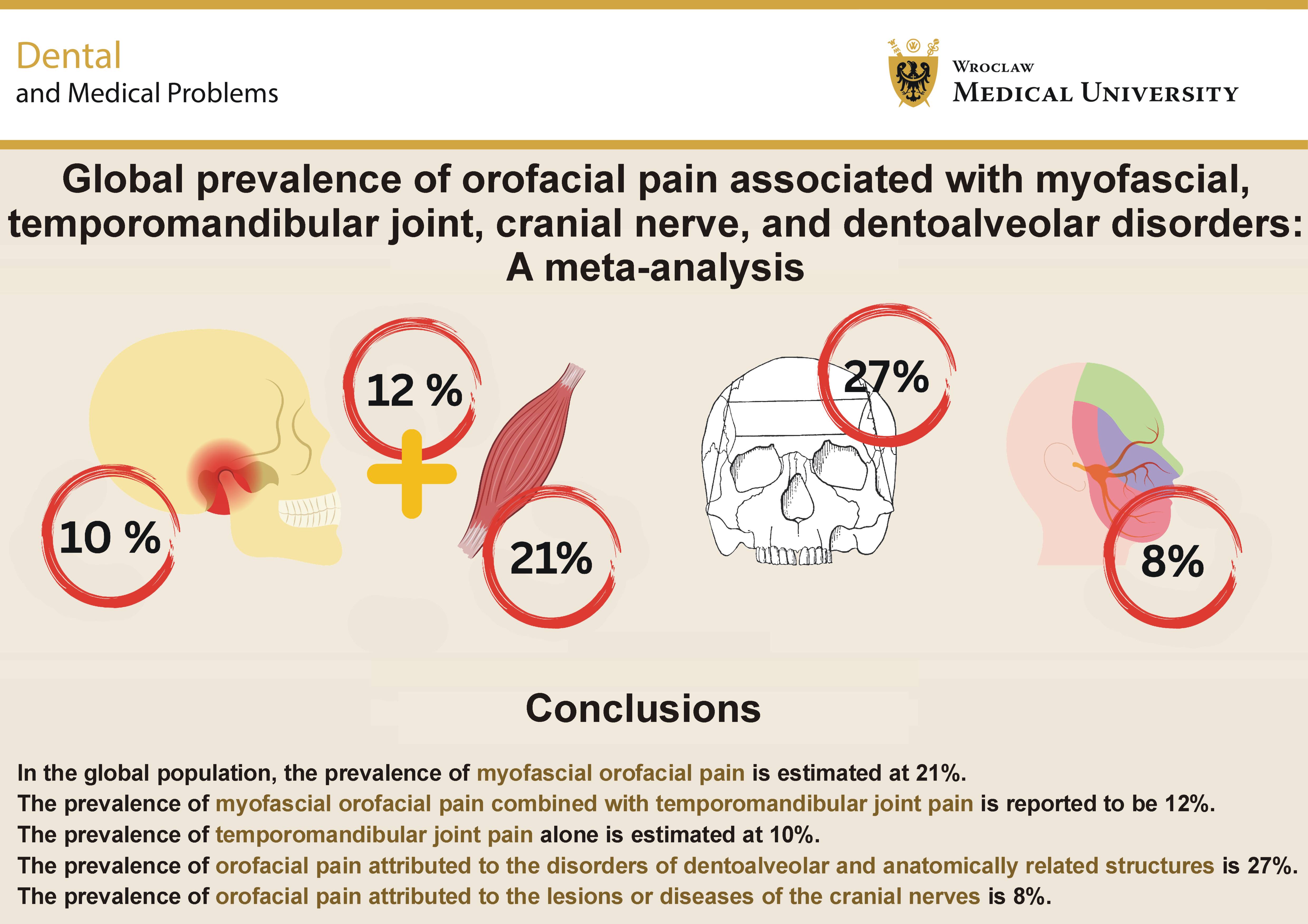
Conclusions. In the global population, the prevalence of myofascial orofacial pain is estimated at 21%. The prevalence of myofascial orofacial pain combined with TMJ pain is reported to be 12%. The prevalence of TMJ pain alone is estimated at 10%. The prevalence of orofacial pain attributed to the disorders of dentoalveolar and anatomically related structures is 27%. The prevalence of orofacial pain attributed to the lesions or diseases of the cranial nerves is 8%. The findings of this study should be interpreted with caution due to the substantial methodological heterogeneity observed across the included studies. This variability underscores the importance of establishing standardized criteria and reporting guidelines for orofacial pain in scientific research.
Keywords: epidemiology, myofascial pain, temporomandibular disorders, orofacial pain, cranial nerve disorders
Introduction
Orofacial pain, particularly when chronic and unrelated to dental pathology, represents a significant diagnostic and therapeutic challenge in clinical practice.1, 2, 3 The literature emphasizes that orofacial pain may have a multifactorial etiology.4, 5 It is not uncommon for several pathogenic mechanisms to overlap, complicating both diagnosis and effective treatment. Due to the dense innervation and complex anatomy of the craniofacial region, pain perception in this area is characterized by high intensity and has a considerable impact on patient functioning.4, 6, 7 Chronic orofacial pain is associated with a significant deterioration in quality of life (QoL), reduced occupational and social activity, and an increased risk of depressive and anxiety disorders.8, 9, 10 Persistent pain symptoms may also lead to central sensitization, thereby perpetuating the pathological process. The diagnostic management of orofacial pain requires an interdisciplinary approach, encompassing dental, neurological and otolaryngological examinations, as well as, when indicated, psychological or psychiatric consultation. The early identification of the source of symptoms is crucial, as delayed diagnosis increases the risk of acute pain transitioning into a chronic condition.11, 12, 13
To address these challenges, the International Classification of Orofacial Pain (ICOP) was developed. The ICOP, published in 2020, is the first comprehensive, international classification of orofacial pain.14 It was created through collaboration among dentists, temporomandibular disorders (TMD) specialists, neurologists, psychologists, and members of organizations such as the International Headache Society (IHS), the International Association for the Study of Pain (IASP), the American Academy of Orofacial Pain (AAOP), the International Network for Orofacial Pain and Related Disorders Methodology (INfORM), and other leading institutions.15 The aim was to establish a common language for researchers and clinicians, enabling the precise definitions and classifications of pain disorders involving the face and the stomatognathic system.14, 15, 16
The ICOP employs a hierarchical structure modeled after the International Classification of Headache Disorders, 3rd edition (ICHD-3),17 facilitating logical organization and ensuring the ease of reference. The classification identifies major categories, including: orofacial pain attributed to the disorders of dentoalveolar and anatomically related structures; myofascial orofacial pain; TMJ pain; orofacial pain attributed to the lesions or diseases of the cranial nerves; orofacial pain resembling the presentations of primary headaches; and idiopathic orofacial pain.14 For each entity, specific diagnostic criteria are provided based on the available scientific evidence.
For example, in the context of orofacial pain, it should be noted that TMD affect up to 34% of the population.18 The most common form of TMD for which patients seek specialist care is pain.19, 20, 21 Projections for the coming decades suggest that the prevalence of TMD could increase from 34% to as high as 44% by 2050.22 This underscores the public health importance of addressing orofacial pain.
Given the rising prevalence of painful conditions within the oral and facial regions, there is an urgent need for further research into the various types of orofacial pain.14, 22 Expanding the knowledge of pathophysiological mechanisms and identifying risk factors that predispose individuals to chronic pain are essential. Equally important are studies evaluating the effectiveness of diverse therapeutic strategies, including both pharmacological and non-pharmacological approaches. Advancing such research may substantially improve patients’ QoL and healthcare effectiveness in this domain.
Although numerous meta-analyses and systematic reviews have examined various aspects of orofacial pain, most have concentrated on selected subtypes, such as TMD, neuropathic pain or headache-related facial pain, rather than providing a comprehensive overview based on the ICOP framework. As a result, the overall epidemiological profile of orofacial pain, organized according to the ICOP taxonomy, remains only partially characterized. Clarifying this issue may contribute to a more consistent understanding of orofacial pain patterns, and support future diagnostic and therapeutic advances.
Accordingly, the present study was designed and conducted. The aim of this study was to determine the global prevalence of orofacial pain of different origin in accordance with the classification defined in ICOP.
Methods
The present methodology was preregistered on the Open Science Framework (OSF) platform on February 19, 2025 (registration link: https://osf.io/c8x2d). Supplementary materials related to the project were also deposited in the OSF repository (link: https://osf.io/k7eds). Registration in OSF is an accepted and recognized practice, consistent with various literature sources.22, 23, 24, 25
Due to differences in subject matter and analytical methods, the collected data were divided into 2 separate publications. This division enabled a more detailed discussion of the results, ensured the consistency of interpretation, and prevented the excessive abbreviation of essential content. A comprehensive description of the database is provided in the supplementary materials.
This paper focuses on the global prevalence of disorders classified according to ICOP.14 Data on the overall prevalence of oral and facial pain, including continental, age- and sex-specific analyses, are presented in the accompanying publication.
The systematic literature review was conducted in accordance with the PRISMA (Preferred Reporting Items for Systematic reviews and Meta-Analyses) 2020 guidelines,26 using the MEDLINE and Scopus databases. The search strategy combined terms such as “orofacial pain”, “facial pain” and “oral pain” with “prevalence”, “epidemiology” and “population”. An additional searche of gray literature was conducted on Google, supplemented by snowballing, following Wohlin’s recommendations.27
A 20-year publication window (January 1, 2004 – December 31, 2024) was applied.4, 27 Detailed inclusion and exclusion criteria are summarized in Table 1. Only original studies published in English that confirmed the presence of pain among participants were eligible for analysis.
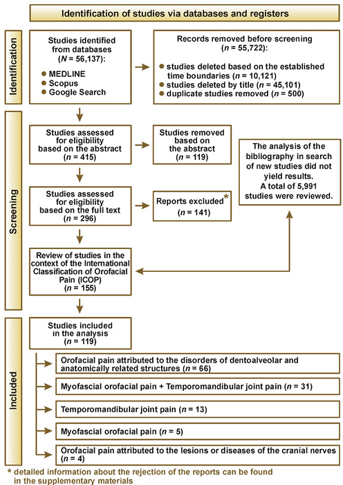
Study selection was performed independently by 2 reviewers (A.G. and M.W.), with disagreement resolved by the first author (G.Z.). Screening was conducted sequentially at the title, abstract and full-text levels. The PRISMA flow diagram illustrates the selection process (Figure 1), and detailed exclusion reasons are available in the supplementary materials.
Statistical analysis
The approach to statistical analysis was derived from previously published studies.18, 22, 28 To investigate the prevalence of orofacial pain, we conducted a meta-analysis of proportions.29 In this framework, each study contributed a proportion, defined as the number of individuals reporting pain divided by the study’s total sample size. To deal with the statistical challenges associated with proportions, we applied a logit transformation, which helps stabilize the variance and ensures that the pooled values, once back-transformed, remain within the [0, 1] range.30 For the main analysis, we relied on a generalized linear mixed model (GLMM) with a logistic link.31 This approach was chosen, as it can handle both within- and between-study variability without requiring artificial continuity corrections, making it well suited for the present dataset.32 Between-study variance (τ2) was estimated using maximum likelihood (ML).33 To compute confidence intervals (CI) for study-specific proportions, we used the Wilson score method.34 To check robustness, we repeated the analysis using a random-effects model based on the inverse variance method35 with the restricted maximum likelihood (REML) estimation.36 This alternative specification produced similar prevalence estimates and consistent measures of heterogeneity. We also conducted subgroup analyses to explore prevalence by pain type, calculating pooled proportions separately for each subgroup.33 Heterogeneity was assessed using τ2 and the I2 statistic,37 and significance was evaluated with the Wald test and the likelihood ratio test (LRT). Notably, the subgroup models incorporated group-specific variances rather than assuming a common effect.33 To aid interpretation, subgroup estimates were back-transformed to the original proportion scale.30 Forest plots were used to present overall prevalence, subgroup comparisons and heterogeneity statistics with 95% CIs. The influence of individual studies was further examined with diagnostic tools such as standardized residuals, DFFITS (difference in fits), Cook’s distance, covariance ratios, leave-one-out heterogeneity checks, and DFBETAS (difference in betas).38 Finally, we assessed publication bias using funnel plots, in which study-specific prevalence was plotted against precision.39 Funnel plot asymmetry was evaluated both visually and statistically. In addition, we applied the regression-based method proposed by Thompson and Sharp,40 which is particularly appropriate for meta-analyses of proportions.
Analyses were conducted using the R statistical language (v. 4.3.3; R Core Team, 2024; https://www.r-project.org) on Windows 11 Pro 64-bit (build 26100), using the packages meta,41 dmetar,42 report,43 and dplyr.44
Results
Due to the number of analyses, their full versions together with detailed descriptions and funnel plots, the results for the influential studies are provided in the supplementary materials.
The current analysis synthesizes data from 119 observational studies published between 2004 and 2024, with excluding 2 influential studies classified as “orofacial pain attributed to the lesions or diseases of the cranial nerves”.45, 46 The collected data were classified by continent and by disorder according to ICOP into the following categories:
– orofacial pain attributed to the disorders of dentoalveolar and anatomically related structures;
– myofascial orofacial pain;
– TMJ pain;
– orofacial pain attributed to the lesion or diseases of the cranial nerves;
– orofacial pains resembling the presentations of primary headaches; and
– idiopathic orofacial pain.14
However, after analyzing the collected publications, it was not possible to divide them according to the abovementioned categorization. The gathered publications were divided as described in Table 2.
Pooled effect estimation of myofascial orofacial pain
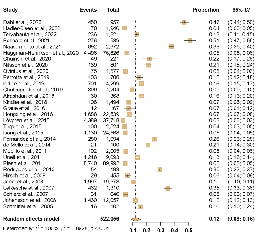
A meta-analysis encompassing 5 studies, comprising 3,395 observations and 799 reported events, determined the pooled prevalence of myofascial orofacial pain to be 20.60% (95% CI: 9.71–38.50), as presented in the forest plot (Figure 2). This estimate indicates a notable health burden associated with this specific type of orofacial pain across the included studies.
Substantial heterogeneity was observed across the studies, with I2 = 99.3% (95% CI: 99.0–99.5) and a τ2 value indicating considerable between-study variability, further supported by a Higgins H statistic of 11.79 (95% CI: 10.07–13.80). The statistical tests for heterogeneity confirmed this variability as highly significant, with a Wald test Q-value of 556.20 (df (degrees of freedom) = 4; p < 0.001) and LRT p < 0.001.
Pooled effect estimation of myofascial orofacial pain combined with temporomandibular joint pain
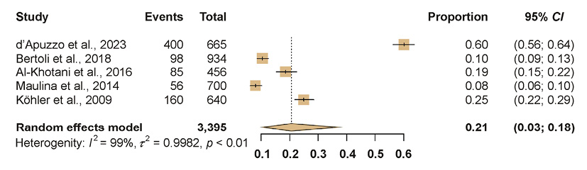
A meta-analysis encompassing 31 studies, comprising 522,056 observations and 29,823 reported events, determined the pooled prevalence of myofascial orofacial pain combined with TMJ pain to be 11.95% (95% CI: 8.85–15.96), as presented in the forest plot (Figure 3). This estimate highlights a significant health burden associated with this combined pain condition across the included studies.
Substantial heterogeneity was observed across the studies, with I2 = 99.8% and a τ2 value indicating considerable between-study variability, further supported by a Higgins H statistic of 21.50. The statistical tests for heterogeneity confirmed this variability as highly significant, with a Wald test Q-value of 13,862.48 (df = 30; p < 0.001) and LRT p < 0.001.
Pooled effect estimation of temporomandibular joint pain
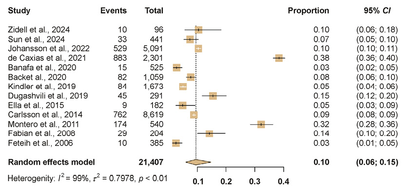
A meta-analysis encompassing 13 studies, comprising 21,407 observations and 2,665 reported events, determined the pooled prevalence of TMJ pain to be 9.51% (95% CI: 6.01–14.73), as presented in the forest plot (Figure 4). This estimate indicates a notable health burden associated with TMJ pain across the included studies.
Substantial heterogeneity was observed across the studies, with I2 = 99.2% (95% CI: 99.1–99.4) and a τ2 value indicating considerable between-study variability, further supported by a Higgins H statistic of 11.49 (95% CI: 10.47–12.61). The statistical tests for heterogeneity confirmed this variability as highly significant, with a Wald test Q-value of 1,584.78 (df = 12; p < 0.001) and LRT p < 0.001.
Pooled effect estimation of orofacial pain attributed to the disorders of dentoalveolar and anatomically related structures
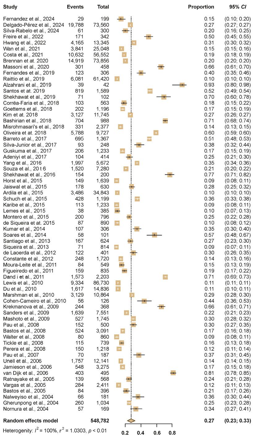
A meta-analysis encompassing 66 studies, comprising 548,782 observations and 104,567 reported events, determined the pooled prevalence of orofacial pain attributed to the disorders of dentoalveolar and anatomically related structures to be 27.46% (95% CI: 22.83–32.63), as presented in the forest plot (Figure 5). This estimate indicates a substantial health burden associated with this specific category of orofacial pain across the included studies.
Substantial heterogeneity was observed across the studies, with I2 = 99.8% and a τ2 value indicating considerable between-study variability, further supported by a Higgins H statistic of 22.21. The statistical tests for heterogeneity confirmed this variability as highly significant, with a Wald test Q-value of 32,071.85 (df = 65; p < 0.001) and LRT p < 0.001.
Pooled effect estimation of orofacial pain attributed to the lesions or diseases of the cranial nerves
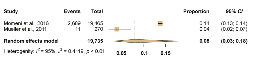
A meta-analysis encompassing 2 studies, comprising 19,735 observations and 2,700 reported events, determined the pooled prevalence of orofacial pain attributed to the lesions or diseases of the cranial nerves to be 7.98% (95% CI: 3.28–18.18), as presented in the forest plot (Figure 6). This estimate demonstrates a notable health burden associated with this specific type of orofacial pain across the included studies.
Substantial heterogeneity was observed across the studies, with I2 = 94.6% (95% CI: 83.4–98.2) and a τ2 value indicating considerable between-study variability, further supported by a Higgins H statistic of 4.30 (95% CI: 2.45–7.55). The statistical tests for heterogeneity confirmed this variability as highly significant, with a Wald test Q-value of 18.53 (df = 1; p < 0.001) and LRT p < 0.001.
To investigate potential publication bias and the effects of small studies, a linear regression test was performed using the method outlined by Thompson and Sharp.40 This procedure evaluated the extent of asymmetry in the funnel plots for the included studies. A p-value above 0.05 indicated that there was not enough evidence to reject the null hypothesis of symmetry, implying that the data did not exhibit notable asymmetry and, therefore, showed no signs of publication bias (Table 3).
Discussion
The aim of this study was to determine the global prevalence of pain according to the classification set forth in the ICOP.
The first set of results concerns myofascial pain in the orofacial region, with a prevalence estimate of 20.60%. Despite the relatively small number of studies (n = 5), this proportion suggests a considerable burden of this condition. The wide CI reflects high heterogeneity. Previous estimates suggest myofascial pain affects 30–93% of patients, which aligns with our findings.47 In light of these findings, the estimate obtained in the present analysis – focused on the orofacial region as a particular anatomical area – falls within the expected range and aligns with prior knowledge. Differences between these results and earlier, more general prevalence estimates should be considered acceptable, as they may reflect both the unique characteristics of the anatomical region under investigation and methodological variability across studies.
The next category concerns myofascial orofacial pain combined with TMJ pain. The prevalence of 11.95% appears lower than that of isolated myofascial pain; however, it should be noted that these data are derived from a substantially larger evidence base (n = 31, >500,000 observations). The large sample size improves reliability despite high heterogeneity.
Another category, TMJ pain alone, was estimated at 9.51%. This is a moderate value, but it pertains to a specific pain subtype that often coexists with other orofacial pain conditions. As in other categories, the results are affected by very high heterogeneity, which limits the ability to draw definitive conclusions about population-level burden, but nonetheless provides a broad outline of the global epidemiological situation.18, 22
The epidemiology of myofascial orofacial pain combined with TMJ pain and TMJ pain alone indicates that both conditions occur in fewer than 12% of the global population. Considering the most recent meta-analysis from 2024 on TMD prevalence, estimates suggest a rate of 34%, though without differentiation between painful and non-painful forms.18 LeResche noted in her review that the painful form of TMD affected 10% of adults over the age of 18.48 Similarly, Yap et al., in their meta-analysis, reported that TMD pain occured in 8% of the global population,49 which corroborates the findings of the present study.
Even higher prevalence values are observed for orofacial pain attributed to the disorders of dentoalveolar and anatomically related structures, estimated at 27.46%. This finding underscores the dominant role of dental pathologies in the etiology of facial pain. The large number of studies (n = 66) and over half a million observations make this estimate appear robust at the population level. Orofacial pain attributed to the disorders of dentoalveolar and related structures represents the most prevalent category in this study, given its direct association with highly common conditions such as dental caries, pulpitis and periodontal disease.50, 51, 52 This type of pain is the leading cause of dental visits, as it serves as a clear warning signal of tissue damage.1, 53 Moreover, the high density of anatomical structures within the craniofacial region often results in pain radiation, which further increases the number of reported complaints.1, 53, 54 Unlike rarer causes, such as neuropathic or functional pain, odontogenic pain is more readily recognized and more likely to prompt patients to seek professional care.55, 56, 57
Also of interest are the data on orofacial pain attributed to the lesions or diseases of the cranial nerves. In the pooled analysis, the prevalence of these conditions was estimated to be 7.98%. While noteworthy, this result should be interpreted with caution due to the very limited number of studies (n = 2) and the wide CI. It is useful to compare these findings with population-based studies on cranial neuralgias. Haller et al. reported that the incidence of trigeminal neuralgia (cranial nerve V) ranges from 4 to 20 cases per 100,000 per year, corresponding to 0.004–0.020% annually.58 Schwaiger et al. found cranial neuralgias in 1.6% of individuals aged 55–94 years, suggesting a higher prevalence among older populations.59 Wettervik et al. estimated the annual incidence of trigeminal neuralgia at 5.5 per 100,000 person-years, corresponding to 0.0055% annually.60 Taken together, these findings indicate that the high prevalence of cranial nerve-related pain observed in the meta-analysis is not fully consistent with epidemiological data on trigeminal neuralgia. Such discrepancies may arise from differences in the definition of study entities (broader neuropathic pain vs. narrowly defined trigeminal neuralgia), study methodologies and population characteristics. This warrants further research.
The most consistent finding across all categories is the extremely high level of heterogeneity, which poses both methodological and practical challenges. This highlights significant differences between studies in design, diagnostic criteria, population characteristics, and potential cultural or regional influences, as demonstrated in other analyses.18, 28, 61 Uniform ICOP standards introduced in 2020 may improve future consistency.14 For TMD, the diagnostic criteria (DC/TMD) were developed as early as in 2014,62 and have since been translated and validated in multiple languages.63 The consistent application of standardized frameworks is essential for reliable meta-analyses. The present review includes studies conducted between 2004 and 2024, meaning that most of the earlier analyses preceded the introduction of current diagnostic criteria, which may further explain the observed heterogeneity.
Heterogeneity arises from differences in diagnostic criteria, population characteristics, and study methodologies.64, 65 The ICOP standardization improves interdisciplinary communication and diagnostic accuracy. Future research should address regional data gaps, explore the determinants of pain heterogeneity, and assess the clinical value of ICOP-based diagnostic pathway.56, 57, 64, 66
Conclusions
In the global population, the prevalence of myofascial orofacial pain is estimated at 21%.
The prevalence of myofascial orofacial pain combined with TMJ pain is reported to be 12%.
The prevalence of TMJ pain alone is estimated at 10%.
The prevalence of orofacial pain attributed to the disorders of dentoalveolar and anatomically related structures is 27%.
The prevalence of orofacial pain attributed to the lesions or diseases of the cranial nerves is 8%.
The findings of this study should be interpreted with caution due to the substantial methodological heterogeneity observed across the included studies. This variability underscores the importance of establishing standardized criteria and reporting guidelines for orofacial pain in scientific research.
Ethics approval and consent to participate
Not applicable.
Data availability
The data related to this article, including additional materials, are available in the Open Science Framework database at the following link: https://osf.io/k7eds/files. The script used in the analysis is available from the corresponding author upon reasonable request.
Consent for publication
Not applicable.
Use of AI and AI-assisted technologies
Not applicable.