Abstract
Background. The susceptibility of glass-ceramics to surface defects can weaken their mechanical performance.
Objectives. The aim of the present study was to investigate the effect of cementation on the load-to-failure of 2 glass-ceramic materials in the presence and absence of an induced surface defect.
Material and methods. Bars of lithium disilicate – LD (Initial LiSi Block; GC Dental) and feldspathic – F (VITA Mark II; VITA Zahnfabrik H. Rauter) ceramics, as well as a dentin analog, were prepared for cementation. The ceramic specimens were assigned to 5 groups: untreated (no cementation); indented (I); cemented (C); indented and cemented with defect filling (CI); and indented and cemented without defect filling (CIN). Flexural strength was measured for the non-cemented specimens, whereas the load-to-failure was evaluated for all cemented groups, using the ball-in-hole test on bar specimens (1 × 1 × 12 mm). Finite element analysis (FEA) was performed for the groups without induced defects. Data was analyzed using the one-way analysis of variance (ANOVA) and Tukey’s post hoc test (α = 0.05).
Results. One-way ANOVA revealed significant differences among the groups for both materials (p < 0.001). For LD, the load-to-failure values were: LDC (16.7 ±2.9 N)A > LD (14.6 ±2.6 N)B = LDCI (13.9 ±3.0 N)B > LDCIN (10.5 ±1.7 N)C > LDI (6.9 ±1.4 N)D. For F, the values were: FC (13.7 ±3.7 N)A = FCIN (13.1 ±3.3 N)A = FCI (11.8 ±3.3 N)A > F (8.1 ±1.3 N)B > FI (5.1 ±0.4 N)C. Indentation significantly reduced the flexural strength of the non-cemented ceramics (p < 0.001). Finite element analysis showed that cementation reduced the maximum principal tensile stress by 44% for LD and by 62% for F.
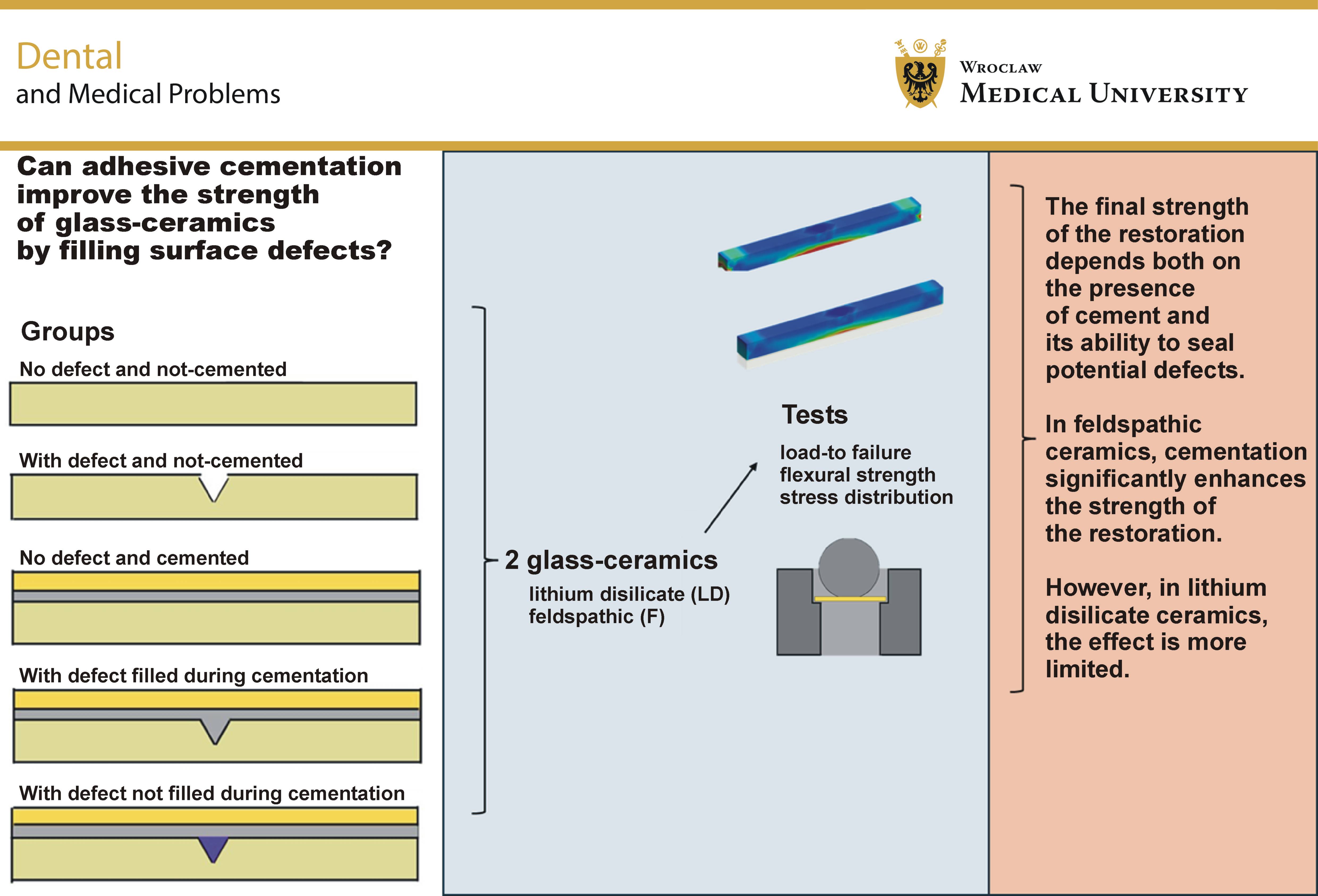
Conclusions. Filling surface defects with cement increased the load-to-failure of LD, but did not significantly affect F. Overall, the mechanical performance of both glass-ceramics was influenced by the cementation process, whereas the effect of defect filling was material dependent.
Keywords: cementation, flexural strength, glass-ceramics
Introduction
Ceramic materials, particularly monolithic restorations, were developed to address the limitations associated with porcelain-fused-to-metal (PFM) prostheses, including esthetic concerns, potential biocompatibility issues, and technical complications such as porcelain chipping.1, 2 Over the past decades, the use of dental ceramics has increased substantially owing to their favorable properties, including excellent esthetics, biocompatibility, chemical durability, low thermal conductivity, high strength, wear resistance, and translucency.1, 3 These materials are widely used for the fabrication of single crowns, veneers, inlays, onlays, and other indirect restorations because of their ability to be predictably bonded to the tooth structure and other glass-containing materials.4, 5
Lithium disilicate (LD) is a glass-ceramic in which the base glass consists of varying SiO2/Li2O ratios. Most commercially available LD materials differ considerably in composition and should therefore be regarded as lithium-based glass-ceramics.6, 7 These ceramics exhibit flexural strength values ranging from 177 to 510 MPa.8 Clinical studies have reported success rates of 88% at 5 years and 77% at 10 years for LD inlays and onlays.9 Other long-term studies have demonstrated a survival rate of 81.9% for single crowns after 15 years10 and 48.6% for three-unit fixed dental prostheses.11 Fracture is the primary cause of failure in LD inlays and onlays, accounting for 61% of failures after a 10-year follow-up.9
Feldspathic ceramic (F) is another commonly used glass-ceramic composed primarily of feldspar, quartz, silica, and alumina as the reinforcing phase.12 In comparison with LD, its mechanical properties are less favorable for the oral environment because of its relatively low flexural strength (60–70 MPa), which limits its clinical indications.13 Feldspathic onlays have a 7-year survival rate of 60.5% and annual failure rates of 6.3–6.7%, with fracture accounting for 16% of failures.14 Feldspathic veneers demonstrate a 5-year survival rate of 92% and a 10-year survival rate of 64%, with fracture (11%) and marginal defects (20%) being the most common reasons for failure.15
Both LD and F can be adhesively cemented using resin cement, which significantly reduces the risk of fracture and increases the failure load of ceramic restorations.16 To enhance bond strength, various kinds of surface treatment are employed, including hydrofluoric acid etching, silica coating, and the application of ceramic primers.3, 11, 17, 18 Although these procedures improve adhesion, they may also introduce surface defects and microcracks that significantly reduce the flexural strength of LD.19, 20 Additional defects may be introduced during the milling process or clinical adjustments,21, 22 resulting in increased surface roughness, greater biofilm accumulation,23, 24 and reduced fatigue strength and mechanical reliability.25 Clinical adjustments performed with diamond burs during grinding can also produce superficial surface defects. These defects may be filled by glazing the ceramic surface, thereby preserving the flexural strength of the restorative material through sealing surface flaws.26
The cementation of dental ceramics has been shown to increase their strength.27 However, it remains unclear whether cementation can protect flexural strength against the incorporated defects and whether this effect depends on defect filling. Considering the adhesive interface, these findings suggest that the resin cement used during cementation may penetrate and fill surface defects, thereby protecting the restoration against failure. Lithium disilicate and F are widely used glass-ceramics that can be adhesively bonded to tooth structures. Although these materials have different mechanical properties, they share similar failure mechanisms; therefore, investigating their mechanical behavior under stress and cemented conditions may contribute to reducing clinical failure rates.
Therefore, this study investigated the effect of cementation on the load-to-failure of 2 different glass-ceramics with compromised structural integrity due to an induced defect in comparison with structurally sound specimens. The null hypothesis was that cementation, with or without defect filling, would not affect the load-to-failure of lithium disilicate and feldspathic ceramics.
Material and methods
Specimen preparation
To simulate indirect restorations, ceramic blocks of LD (Initial LiSi Block; GC Dental, Lucerne, Switzerland) and F (VITA Mark II; VITA Zahnfabrik H. Rauter, Bad Säckingen, Germany) were sectioned into bars measuring 1 × 1 × 12 mm, using a water-cooled diamond-coated saw (IsoMet 1000; Buehler, Lake Bluff, USA). The irregularly shaped bars were polished using EcoMet™ polishing sheets (Buehler) with grit sizes ranging from P320 to P2400 until the specified dimensions were achieved (±15%). The tooth substrate was simulated using dentin analog sheets (G10 plates; Carbotec GmbH & Co. KG, Aachen, Germany), which were sectioned into specimens measuring 1 × 12 mm with a standard thickness of 0.5 mm. Surface irregularities were removed following the same polishing procedure described above.
To simulate the introduction of a structural defect on the bonding surface, an indentation was created at the midpoint of the tensile surface of each ceramic bar.26 A controlled defect was introduced in 3 of the 5 subgroups for each ceramic material. The defects were produced using a Vickers indenter (HM-124 hardness-testing machine; Mitutoyo Corp., Kanagawa, Japan), applied to the fracture surface with a load of 19.6 N for 15 s. The indentation parameters were established through pilot testing and the optimization of surface morphology through high-grit polishing. In one indented group for each material, the defect was intentionally filled with wax to prevent resin cement penetration into the defect (subgroup CIN).
The distribution of groups according to the restorative material, the presence of defect, and the cementation condition is presented in Table 1. Table 2 summarizes the materials used and their compositions.
The specimens assigned to the cemented groups received surface treatment on the bonding surface prior to cementation. The treatment consisted of etching with 9% hydrofluoric acid (Ultradent® Porcelain Etch; Ultradent Products Inc., South Jordan, USA) for 20 s for LD and 60 s for F, followed by the application of a ceramic primer (Clearfil™ Ceramic Primer; Kuraray Noritake, Tokyo, Japan) according to the manufacturer’s instructions. The dentin analogs were also etched with 9% hydrofluoric acid for 60 s, followed by the application of a bonding system (Tooth Primer; Kuraray Noritake). Subsequently, cementation was performed for 6 groups (n = 90; 15 specimens per group), using a dual-curing resin cement (Panavia™ V5; Kuraray Noritake) according to the manufacturer’s instructions. The 4 non-cemented groups (n = 60; 15 specimens per group) did not receive any surface treatment. The final specimens, as illustrated in Table 1, were stored in water at 37°C for 1 week prior to testing.
Load-to-failure test and flexural strength analysis
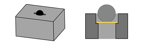
The load-to-failure test was performed using the ball-in-hole test (Figure 1) with a universal testing machine (Instron 6022; Instron Ltd., High Wycombe, UK) on 15 specimens (n = 15) from each experimental group. The load was applied at a crosshead speed of 0.5 mm/min until failure. The load-to-failure [N] was recorded for all specimens. For the non-layered groups (LD, LDI, F, and FI), flexural strength [MPa] was calculated according to ISO 6872:2015, using the following formula:
where:
σ – flexural strength [MPa];
F – maximum load applied load [N];
l – distance between the 2 points of support [mm];
b – width of the specimen [mm]; and
d – thickness of the specimen [mm].
To evaluate the maximum principal stress, a three-dimensional (3D) finite element analysis (FEA) was conducted, replicating the dimensions of the in vitro specimen while maintaining a standardized cement layer thickness of 100 μm. The geometries were created using computer-aided design (CAD) software (Rhinoceros, v. 5.0 SR8; McNeel North America, Seattle, USA) and subsequently exported in the STEP format to the analysis software (ANSYS, v. 17.2; ANSYS Inc., Canonsburg, USA). A 10% convergence criterion was applied, and the mesh was generated using tetrahedral elements. The mechanical properties of each material were defined assuming isotropic behavior. The maximum principal tensile stress at the interface was analyzed for the defect-free groups, using the elastic modulus and Poisson’s ratio (ν) of the materials (LD: 95 GPa, ν = 0.25; F: 48.7 GPa, ν = 0.23),27 under an applied load of 10 N.
Statistical analysis
Statistical analysis of the data obtained from the three-point bending test was performed using the one-way analysis of variance (ANOVA) with a 95% confidence level, followed by Tukey’s post hoc test. The load-to-failure data was analyzed to identify differences among the groups for each ceramic separately. The flexural strength values were compared between the non-cemented groups of each ceramic to validate the experimental method and support the discussion in relation to the available literature. Stress distribution was qualitatively analyzed to describe the maximum tensile strength in the defect-free groups.
Results
One-way ANOVA revealed significant differences in the load-to-failure among the groups for both LD (p < 0.001; F = 34.3) and F (p < 0.001; F = 25.8) (Table 3). As LD and F are distinct materials, this study was not designed to compare them directly. Instead, the experimental conditions were evaluated independently for each ceramic material.
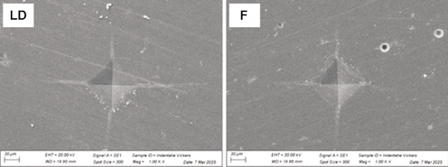
The presence of a single defect reduced the load-to-failure. Representative images of the indentations are shown in Figure 2. Based on this reference, the cemented LD group without defects (LDC) exhibited the best mechanical performance (Table 3). Therefore, the cementation procedure increased the load-to-failure values of LD under all evaluated conditions, including specimens without defects and those with defects (whether filled with cement or not). Moreover, filling the defect with cement resulted in higher load-to-failure values than leaving the defect unfilled.
For F, the presence of a defect reduced the flexural strength by 41%. Cementation improved its mechanical performance regardless of the presence of a defect, or whether the defect was filled with cement or not (Table 3).
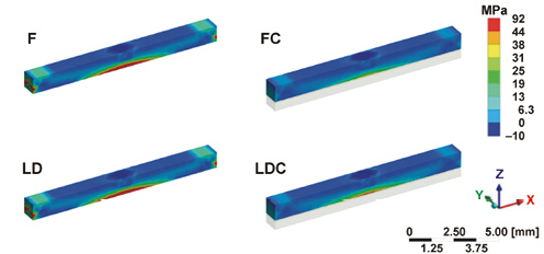
Finite element analysis revealed that the cemented groups exhibited lower stress concentrations at the interface than the non-cemented groups (LDC: 51 MPa and FC: 35 MPa vs. LD: 91 MPa and F: 92 MPa, respectively). These findings suggest that cementation reduced the maximum principal tensile stress by 44% for LD and by 62% for F. Figure 3 presents the stress distribution color maps for the LD, LDC, F, and FC groups.
Discussion
This study investigated the influence of cementation on the load-to-failure of 2 types of glass-ceramics by comparing defect-free specimens with specimens containing induced structural defects. The aim was to evaluate the effect of cementation on the load-to-failure when the defect was either filled with cement or left unfilled.
The defect simulated in this study has been used in previous investigations. It was created by a Vickers indentation positioned at the center of the tensile surface of the bar. To determine whether the indentation produced a significant effect on the flexural strength of the materials, both ceramics were evaluated with and without the induced defect. The results showed that the indentation weakened the specimens and provided a reproducible defect model.26 Consistent with the present findings and previous studies, the reduction in flexural strength demonstrates the susceptibility of glass-ceramics to structural defects.3, 8, 13, 26
According to the results, the cementation procedure increased the load-to-failure of the intact ceramic bars. This improvement in mechanical strength following cementation is consistent with the current understanding of the adhesive properties of ceramic materials.28
Lithium disilicate with an induced defect exhibited compromised strength. However, after cementation, the reduction in the load-to-failure was partially compensated when the defect was completely filled with resin cement. In this condition, the increase in the load-to-failure can be attributed to the adhesive procedure.29 Although cementation counteracted part of the reduction in the load-to-failure observed in the LD specimens with defects, their strength remained lower than that of the cemented specimens without defects. The presence of elongated crystals in LD contributes to the inhibition of microcrack propagation12 and results in high fracture strength. Therefore, after cementation, the indentation remains the weakest region within the specimen structure. Although the specimen is reinforced by resin cement within the indentation, fracture initiation and propagation still occur from this location.
For F, the reduction in performance caused by the indentation was completely counteracted after cementation, regardless of whether the defect was filled with cement or not. After cementation, all groups exhibited higher load-to-failure values as compared to the non-cemented specimens, which can be attributed to the positive effect of adhesive cementation.29 Poor mechanical properties and the presence of porosities in the microstructure of F suggest that, after cementation, the indentation is no longer the weakest region within the specimen structure.12 Instead, fracture initiation may occur from another mechanically vulnerable area within the material. Since no difference in the load-to-failure was observed between the F specimens cemented with or without a defect, the null hypothesis can be partially accepted.
The increase in strength resulting from the cementation procedure has been demonstrated in the literature,29 as has the influence of surface topography on the flexural strength of glass-ceramics. These findings corroborate previous studies showing that the surface microstructure plays a critical role in the fatigue strength of cemented glass-ceramics.30 However, no previous research has investigated whether the presence of cement within a defect affects the strength of glass-ceramics.
Clinicians should recognize that glass-ceramics differ in composition and microstructure, which influence their crystal structure, fracture toughness, and mechanical behavior under stress, ultimately affecting clinical recommendations.8 This study demonstrates that filling a defect with cement plays a critical role in increasing the fracture strength of LD, but not of F. The strength of LD specimens depends not only on the presence of cement, but also on the effective filling of surface defects. These findings highlight the importance of precise cementation protocols, as both factors influence the overall mechanical performance. Since cementation cannot fully compensate for the loss of strength caused by surface defects, clinicians should minimize their occurrence whenever possible.
Limitations
As a limitation of this study, the non-cemented groups did not receive surface treatment with hydrofluoric acid etching, whereas the cemented groups underwent hydrofluoric acid etching. This difference should be considered when interpreting the results. Flexural strength is not the only mechanical factor determining the performance of dental ceramics, as fatigue strength may also play a crucial role in their mechanical behavior. Therefore, further studies are needed to investigate the effects of these conditions on other dental ceramics, as well as to evaluate the influence of multiple defects, defect size, and different types of cements on the mechanical properties of these materials.
Conclusions
In conclusion, the presence of defects significantly weakened specimens made of both LD and F, although cementation substantially improved their strength. Feldspathic ceramic was less sensitive to defects, further highlighting the beneficial effect of cementation on its mechanical performance. In contrast, the strengthening effect of cementation was more limited for LD. Therefore, clinicians and technicians should prioritize minimizing surface defects during adjustments, as cementation alone cannot fully compensate for the reduction in strength caused by these flaws.
Ethics approval and consent to participate
Data availability
The datasets supporting the findings of the current study are available from the corresponding author on reasonable request.
Consent for publication
Not applicable.
Use of AI and AI-assisted technologies
Artificial intelligence (Grammarly) was used to check the English language.