Abstract
Background. Surgical injury triggers a stereotypical cascade of responses known as the systemic inflammatory response (SIR). To the best of our knowledge, the SIR following palatal graft harvesting has not been previously investigated.
Objectives. The aim of the study was to examine the magnitude of the SIR following palatal graft harvesting in Wistar rats by assessing serum levels of tumor necrosis factor (TNF)-α, interleukin (IL)-6 and IL-18, peripheral white blood cell (WBC) counts (differential and absolute counts of lymphocytes, neutrophils, monocytes, eosinophils, and basophils); and anemia-related parameters (red blood cell (RBC) count, hemoglobin (Hgb), hematocrit (HCT), mean corpuscular volume (MCV), mean corpuscular hemoglobin (MCH), and mean corpuscular hemoglobin concentration (MCHC)).
Material and methods. A total of 48 male Wistar rats were included in the study. A 4.2-mm palatal wound was created using a biopsy punch in the middle of the palate. Blood samples were collected before surgery and at 24, 48, 72, and 96 h postoperatively. Serum levels of TNF-α, IL-6 and IL-18 were measured using enzyme-linked immunosorbent assay (ELISA), and complete blood counts were analyzed with an automated blood cell counter on the day of sacrifice.
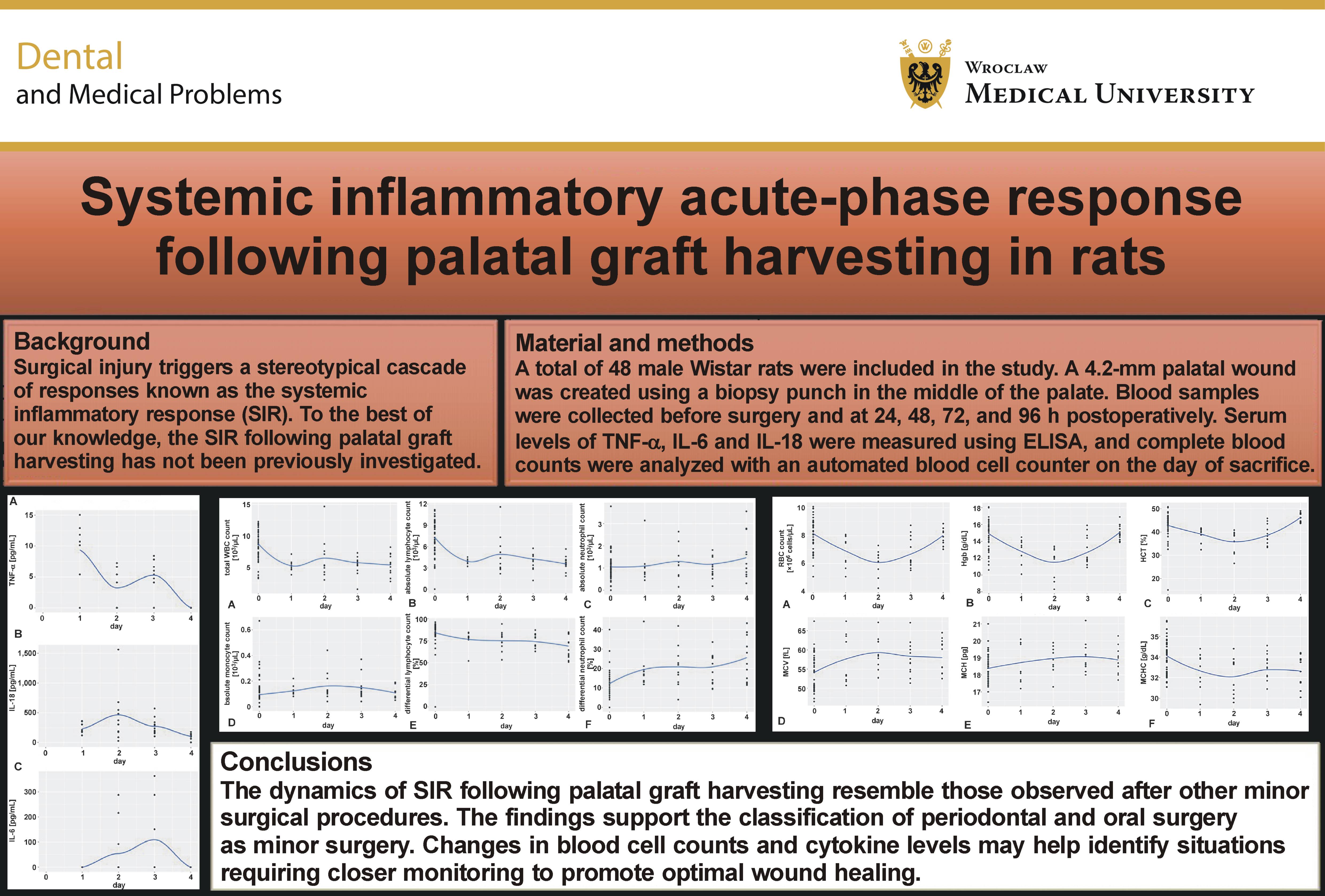
Results. TNF-α levels peaked on day 1, IL-18 on day 2, and IL-6 on day 3. All cytokine levels returned to baseline by day 4. A significant 40% decrease in WBC count was observed on day 1, primarily due to a 47% reduction in lymphocyte count despite a significant 50% increase in neutrophil count. Red blood cell count, Hgb and HCT decreased significantly by 25%, 23% and 23%, respectively, with the greatest reductions observed on days 1, 1 and 2. All values returned to baseline by day 4.
Conclusions. The dynamics of SIR following palatal graft harvesting resemble those observed after other minor surgical procedures. The findings support the classification of periodontal and oral surgery as minor surgery. Changes in blood cell counts and cytokine levels may help identify situations requiring closer monitoring to promote optimal wound healing.
Keywords: IL-6, TNF-α, IL-18, hemoglobin, systemic immune response
Introduction
Palatal tissue harvesting is routinely performed in oral, periodontal and systemic surgery. The most common drawback of this procedure is postoperative patient morbidity, although other complications may also occur. Therefore, elucidating the local and systemic factors, biological agents and genes involved in palatal wound healing is essential.1
Surgical injury triggers a well-defined cascade of systemic inflammatory responses.2 This response is characterized by increased production of pro-inflammatory cytokines, including tumor necrosis factor (TNF)-α, interleukin (IL)-18, and, in particular, IL-6.3 Together, these cytokines constitute a complex signaling system referred to as the systemic inflammatory response (SIR).4 In the oral cavity, changes in the levels of inflammatory mediators involved in the SIR are induced not only by surgical procedures but also by prosthetic rehabilitation and its chronic inflammatory effects on the peri-implant soft tissues.5, 6, 7, 8, 9, 10, 11 The systemic inflammatory response promotes skeletal muscle catabolism to provide energy and substrates for the liver, maintains fluid and cardiovascular homeostasis, and supports wound healing at the surgical site. The magnitude of the increase in these markers is considered to reflect the intensity of the SIR.2
The cellular hallmark of the SIR is the response of white blood cells (WBCs), including lymphocytes, neutrophils, monocytes, eosinophils, and basophils, each type having a distinct role in the immune response. The primary function of the immune system is to protect the host against pathogenic microorganisms and foreign substances.12 Following surgery, which represents a sterile injury, this response is reflected by acute changes in WBC counts despite the absence of microorganisms or foreign material. These changes are thought to be triggered by either physical or psychological stress. Accordingly, WBC parameters have been widely used as indicators of the physiological stress response.13
Postoperative acute-phase anemia has also been reported following surgery. Surgical anemia is usually subclinical and associated with iron deficiency, and it may persist for up to 8 weeks.14
A better understanding of the SIR associated with oral surgical procedures may improve postoperative patient management. Several strategies have been proposed. Refinement of surgical techniques, particularly the adoption of minimally invasive approaches, may reduce the inflammatory response. Laparoscopic surgery, for example, has been associated with faster postoperative recovery. Nutritional interventions, including glutamine, arginine and omega-3 fatty acid supplementation, may also improve outcomes. In addition, anabolic steroids and insulin infusions, with or without glucose supplementation, may also minimize muscle wasting. Maintenance of normothermia has also been suggested as a strategy to reduce the SIR.15
The rat is a well-established experimental model for investigating systemic responses to surgical injury.16 Metabolic studies have demonstrated that surgery induces skeletal muscle catabolism as part of a generalized adaptive response aimed at promoting survival and tissue repair. Because injury reduces activity and food intake, the body relies on its internal sources of energy (protein and fat) to enable wound healing and physiological maintenance.
The palate is the preferred donor site for harvesting autogenous grafts, and the development of strategies to minimize postsurgical morbidity remains of considerable clinical interest.17 Therefore, the present study aimed to evaluate the magnitude of the SIR following palatal graft harvesting in Wistar rats by assessing serum levels of TNF-α, IL-6 and IL-18, peripheral WBC counts and anemia-related parameters.
Material and methods
The study protocol was approved by the Ethics and Institutional Animal Care and Use Committee of Tel Aviv University, Israel (approval No. TAU – MD–IL-2402-109-3; February 1, 2024). All animals received humane care throughout the study. The experimental design complied with the Animal Research Reporting of In Vivo Experiments (ARRIVE) guidelines.18 A total of 48 male Wistar rats were randomly allocated to 4 equal groups (n = 12 per group). The groups were categorized according to the postoperative day on which the animals were euthanized (days 1–4).
General anesthesia was induced on the day of surgery by intraperitoneal injection of 10% ketamine (90 mg/kg; Kepro, Deventer, the Netherlands) and 2% xylazine (10 mg/kg; Medical Market, Tel Izhak, Israel).
Immediately before surgery (day 0), baseline blood samples were collected from all animals by independent laboratory personnel.
All surgical procedures were performed by the same experienced operator (LC). Following blood collection, a 4.2-mm-diameter circular excisional wound was created in the center of the palatal mucosa using a tissue punch (MIS Implants Technologies, Bar-Lev Industrial Park, Israel). This procedure resulted in a circular area of denuded bone left for secondary healing. Gentle pressure was applied with gauze until hemostasis was achieved.
A 2-h postoperative feeding break was advocated to minimize mechanical trauma to the surgical site. Soft food was then provided for the following 24 h. Tramadol was administered for postoperative analgesia, with no chemical effect on wound healing.
Animals were euthanized on postoperative day 1 (G1), day 2 (G2), day 3 (G3), or day 4 (G4), corresponding to the stages of the acute inflammatory response. General anesthesia was induced as described above. Blood samples were obtained immediately before euthanasia, which was performed through transection of the heart arteries and veins. Cardiac blood collection was selected because it provides increased blood volume at the time of euthanasia and yields hematological results comparable to those obtained from other sampling sites.19, 20, 21 Because blood collection site and sampling technique may influence hematological measurements, the same sampling protocol was used for all animals throughout the study.19, 20, 21 The blood samples were collected into ethylenediaminetetraacetic acid (EDTA) test tubes, with a minimum sample volume of 250 µL.
Each test tube was marked with the animal identification number and stored in a refrigerator at 2–8°C until delivery to the laboratory. Before shipping, the samples were placed in a Styrofoam container with thermal packs. There was no direct contact between the models and the thermal packs.
A few days before dispatching the samples, the laboratory manager issued an e-mail with a form containing details such as the scheduled shipment date, the anticipated quantity of samples and the preferred collection time. On the day of shipment, a laboratory representative sent the referral form via text message for the specific samples being dispatched. A representative of the veterinary department entered the sample information into the laboratory software (LIRIS).
Blood samples for enzyme-linked immunosorbent assay (ELISA) were collected into serum tubes provided by the laboratory. Serum concentrations were measured using a MILLIPLEX® Rat Cytokine/Chemokine Magnetic Bead Panel on a Luminex device. The test was designed for 40 samples, which were analyzed in duplicate. All samples were analyzed in a single run because each run required calibration of the MILLIPLEX® kit.
Complete blood counts were analyzed using an Advia 2120 hematology analyzer (Siemens Healthcare Diagnostics Inc., Tarrytown, USA), and differential counts were obtained automatically. The results were provided electronically in an Excel report containing data for all animals. To validate the results obtained from the hematology analyzer and manual differential counts, the director of the veterinary department or a designated deputy reviewed and verified the results using the laboratory software (LIRIS). The laboratory retained the data for 1 month before archiving it according to laboratory’s standard procedures, under which data will be stored for approx. 15 years.
Statistical analysis
Categorical variables were summarized as frequencies and percentages, whereas the continuous variables were presented as ranges, means, and standard deviations. Kernel density estimation (KDE) plots were used to visualize the distribution of the data. Statistical analysis was conducted using R software (v. 4.1.1; R Foundation for Statistical Computing, Vienna, Austria) in RStudio (v. 2023.09.1). Linear mixed models with random intercepts were used. In medicine, dependent variables are often analyzed on a logarithmic scale because this transformation may improve the approximation to a normal distribution and reduce heteroscedasticity (non-constant variance across the range of the independent variable). Additionally, observations may belong to the same spatial cluster. Under these circumstances, random-intercept models provide a more appropriate analytical approach. P-values were adjusted for multiple comparisons using the Benjamini–Hochberg procedure to control the false discovery rate.
Results
Cytokines
TNF-α
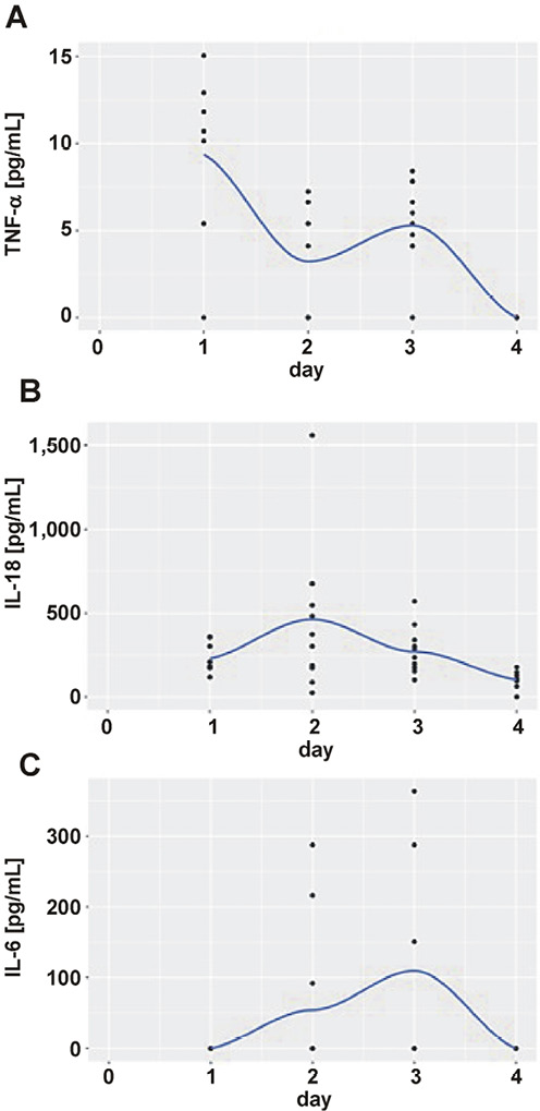
Serum TNF-α concentrations (Figure 1A) peaked on postoperative day 1 (9.2 pg/mL), decreased to 3.4 pg/mL on day 2, increased again to 5.3 pg/mL on day 3, and were undetectable on day 4. The difference between the maximum and minimum mean daily values was statistically significant (p < 0.001).
IL-18
Serum IL-18 concentrations (Figure 1B) reached 224 pg/mL on postoperative day 1, peaked at 448 pg/mL on day 2, declined to 270 pg/mL on day 3, and remained detectable on day 4 (69 pg/mL). The difference between the maximum and minimum mean daily values was statistically significant (p < 0.01).
IL-6
Serum IL-6 concentrations (Figure 1C) were undetectable on postoperative day 1, reached 54 pg/mL on day 2, peaked at 110 pg/mL on day 3, and were not detected on day 4. The difference between the maximum and minimum mean daily values was statistically significant (p < 0.05).
Absolute cell counts
White blood cells
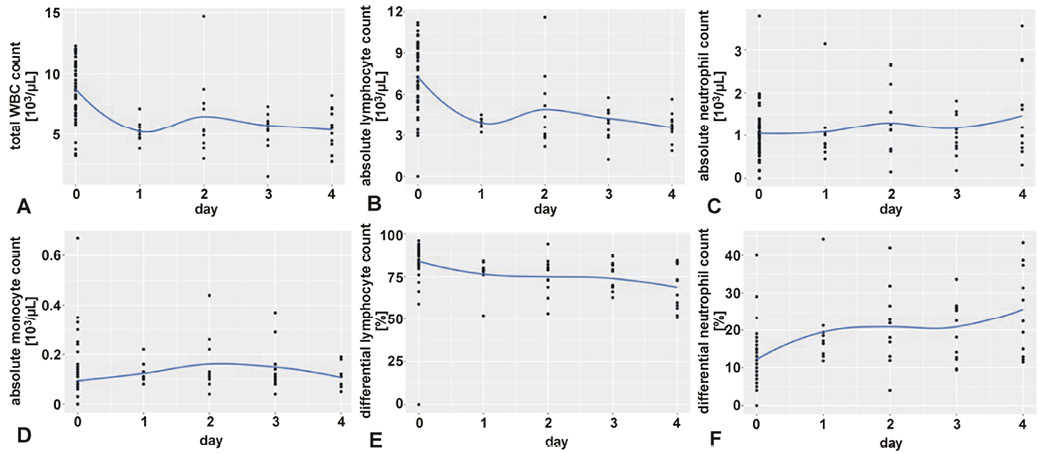
The total white blood cell (WBC) count decreased significantly by 40% (p < 0.001), from 8,700 ±2,500 cells/µL at baseline to 5,200 ±900 cells/µL on postoperative day 1. The greatest reduction occurred on postoperative day 1, followed by partial recovery on day 2, a subsequent decline on day 3, and a further decrease on day 4 (Figure 2A).
Lymphocytes
Absolute lymphocyte count decreased significantly by 47% (p < 0.001), from 7,300 ±2,500 cells/µL at baseline to 3,900 ±400 cells/µL on postoperative day 1. Lymphopenia persisted throughout the 4-day observation period (Figure 2B).
Neutrophils
Absolute neutrophil count increased gradually by 50% during the postoperative period, rising from 1,000 ±600 cells/µL to 1,500 ±1,000 cells/µL. This increase was statistically significant (p = 0.002) (Figure 2C).
Monocytes
Absolute monocyte count increased by approx. 100%, from 100 ±100 cells/µL to 200 ±100 cells/µL on postoperative day 2; however, this increase was not statistically significant (p = 0.30). Monocyte counts subsequently returned to baseline on the 4th postoperative day (Figure 2D).
Eosinophils
No eosinophils were detected at baseline. Postoperatively, eosinophil counts increased significantly (p < 0.001) to 100 ±100 cells/µL on day 2 and reached 200 ±200 cells/µL by day 4.
Basophils
Absolute basophil counts remained very low and were barely detectable throughout the observation period.
Differential cell counts
Lymphocytes
The percentage of lymphocytes decreased by 18%, from 84 ±15% at baseline to 68.5 ±13% during the postoperative period (Figure 2E).
Neutrophils
Differential neutrophil count increased gradually and significantly (p < 0.001) by 112%, from 12.2 ±6.8% on day 0 to 25.9 ±11.8% on postoperative day 4 (Figure 2F).
Monocytes
Differential monocyte count increased significantly (p = 0.03) by 116%, from 1.2 ±1.7% at baseline to 2.6 ±1.1% during the 3 postoperative days. On day 4, the value declined to 2.0 ±0.5%, remaining above baseline.
Eosinophils
Differential eosinophil count increased significantly (p < 0.001), from 0.3 ±0.6% at baseline to 2.8 ±2.6% during the postoperative period.
Basophils
Differential basophil count increased significantly (p < 0.001) from 0.0 ±0.1% to 0.4 ±0.2%.
Anemia-related parameters
Red blood cells
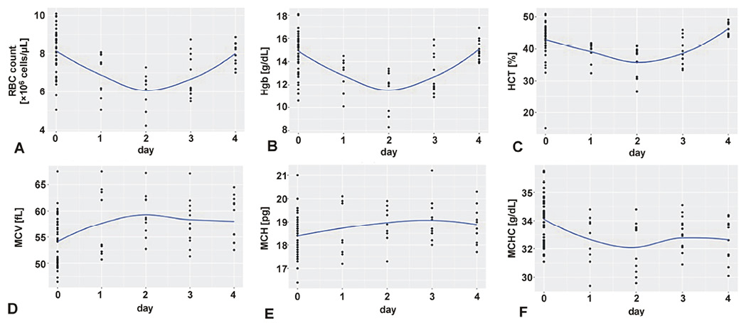
Red blood cell (RBC) count showed a transient postoperative decrease followed by recovery. A statistically significant reduction of 25% (p < 0.001) was observed, from 8.1 ±1.2 × 10⁶ cells/µL at baseline to 6.1 ±0.9 × 10⁶ cells/µL on day 2. The greatest decrease was observed on postoperative day 2, after which RBC count gradually recovered to baseline values (8.0 ±0.7 × 10⁶ cells/µL) on postoperative day 4 (Figure 3A).
Hemoglobin
Hemoglobin (Hgb) concentration also demonstrated a transient postoperative decline followed by recovery. Hemoglobin decreased significantly by 23% (p < 0.05), from 14.9 ±1.7 g/dL at baseline to 11.5 ±1.8 cells/µL on postoperative day 2, and returned to baseline values (15.0 ±0.9 g/dL) on the 4th day (Figure 3B).
Hematocrit
Hematocrit (HCT) exhibited a transient postoperative decrease followed by recovery. The values decreased significantly by 23% (p = 0.035), from 42.8 ±5.8% at baseline to 35.8 ±4.8% on postoperative day 2, and returned to baseline by postoperative day 4 (46.2 ±2.0%) (Figure 3C).
Mean corpuscular volume
Mean corpuscular volume (MCV) increased initially and then gradually declined. The value increased significantly by 10% (p < 0.001), from 54.1 ±4.8 fL at baseline to 59.3 ±4.3 fL on postoperative day 2, and remained elevated on postoperative day 4 (58.2 ±4.4 fL) (Figure 3D).
Mean corpuscular hemoglobin
Mean corpuscular hemoglobin (MCH) showed a slight but statistically significant increase (p < 0.001) throughout the 4-day postoperative observation period (Figure 3E).
Mean corpuscular hemoglobin concentration
Mean corpuscular hemoglobin concentration (MCHC) decreased slightly but significantly (p < 0.001) by 6%, from 34.1 ±1.6 g/dL at baseline to 32.1 ±1.9 g/dL on postoperative day 2. Subsequently, the value increased by 1 unit on postoperative days 3 and 4 (Figure 3F).
Discussion
To the best of our knowledge, this study is the first to characterize the SIR following palatal graft harvesting.
Serum cytokine concentrations peaked during the acute phase: TNF-α at 12–24 h, IL-18 at 24–48 h, and IL-6 at 48–72 h. Following elective surgery, TNF-α is among the earliest and most potent mediators of the subsequent host inflammatory response. The primary sources of TNF-α synthesis are monocytes/macrophages and T cells.22 The half-life of TNF-α is less than 20 min. Despite its brief half-life, TNF-α can induce marked metabolic and hemodynamic changes and activate mediators in the cytokine cascade.23
The prolonged elevation of serum TNF-α concentrations observed in the present study (3 days), despite its short half-life, may be attributed to the secondary healing characteristics of palatal graft harvesting. Exposure of the underlying bone and disruption of epithelial continuity in the middle of the palate may result in sustained cellular injury and prolonged TNF-α production.
IL-18 is a proinflammatory cytokine produced by activated macrophages. It promotes the early resolution of bacterial infections.24 Bacterial products can stimulate IL-18 production from monocytes.25 Previous studies have demonstrated significantly elevated circulating IL-18 concentrations during sepsis for up to 21 days, with particularly pronounced increases in gram-positive sepsis.26 Consequently, IL-18 may serve as a systemic marker of the bacterial challenge associated with the surgical site. The high circulating levels of IL-18 observed in the present study may be attributed to the secondary healing nature of the oral wound, exposing the rats to a greater bacterial challenge.
A review demonstrated that the serum IL-6 concentrations typically peak on the first postoperative day following elective surgery.27 In contrast, IL-6 levels in the present study peaked on postoperative day 3. This delayed peak may be attributed to the secondary healing pattern, which could sustain IL-6 production through the prolonged release of TNF-α during postoperative days 1–3. TNF-α induces IL-6 production.
The same review also demonstrated that IL-6 levels are consistently associated with the magnitude of surgical trauma.27 Peak IL-6 concentrations following minor surgical procedures (inguinal hernia repair and cholecystectomy) were 13 pg/mL and 77 pg/mL, respectively. Corresponding peak concentrations following moderate surgical procedures (prostatectomy, total hip replacement, colorectal cancer resection, and total knee replacement) were 62 pg/mL, 140 pg/mL, 161 pg/mL, and 321 pg/mL, respectively. In contrast, major surgical procedures (abdominal aortic aneurysm repair, major liver resection, open cardiac surgery) were associated with peak IL-6 levels of 248 pg/mL, 345 pg/mL and 428 pg/mL, respectively.27
A difference in serum IL-6 concentrations was also observed between minimally invasive and open surgical procedures.27 This difference was particularly evident when comparing laparoscopic and open rectopexy (21 pg/mL vs. 111 pg/mL), laparoscopic and open cholecystectomy (62 pg/mL vs. 95 pg/mL), laparoscopic and open gastrectomy (44 pg/mL vs. 129 pg/mL), laparoscopic and open abdominal hysterectomy (19 pg/mL vs. 166 pg/mL), laparoscopic and open miscellaneous colorectal resection (140 pg/mL vs. 393 pg/mL), and endovascular and open aneurysm repair (116 pg/mL vs. 332 pg/mL).
Serum IL-6 levels are associated with both the magnitude and invasiveness of the surgical procedure. In the present study, the peak serum IL-6 concentration (110 pg/mL) was comparable to those reported for moderate open surgical procedures. These findings may have implications for postoperative patient management.
Therefore, IL-6 appears to be a useful marker for assessing the magnitude of the SIR after elective surgery. Based on the previous findings, it can be hypothesized that, following oral and periodontal surgery, prolonged serum TNF-α elevation reflects persistent cellular damage (primary vs. secondary healing), IL-18 indicates the degree of bacterial challenge, and IL-6 reflects the magnitude of the surgical intervention.
Monitoring postoperative serum concentrations of TNF-α, IL-6 and IL-18 may therefore be clinically valuable. Assessment of these cytokines may be predictive of the onset of infection or the need for urgent attention to ensure optimum wound healing after oral and periodontal surgery.
The present study revealed changes in WBC counts following palatal graft harvesting. A significant postoperative reduction in total WBC count was observed, which was primarily attributable to a marked decrease in circulating lymphocytes. The linear decrease continued for the 96-h observation period. In contrast, previous studies involving cardiovascular, orthopedic, abdominal, gynecological, and urological surgeries have generally reported postoperative increases in total WBC counts.28 One possible explanation for this discrepancy is the extent of surgical trauma. Major surgical procedures induce a pronounced cortisol-mediated stress response, resulting in increased neutrophil release from the bone marrow and marked peripheral neutrophilia, which increases the total WBC count. Oral and periodontal surgical procedures are substantially less invasive than these major operations. Consequently, the relatively limited surgical trauma associated with palatal graft harvesting may not elicit a marked increase in neutrophils. Instead, the postoperative reduction in total WBC count appears to result primarily from the marked decrease in absolute lymphocyte count.
In the previous studies, we evaluated the local time-related changes in the density of inflammatory infiltrates following palatal graft harvesting in rats.29, 30 Three weeks after the surgery, the density of inflammatory infiltrates remained high in the central part of the wound. The differences between the SIR response observed in the present study and those reported after major surgical procedures may therefore be explained by the secondary wound healing characteristic of palatal graft harvesting. In the present procedure, the time frame of the inflammatory response was extended compared to primary healing in evaluated major surgeries. The use of wound dressings may help attenuate the SIR associated with secondary wound healing.
The effects of trauma on absolute WBC counts have previously been investigated in mice.31 The results showed that both major trauma (nephrectomy) and minor trauma (e.g., injection, bleeding), as well as surgical stress, induced a significant decrease (50–70%) in absolute lymphocyte count. The decline was gradual, with the greatest reduction occurring within the first postoperative day. These findings are consistent with those of the present study. Major trauma resulted in decreases in both B and T lymphocytes. Minor trauma affected only B lymphocytes. In addition, major trauma induced a three- to four-fold increase in absolute neutrophil counts, whereas no significant neutrophil response was observed following minor trauma. In both major and minor trauma models, hematological parameters generally returned to baseline within 24 h. Repeated exposure to trauma after recovery elicited a renewed trauma response. Bilateral adrenalectomy abolished the lymphocyte response to both major and minor trauma and reduced the neutrophil response to major trauma by more than 50%, indicating that stress hormones play an important role in mediating these changes. The authors proposed that reduced migration of lymphocytes into the circulation represented the most likely underlying mechanism.31
Another study evaluated the potential causes of postoperative lymphopenia by comparing the effects of major surgery, blood loss and psychological stress. Major surgery resulted in a 30–60% reduction in absolute lymphocyte counts and a 165–350% increase in absolute neutrophil counts within the 1st postoperative day. In contrast, blood loss and psychological stress (e.g., tooth extraction, blood donation, important examination) did not alter absolute lymphocyte counts during the same period. Shortly after these events, an increase in absolute neutrophil counts was observed following minor tissue trauma, such as blood donation or tooth extraction. However, no increase was detected after psychological stress alone, and the neutrophil response to minor trauma (20–55%) was substantially smaller than that observed after major surgery. It was concluded that the degree of tissue trauma is a key determinant of postoperative changes in circulating cellular immune hallmarks.32
The marked reduction in absolute lymphocyte counts and the moderate increase in absolute neutrophil counts observed in the present study are consistent with the response to minor surgical trauma. However, unlike previous reports in which these changes resolved within 24 h, the alterations persisted throughout the 4-day observation period, resembling continuous trauma creation. To the best of our knowledge, this is the first study to demonstrate the cellular systemic immune response following palatal graft harvesting. The observed stress response is not only psychological but also physical. Future studies should investigate the SIR associated with different soft tissue harvesting techniques and evaluate the effects of different agents that may modify this response.
The present study also revealed the postoperative changes in RBC count, HCT and Hgb levels following palatal graft harvesting. Significant postoperative reductions in all 3 parameters were observed during the first 48 h after surgery, with complete recovery to baseline values by postoperative day 4.
Previous studies have reported a longer duration of postoperative anemia following major surgeries.33 After open aortic surgery, the acute-phase response (APR) has been shown to induce anemia, with RBC counts returning to baseline only after 45 days, whereas Hgb concentrations remained reduced for an even longer period.33 It may be speculated that iron deficiency is more involved in Hgb formation than in blood cell production.34 Acute-phase response has been well documented following lower limb revascularization and aortic surgery.35 Functional iron deficiency is considered the principal underlying mechanism and may persist for more than 6 weeks. Cytokines stimulate macrophages, blocking iron metabolism and blood cell production.36
Mohammed et al. reported postoperative anemia following minor surgical procedures, including herniorrhaphy and arthroscopy.37 Anemia peaked on postoperative day 3 and returned to baseline by day 7. These findings suggest that the magnitude of anemia is related to the degree of associated stress. Factors influencing postoperative anemia include operative time, the extent of tissue injury (superficial vs. major intra-abdominal surgery), and the occurrence of postoperative complications.37
Compared with major surgical procedures, oral and periodontal surgery is considerably less invasive. Therefore, the shorter duration of postoperative anemia observed in the present study (approx. 2 days), its lower magnitude (25%), and the rapid recovery to baseline within 4 days may be explained by the relatively limited surgical trauma associated with palatal graft harvesting.
Serial assessment of complete blood counts and inflammatory cytokines may also be useful for evaluating the efficacy of different surgical harvesting techniques and patient-related treatment approaches. Future studies combining serial hematological and cytokine measurements with new technologies may provide additional insight into the wound healing process following palatal graft harvesting. A pilot study used digital imaging technology and three-dimensional image analysis software to evaluate healing dynamics after free palatal graft harvesting and demonstrated progressive reductions in palatal soft tissue volume and thickness.38 Combining serial hematological and cytokine analyses with 3D digital assessment may provide a reproducible approach for investigating the cellular and humoral mechanisms underlying changes in soft tissue thickness and volume after graft harvesting. Future studies should also evaluate cytokine-modulating therapies and compare alternative graft harvesting techniques.
Limitations
To the best of our knowledge, this study is the first to characterize the SIR following palatal graft harvesting. The observed response resembles that reported after other minor surgical procedures. Nevertheless, several limitations should be acknowledged. Although the rat model provides valuable insights into postoperative inflammatory responses, inherent differences exist between rodents and humans. Only male Wistar rats were used in the present study; therefore, future investigations should include female rats to evaluate potential sex-related differences in the inflammatory response. In addition, the number of animals in each experimental group was relatively small, age-related differences were not investigated, and other potentially relevant variables, such as metabolic parameters, were not evaluated. Consequently, extrapolation of these findings from a small-animal model to humans should be undertaken with appropriate caution.
Conclusions
Based on the observed systemic inflammatory response following palatal graft harvesting, oral and periodontal surgical procedures may generally be considered minor surgical interventions. Serial assessment of complete blood counts and inflammatory cytokines may provide valuable information for postoperative monitoring and may help optimize wound healing following oral surgery.
Ethics approval and consent to participate
The study protocol was approved by the Ethics and Institutional Animal Care and Use Committee of Tel Aviv University, Israel (approval No. TAU – MD–IL-2402-109-3; February 1, 2024). All animals received humane care throughout the study. The experimental design complied with the Animal Research Reporting of In Vivo Experiments (ARRIVE) guidelines.
Data availability
The datasets generated and/or analyzed during the current study are available from the corresponding author on reasonable request.
Consent for publication
Not applicable.
Use of AI and AI-assisted technologies
Not applicable.