Abstract
Background. Chronic inflammation in the oral cavity has a negative effect on the course of chronic mucosal lesions.
Objectives. The present study aimed to compare the volume and proportional presence of aggressive periodontal pathogens in patients with oral lichen planus (OLP) accompanied by desquamative gingivitis (DG) vs. those with OLP without gingival involvement. All patients in the cohort were also diagnosed with moderate chronic periodontitis. The observed differences may help explain the higher risk associated with gingival involvement in OLP.
Material and methods. The presence of periodontal pathogens was evaluated in 50 biopsy-confirmed OLP with DG cases and 50 OLP cases without gingival manifestations. All participants presented with chronic periodontitis of comparable severity (moderate: periodontal pocket depth (PPD) up to 6 mm). Aggressive periodontal pathogens were identified and quantified using DNA testing (VariOr®-Dento). The periodontal status was assessed using orthopantomography (OPG) or cone-beam computed tomography (CBCT), in combination with the clinical examination of periodontal pockets.
Results. In comparison with the non-gingival OLP cases, the OLP cases with gingival involvement exhibited a significantly higher proportional presence of aggressive periodontal pathogens (p = 0.0009) and a higher bacterial load (n/µL; p = 0.001). Desquamative gingivitis associated with OLP was also linked to an increased risk of subsequent periodontal resorption. Given the unstable chronic inflammatory environment, careful monitoring of periodontal tissues in OLP cases appears to be essential, as persistent inflammation may contribute to an elevated risk of malignant transformation.
Conclusions. Our findings demonstrate a significant difference in the presence of aggressive periodontal pathogens between OLP cases with and without gingival involvement. The median pathogen volume in the OLP with DG cases was over 7.4 times higher. Chronic inflammation and bacterial by-products may act as cofactors in the development of dysplasia and the malignant transformation of oral lichen with dysplasia (OLD), which may present clinically as DG.
Keywords: oral lichen planus, periodontal pathogens, oral potentially malignant disorder, desquamative gingivitis, DNA testing
Introduction
Oral lichen planus (OLP) is a common chronic inflammatory disease of the oral mucosa, with an incidence of approx. 2%.1, 2, 3, 4 It is characterized by white reticular (lace-like) patches and atrophic or erosive (ulcerative) lesions.1, 2, 3 Oral lichen planus, which typically affects females over 40 years of age, with a female-to-male ratio of 1.4:1, was first described by Wilson in 1869.2 Its clinical course is often unstable, featuring multiple remissions and relapses. Gingival involvement may present as desquamative gingivitis (DG), while lesions on the dorsum of the tongue often appear as plaque-like patches. Desquamative gingivitis is characteristic gingival involvement in OLP, both localized and generalized. Gingival lesions exhibit a spectrum of clinical manifestations, ranging from hyperkeratosis and atrophy to the erosive destruction of the gingival epithelium.5, 6, 7, 8
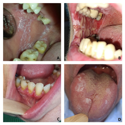
Various clinical manifestations of OLP are shown in Figure 1. White lesions are usually asymptomatic, whereas ulcerative lesions may cause a burning sensation and pain.3, 4 Mucosal alterations are the result of the disrupted epithelial regeneration following basal cell destruction.5 This process is associated with a lichenoid subepithelial infiltrate composed of cytotoxic T-lymphocytes (cluster of differentiation: CD8+).5, 9 The clinical severity of OLP is often assessed using the reticular-erythematous-ulcerative (REU) index,10 and subjective symptoms are measured using the visual analog scale (VAS).10, 11 In contrast to bilateral OLP, oral lichenoid lesions (OLL) are typically unilateral or asymmetrical.1, 6, 12 Oral lichenoid lesions include oral contact lesions, lichenoid drug eruptions, and the oral manifestations of graft-versus-host disease (GvHD).6, 13, 14
Histopathological criteria for OLP include a subepithelial lymphocytic infiltrate, parakeratosis and acanthosis.1, 4, 6, 13, 15, 16 Biopsy is essential to distinguish non-dysplastic OLP from OLP with dysplasia (OLD).1, 9, 17 Direct and indirect immunofluorescence are valuable tools for differentiating OLP from autoimmune bullous diseases.8, 18, 19, 20, 21 Immunohistochemistry further enhances diagnostic accuracy, often through the analysis of proteins such as p53 and Ki-67.7, 22, 23 Mutated p53 protein is typically found in the nuclei of dysplastic or neoplastic epithelial cells.23, 24, 25 In differentiated oral intraepithelial neoplasia (dOIN), p53 is detected along a continuous row of nuclei in the epithelial basal layer. Ki-67 serves as a marker for cellular proliferation.26
Oral lichen planus has been classified as a potentially precancerous condition since 1978. More than a century has passed since the first documented case of malignant transformation to oral squamous cell carcinoma (OSCC) in OLP.7 Malignant transformation is driven by increased epithelial proliferation and inflammation-mediated carcinogenic processes.27, 28 A 2019 systematic review estimated the risk of transformation at 0–3.5%.29 However, a more recent one reported a lower risk of 0.44% after excluding cases with less than 6 months between OLP biopsy and carcinoma diagnosis.27 Risk factors for OLP progression include the presence of oral epithelial dysplasia (OED) and Candida albicans infection.7, 30 Clinically, the erosive form carries the highest risk.13
Since early lesions may resemble flat leukoplakia, and lichenoid infiltrates are present in biopsy, the more serious diagnosis of proliferative verrucous leukoplakia (PVL) may be mistakenly identified as OLP.7 The rate of malignant transformation in PVL ranges from 50% to 100%.29 If lesions meet the Cerero-Lapiedra criteria for PVL, a more intensive follow-up is warranted to minimize the risk of malignant transformation (Figure 1).31
The etiology of OLP is usually unknown.1 A time delay of 6–12 months between the triggering factor and the clinical manifestation is often observed.32, 33 Common comorbidities of OLP include immune-mediated liver and thyroid gland diseases and cutaneous lichen planus (CLP).34, 35, 36, 37, 38
Topical treatment for OLP includes corticosteroids in gel form, mucosal adhesive patches and laser therapy.39, 40, 41, 42 The surgical excision of the most clinically severe lesions is often a necessary part of the therapeutic protocol. In suspected cases of drug-induced lichenoid reactions, clinicians are advised to modify the patient’s medication regimen.
The relationship between periodontal tissues and OLP manifestations remains unclear. The inflammatory process in periodontitis is strongly associated with aggressive periodontal pathogens.43 In the oral cavity, anatomical proximity allows even localized infections to exert widespread pathological effects. The most aggressive bacteria involved in periodontitis are members of the so-called “red complex”. These pathogens – Porphyromonas gingivalis (Pg), Tannerella forsythia (Tf), Treponema denticola (Td), and Filifactor alocis (Fa) – are implicated in alveolar bone resorption through inflammatory mechanisms.43 The virulence of these pathogens is directly correlated with their concentration. The members of the red complex are often accompanied by other aggressive species belonging to the orange complex, including Parvimonas micra (Pm), Prevotella intermedia (Pi) and Fusobacterium nucleatum (Fn).43, 44 These bacteria produce proteases and other cytotoxic by-products.
Numerous studies have demonstrated that such bacteria not only induce inflammation, but also contribute to the development of epithelial dysplasia, thereby increasing the risk of malignant transformation. These microorganisms can inhibit apoptosis in atypical cells, further contributing to carcinogenesis.45, 46 Periodontal pathogens, such as Fn, have been implicated in the development of the cancers of the pancreas and the intestine by promoting inflammation, cellular proliferation and immune evasion.47 Similarly, Tf and Pg have been linked to esophageal cancer.45, 48 Biopsies of OSCC have frequently been found to contain high levels of Pg contamination.45 Bacterial by-products include matrix metalloproteinases (MMPs) – zinc-dependent peptidases that degrade the extracellular matrix and contribute to the disruption of the basement membrane.49, 50 Antibiotics such as amoxicillin, in combination with metronidazole or tetracycline, are often prescribed as adjunctive therapy for periodontitis.44, 51 Differences in antibacterial and anti-MMP efficacy depend on dosage and treatment duration.52, 53, 54
The aim of this study was to compare the periodontal pathogen profile in OLP patients with DG and in OLP cases without DG, matched by sex, age and periodontitis severity (the same periodontal pocket depth (PPD), the same periodontitis stage and grade). This comparison may help elucidate the differential interaction of aggressive microorganisms and their by-products with the host immune system. Such mechanisms could explain the higher rate of malignant transformation observed in cases of OLD in alveolar localization. Chronic inflammation, the presence of cytotoxic bacterial by-products and the formation of carcinogenic compounds may drive excessive epithelial proliferation, ultimately contributing to the development of OSCC within OLP lesions.
Material and methods
Between 2004 and 2025, a total of 454 biopsy-verified cases of OLP or OLD were diagnosed and managed at the Department of Stomatology-Oral Medicine, University Hospital Pilsen, Czechia. Each case underwent a subsequent immunohistochemical examination. Clinical evaluations included radiographic examinations, the assessment of tobacco use, REU scoring, and VAS profiling. Regular swab cultures for Candida albicans from the oral mucosa were also performed.
Oral lichen planus/OLD was diagnosed through routine biopsy, supplemented by direct immunofluorescence, which identified fibrinogen deposits along the basement membrane. Immunohistochemistry was used to evaluate p53 and Ki67 expression.
The inclusion criteria were as follows: OLP cases verified by a clinical examination with an REU evaluation and differentiation by the affected mucosal sites, with mandatory confirmation via biopsy. All biopsies were followed by direct immunofluorescence. The patients who had not taken antibiotics or undergone oral surgery in the previous 4 months were included. Only OLP cases with the co-existing chronic periodontitis were included (defined by the 1999 classification as periodontal pockets with an average depth of 6 mm and radiographic evidence of bone resorption, or Stage III, Grade B periodontitis by the 2017 classification). The cases of OLP with DG were sex- and age-matched to the patients with OLP without gingival involvement.
The exclusion criteria comprised OLP cases not confirmed by biopsy; the cases showing features of aggressive periodontitis or the acute exacerbations of chronic periodontitis (e.g., periodontal abscess or excessive bleeding on probing, Stage I, II, IV and Grade A, C); the use of antibiotics or oral surgery within the last 4 months; and OLP cases not matched by sex and age to the study cohort.
The study compared 50 patients with OLP with DG to 50 patients with OLP without gingival involvement. The sex ratio matched the overall clinic population (2.2:1), resulting in 34 females and 16 males in each group. The average age of the participants was 62.55 years (range: 31–85 years). Patients with OLP with DG were classified as group 1 (G1), and those with non-gingival OLP formed group 2 (G2). All participants provided written informed consent. The characteristics of the comparative study cohort are presented in Table 1.
The diagnosis of chronic periodontitis was established clinically using a WHO periodontal probe. Bone resorption was confirmed radiographically via orthopantomography (OPG) or cone-beam computed tomography (CBCT). Periodontal pocket depth ranged from 5.5 to 6 mm. Tooth mobility was assessed through manual palpation, which was a mandatory component of the clinical evaluation.
The detection of aggressive periodontal pathogens belonging to the red and orange complexes was performed using a DNA-based test on the subgingival plaque collected with sterile paper points. The VariOr®-Dento Plus system (Gen-Trend Co., Rudolfov, Czech Republic) was used to identify 12 periodontal pathogens. The results were reported both as proportions (scale 0–4) and as bacterial load (in microliters). A proportional value of 4 corresponds to approx. 106–107 bacteria, while a value of 1 equates to 102–103. Higher values indicate worse prognosis in terms of periodontal tissue resorption. DNA detection was conducted using the real-time quantitative polymerase chain reaction (qPCR) with TaqMan™ probes, with a detection threshold of 101.
Statistical analysis
Statistical analysis was performed using the SigmaXL software (SigmaXL Inc., Kitchener, Canada). Non-parametric tests, including the Kruskal–Wallis test, were applied for group comparisons.
Results
The cohort included in the comparative study comprised 454 patients diagnosed with OLP/OLD. Of these, 60% were classified as OLP (n = 274/454) and 40% as OLD (n = 180/454). The average follow-up period was 63.1 months (5.26 years). The female-to-male ratio was 2.2:1 (n = 314 and n = 140, respectively). The mean age at the time of the study was 63.1 years. Gingival involvement (DG) was observed in 43% of cases (n = 195/454). The mean REU index across the cohort was 10.1, with higher values in the DG cases (13.9) as compared to those without gingival manifestations (7.4). The erosive-ulcerative form of OLP was more frequently associated with gingival involvement (47% vs. 33% in non-gingival OLP). The mean VAS score for pain was slightly higher in the DG cases (1.44) as compared to the non-gingival cases (1.09).
Only 19.6% of the patients were smokers (n = 89/454), with no difference in prevalence between the DG cases and OLP without gingival manifestations. Concurrent thyroid disease was found in 19.8% of the OLP/OLD cases (n = 90/454). The mean colony-forming unit (CFU) count of Candida albicans was notably higher in patients with OLP with DG (17.1) than in the non-gingival OLP cases (10.3).
The immunohistochemical analysis of the p53 protein was performed in 48.2% of cases (n = 219/454). Nuclear positivity for p53 in long rows of basal epithelial cells was present in 50% of the DG cases (n = 44/88) and 59% of the non-gingival cases (n = 77/131). Due to limited sampling (<5% of cases), Ki67 expression did not provide a statistically meaningful diagnostic insight.
Differentiated oral intraepithelial neoplasia was diagnosed with similar frequency in both groups (80% in DG vs. 81% in non-gingival OLD). The malignant transformation rates were also comparable between the OLP cases with DG (2.4%, n = 3/125) and those without (2.0%, n = 3/149). However, among the OLD cases, the rate of malignant transformation was significantly higher in the DG cases (11.4%, n = 8/70) than in non-gingival OLD (7.3%, n = 8/110).
A summary of the clinical-pathological characteristics of both OLP variants is presented in Table 2.
Topical corticosteroids – dexamethasone acetate and triamcinolone acetonide – were used to treat mild to moderate cases of OLP. In severe cases, clobetasol propionate was administered. The formulation was selected based on the location of the lesion; solutions and gels were optimal for the buccal mucosa and the tongue, while mucosal adhesive pastes were used to treat DG. Among the patients without DG, 56.8% (n = 147/259) required corticosteroid therapy as compared to 74.9% (n = 146/195) of those with OLP and DG. Over 90% of topical applications involved dexamethasone, with the remainder using clobetasol.
Surgical treatment included diagnostic excision at the initial OLP follow-up visit, with the re-excision performed in cases of clinical lesion progression. In instances of gingival desquamation, the adjacent teeth were extracted when malignant transformation was suspected. This approach ensured the removal of all at-risk tissue.
In the patients with OLP and DG, OPG or CBCT scans were performed to evaluate alveolar bone resorption and assess the potential for gingival lesion progression, even before surface abnormalities appeared.
To compare the bacterial profiles, the volume of aggressive periodontal pathogens was assessed in 50 patients with OLP and DG and in 50 patients with OLP without gingival involvement. All patients had a comparable periodontal status, with moderate chronic periodontitis confirmed through the clinical examination and CBCT analysis. The mean PPD in the compared cases was 6 mm, and periodontitis was classified as Stage III and Grade B.
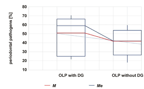
Both the proportional presence and absolute bacterial volumes (in milliliters) of aggressive periodontal pathogens were analyzed. Particular emphasis was placed on pathogens from the red and orange complexes. In both categories, OLP with DG showed a predominance. Specifically, the red complex pathogens – Pg, Tf, Td, and Fa – were significantly more prevalent in the DG group. Similarly, the orange complex bacteria such as Pm and Fn were also more common in OLP with DG. The only pathogen with a slightly lower proportional presence in DG was Pi. These differences were statistically significant (p = 0.0009; Kruskal–Wallis test), with confidence intervals (CI): 2–3 for OLP with DG, and 1–2 for OLP without DG.
Table 3 summarizes the proportional comparison of the red and orange complex pathogens.
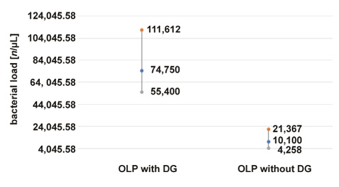
Similar trends were observed for the absolute bacterial volumes. The mean and median bacterial counts (in milliliters) were significantly higher in the OLP with DG group (p = 0.001; Kruskal–Wallis test). The CIs were as follows: 55.4k–111.6k for OLP with DG; and 4.3k–21.4k for OLP without DG. On average, the quantity of aggressive periodontal pathogens in OLP with DG was 7.4 times higher than in those without gingival involvement.
The results of both comparisons are presented in Figure 2 and Figure 3.
Discussion
Oral lichen planus is a common chronic disease of the oral mucosa, with an incidence exceeding 2%.1, 2, 3 It typically affects patients over 40 years of age and shows a slight female predominance, with a female-to-male ratio of approx. 1.4:1.4 In our study, this ratio was even more pronounced at 2.2:1 in favor of women.
Oral lichen planus frequently coexists with thyroid gland disorders. Studies from Northern Europe and Spain report thyroid involvement in over 8% of OLP cases.36, 37 In our cohort, 19.8% of patients with OLP or OLD had concomitant pathologies in the thyroid. This higher prevalence may be attributed to our standard practice of referring all OLP patients to a general practitioner for thyroid function screening.
Oral lichen planus was included among the oral potentially malignant disorders (OPMDs) in the 3rd and 4th editions of the World Health Organization (WHO) classification.7 However, the current 5th edition limits this classification to cases of OLD, reflecting the updated understanding of malignant potential.7
A high incidence of dOIN has been observed in OLP patients. Differentiated oral intraepithelial neoplasia is considered a high-risk lesion in some specialized centers.55 In our study, dOIN was present in 80% of the OLD cases with DG (n = 56/70) and in 81% of the non-gingival OLD cases (n = 89/110).
Data on the expression of p53 and Ki-67 in OLP is conflicting. Some studies link these markers to malignant transformation, while others do not support this association.22 In our study, p53 positivity was more frequent in the OLP cases without DG (59%) than in those with gingival involvement (50%). Other studies have reported p53 and Ki-67 positivity in over 80% of OLP biopsies.9
Historically, the reported rate of malignant transformation in OLP was high, largely due to a failure to distinguish between non-dysplastic OLP and OLD. In 2019, González-Moles et al. estimated the malignant transformation rate for OLP to be between 0% and 3.5%.29 This was further reduced to 0.44% by Idrees et al. in 2021 after excluding cases suspected of concurrent OLP and OSCC.27 Using the same exclusion criteria, we found the malignant transformation rate for OLP with DG to be 2.4% (n = 3/125), and for non-gingival OLP, 2.0% (n = 3/149). Both figures fall at the higher end of recent estimates.27, 29 However, the malignant transformation rate was significantly higher in the OLD patients. Transformation to OSCC occurred in 7.3% of the cases without gingival involvement (n = 8/110) and in 11.4% of those with DG (n = 8/70). This increased rate in the DG cases may be attributed to long-standing, intense chronic inflammation of the surrounding tissues. Such inflammatory processes, driven by microbial activity and their by-products, interact with the immune system, influencing tissue repair pathways and dysplasia progression.
Given the risk of misdiagnosing the early lesions of PVL as OLP, all patients with biopsy-confirmed OLP should remain under continuous clinical follow-up.7, 31 Inexperienced clinicians may overlook the subtle changes associated with malignant transformation.31
Radiographic imaging should be a routine part of comprehensive care for patients with OLP with DG and those suspected of PVL. Malignant transformation in PVL is notably high, ranging from 50% to 100%.29 Our findings – an 11.4% malignant transformation rate in OLD with DG – indicate that this condition also carries significant malignant potential.
The OLP therapy typically includes topical corticosteroids, such as dexamethasone acetate and clobetasol propionate.39, 40 In our study, corticosteroids were required in 56.8% of the OLP cases without gingival involvement. The cases presenting with DG were more challenging to manage, requiring corticosteroid treatment in over 74.9% of instances. Laser therapy is also frequently employed in symptomatic OLP cases,56, 57, 58 particularly for ulcerative lesions that do not respond to corticosteroids. However, therapeutic outcomes in OLP are generally inconsistent across studies,42 and no curative treatment has yet been identified.4, 10
Functional immune surveillance is essential for good oral mucosal and periodontal health. Defense against pathological processes requires a rational lifestyle, ensuring the intake of nutrients and elements necessary for the functioning of the organism.59 Periodontal health can be compromised by comorbidities or genetic predisposition to gum disease.60, 61 The periodontal disease classification system has evolved from the specification of the suspected induction to the determination of the risk of sequelae.62 The association between aggressive periodontal pathogens and diseases or malignancies at both local and distant sites is well-documented.45, 46, 47, 48, 63, 64 The interactions between these bacteria and the human immune system are complex. Their presence and metabolic by-products perpetuate chronic inflammation, leading to tissue destruction, dysplasia development and dysregulated immune responses. In OLP, this process is further complicated by the cytotoxic activity of the subepithelial lichenoid infiltrate.5
The atrophy and erosions associated with DG cause pain and burning sensations, impairing the patient’s ability to maintain proper oral hygiene.10, 11 This creates a bidirectional feedback loop – immune-mediated mucosal damage impedes hygiene, promoting plaque accumulation, which in turn fosters aggressive bacterial colonization and immune system activation.
Our results showing a significant prevalence of aggressive perio-pathogens in the OLP cases with DG confirm the bilateral nature of the pathological process. Chronic inflammation associated with the effects of bacterial activity increases the intensity of OLP manifestations, expressed by the REU index and a higher incidence of the erosive form of OLP. The difficult-to-manage course of these cases of lichen requires more frequent use of corticosteroids, resulting in higher contamination of the oral cavity with Candida species. Pain and reduced oral hygiene options in severe OLP in turn retrospectively impair periodontal tissue management.
In cases of OLP/OLD with DG, both comprehensive diagnostics and targeted treatment are essential. The risk of dysplasia development in clinically diagnosed OLP lesions remains.65, 66 A well-established link exists between periodontal pathogen activity and oral carcinogenesis.67 Some researchers have proposed that the ultimate therapeutic goal in OLP might be to “erase the memory” of the affected oral tissue.68
To optimize patient care, it is essential to integrate histopathological biopsy findings, radiographic imaging, the clinical assessment of periodontal tissues, and microbial profiling.69 These diagnostic tools should guide a multifaceted treatment approach aimed at reducing lichenoid infiltrate activity, promoting periodontal healing, and neutralizing the effects of aggressive periodontal pathogens and their virulence factors.
A major challenge in conducting the present study was minimizing the potential bias. To address this, only biopsy-confirmed cases of OLP were included, and patients were matched by sex, age and the periodontal clinical status. Further large-scale studies exploring the interactions between periodontal bacteria, their by-products and immune responses are needed to improve treatment strategies for chronic oral mucosal lesions.
Conclusions
Our study demonstrates a significant difference in the presence of aggressive periodontal pathogens – specifically those belonging to the red and orange complexes – between cases of OLP with gingival involvement and those with lesions in other oral locations. The median bacterial volume in OLP cases with DG was more than 7.4 times higher. Chronic inflammation, along with the by-products of these aggressive bacteria, may act as cofactors in promoting dysplasia and increasing the risk of malignant transformation in OLD involving DG.
These findings support the need for a comprehensive therapeutic approach that combines agents targeting the cytotoxic activity of the subepithelial lichenoid infiltrate with drugs that have antimicrobial effects. Additionally, cases of OLP and OLD with gingival manifestations should undergo an X-ray examination to reduce the risk of the delayed detection of malignant transformation.
Ethics approval and consent to participate
The present scientific project was approved by the Institutional Review Board (IRB) and Ethics Committee (EC) of the University Hospital Pilsen, Czechia (approval No. 231/24). Written informed consent was obtained from all participants before their inclusion in the study. This study was conducted in accordance with the Declaration of Helsinki.
Data availability
The datasets supporting the findings of the current study are available from the corresponding author on reasonable request.
Consent for publication
Not applicable.
Use of AI and AI-assisted technologies
Not applicable.