Abstract
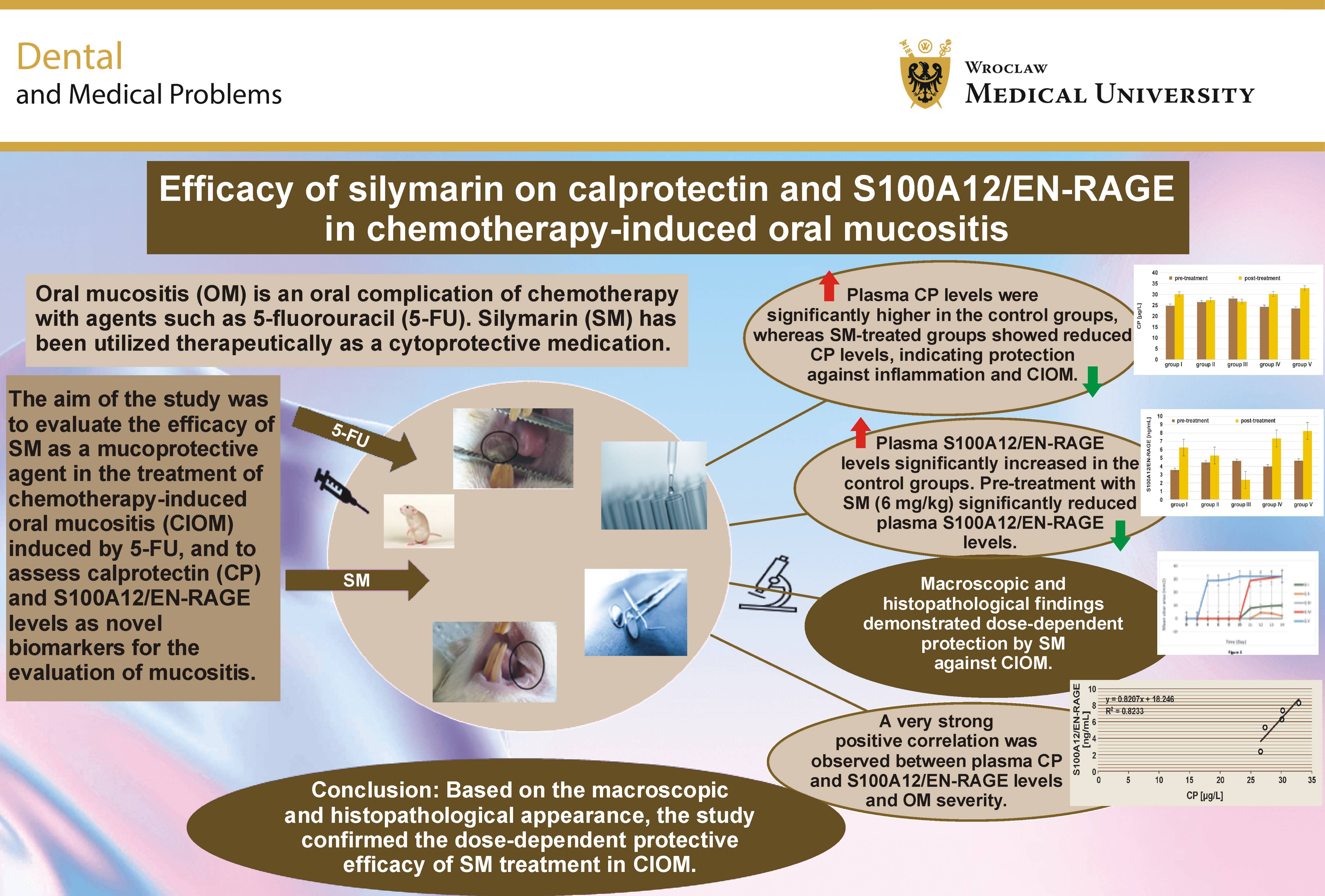
Background. Oral mucositis (OM) is an oral complication of chemotherapy with agents such as 5-fluorouracil (5-FU). Silymarin (SM) has been utilized therapeutically as a cytoprotective medication.
Objectives. The aim of the study was to evaluate the efficacy of SM as a mucoprotective agent in the treatment of chemotherapy-induced oral mucositis (CIOM) induced by 5-FU, and to assess calprotectin (CP) and S100A12/EN-RAGE levels as novel biomarkers for the evaluation of mucositis.
Material and methods. Fifty Wistar albino rats of both sexes were divided randomly into 5 groups. Three groups were treated with different doses of SM (2 mg/kg, 4 mg/kg, 6 mg/kg) for 10 days. Subsequently, the animals from groups I–III received intraperitoneal injections of 5-FU (60 mg/kg) on days 11–14. The 4th group was treated with dimethyl sulfoxide (DMSO) as a control for 10 days, followed by 2 doses of 5-FU, and the 5th group served as a second control and received 5-FU only. The buccal mucosa of the animals was gently injured with a 0.8-mm orthodontic wire (2×6 strokes) under anesthesia. The efficacy of SM treatment on CP and S100A12/EN-RAGE levels in CIOM was evaluated. Macroscopic and histopathological findings were analyzed, and the correlation coefficients between the studied parameters were calculated.
Results. The study demonstrated a significant protective effect of SM in chemotherapy-induced buccal mucositis in a dose-dependent manner. Mucositis ulceration was not observed, in contrast to severe OM lesions noted in the control groups. Analysis using a 95% confidence interval (CI) showed highly significant differences between the groups regarding the levels of CP and S100A12/EN-RAGE, as well as very strong correlations with OM ulcers.
Conclusions. Based on the macroscopic and histopathological appearance, the study confirmed the dose-dependent protective efficacy of SM treatment in CIOM. In addition, the findings suggest a novel mucoprotective mechanism of SM mediated through its effects on CP and S100A12/EN-RAGE levels. A very strong positive correlation was found between plasma CP and S100A12/EN-RAGE levels and the presence of OM ulcers, suggesting that these biomarkers may be useful in the clinical assessment of CIOM.
Keywords: calprotectin, chemotherapy-induced oral mucositis, S100A12/EN-RAGE, silymarin
Introduction
Oral mucositis (OM) is an inflammatory condition of the oral and oropharyngeal mucosa, often resulting from radiotherapy or chemotherapy.1 Preventive strategies for OM are important, as the condition can decrease the effectiveness of chemotherapy.2, 3 Chemotherapy can cause OM, which typically develops within 5–14 days after treatment.4 It can initially present as erythema of the oral mucosa and then progress to ulcers. These ulcers are usually located on the non-keratinized surfaces of the oral cavity, such as the buccal mucosa.5
5-fluorouracil (5-FU) is a major cause of OM because its administration may necessitate dose reduction, treatment delay or discontinuation of chemotherapy, which occurs in more than 15% of cases receiving 5-FU.6 5-fluorouracil is a promising pyrimidine analogue that largely interrupts DNA and RNA synthesis through its incorporation into DNA, thereby inhibiting mitosis and stimulating cell death in the dividing cells.7 Additionally, 5-FU is the most commonly used chemotherapy agent for the treatment of various types of cancer. The interactions between the drug and cancer cells can be influenced by non-coding RNAs involved in the treatment process.8 However, a major adverse effect of 5-FU is OM.9 The intraperitoneal administration of 5-FU combined with superficial mechanical mucosal infusions resulted in ulcerative OM in animals.10
Calprotectin (CP) can induce the production of the provocative cytokines in the macrophages and monocytes throughout the activation of the p38 mitogen-activated protein kinase (MAPK) and nuclear factor κB (NF-κB) pathways.11 Numerous studies have demonstrated that S100P contribute to leukocyte migration and adhesion. Furthermore, the release of S100A8/A9 promotes the migration of neutrophils and monocytes.12 Studies have shown that CP can act as a toll-like receptor 4 (TLR4) agonist.13
The S100 protein family is a group of calcium-binding proteins that play a role in the regulation of inflammation and immune homeostasis. When activated by the receptor for advanced glycation end-products (RAGE), these proteins can trigger the production of pro-inflammatory cytokines.14 S100A12 is overexpressed in inflammatory tissues, and serum S100A12 levels are elevated in numerous neurodegenerative, inflammatory, neoplastic, and metabolic disorders. As a result, the interactions between S100A12 and RAGE or soluble RAGE (sRAGE) receptors are thought to play an important role in disease pathogenesis.15 During the initial phase of inflammation, modified proteins, proinflammatory cytokines and free radicals are released from cells within the oral mucosa, including tumor necrosis factor (TNF), interleukin (IL)-1β and prostaglandins. These mediators can cause further damage either directly or indirectly by enhancing vascular permeability, thereby increasing the absorption of cytotoxic drugs into the oral mucosa.16 S100A12 activates the NF-κB signaling pathway, resulting in the upregulated expression of cytokines and other pro-inflammatory factors, such as IL-1β or TNF-α.17
Previous studies investigating the treatment of OM have evaluated the efficacy of dexamethasone, sucralfate mouthwash and olive leaf extract. However, no treatment has provided complete protection against OM.18 The protective effects of silymarin (SM) on mucous membranes could be beneficial in the treatment of patients with OM caused by radiotherapy or chemotherapy.19
Silymarin is derived from Silybum marianum (milk thistle). It has been utilized therapeutically for many years as a hepatoprotective and cytoprotective agent. Pharmacological evidence shows that SM has a direct influence on cell-managing instruments, characteristics of reactive oxygen species (ROS) and anti-inflammatory activities. These properties may reduce the harmful effects of various drugs, such as cisplatin and amiodarone.20 Additionally, SM acts as an anticancer, immunomodulatory, antiviral, and cardioprotective agent.21, 22
Dimethyl sulfoxide (DMSO) is one of the most widely used pharmaceutical drugs in the life sciences industry. It is a universal solvent capable of dissolving both non-polar and polar compounds. Numerous studies have investigated the properties of DMSO in the treatment of various diseases.23
Material and methods
Reagents
The following reagents were used in the study: 5-FU, 50 mg/mL (Sigma-Aldrich, St. Louis, USA); SM, 87% powder (Sigma-Aldrich); 40% DMSO (Laboratory Rasayan, Anand, India); 95% diethyl ether (Alpha Chemicals, Panvel, India); 37–40% formaldehyde (AZ chem, Berlin, Germany); rat CP enzyme-linked immunosorbent assay (ELISA) kit (AZ chem); rat S100A12 ELISA kit (AZ chem).
Study design
The present study was designed to assess the efficacy and dose-dependent effects of various doses of SM as a mucoprotective agent against chemotherapy-induced oral mucositis (CIOM). Outcome measures included macroscopic evaluation, ulcer size measurement, histopathological examination, and the assessment of CP and S100A12/EN-RAGE levels. In addition, correlation analyses were performed between CP and S100A12/EN-RAGE levels, ulcer size, the methods of their reduction or prevention, and the other studied parameters.
Rats and housing
Adult Wistar albino rats of both sexes, aged more than 2 months and weighing 250–350 g, were obtained from the Faculty of Pharmacy at Isra University, Amman, Jordan. The animals were housed under standard laboratory conditions in polycarbonate cages containing a wood-shaving layer. Four rats were housed per cage. The animals were maintained at 20–25°C, with a relative humidity, under a 12-h light/12-h dark cycle. All rats were identified by tail markings, weighed and randomly allocated into 5 groups using Microsoft Excel (Microsoft Corp., Redmond, Washington). The study protocol was approved by the Institutional Committee for Ethics in Animal Use at the Faculty of Pharmacy, Isra University (registration No. 3-24/2019), and was conducted in accordance with the National Institutes of Health Guide for the Care and Use of Laboratory Animals.
Experimental design and study groups
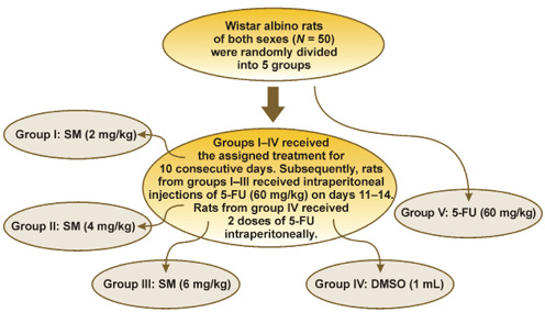
Wistar albino rats of both sexes were randomly assigned to 5 groups (n = 10/group). The first 3 groups received intraperitoneal injections of SM at doses of 2 mg/kg, 4 mg/kg and 6 mg/kg, respectively. Silymarin was dissolved in DMSO and administered in a total injection volume of 1 mL once daily for 10 consecutive days. Beginning on day 11, all 3 groups received 5-FU (60 mg/kg) once daily for 4 consecutive days (days 11–14). The 4th group received DMSO (1 mg/kg) for 10 days, followed by the intraperitoneal administration of 2 doses of 5-FU. The 5th group received 5-FU (60 mg/kg) only and served as the positive control for the induction of OM. The animals were anesthetized with diethyl ether, after which their left cheeks were exposed and gently injured using a 0.8-mm steel orthodontic wire (2×6 strokes). The rats were weighed weekly to adjust the administered dose and to monitor their general health. The flowchart of the study design is presented in Figure 1.
Preparation of the dosage and samples
Silymarin and 5-FU solutions were prepared daily under sterile conditions immediately before administration. The administered dose was adjusted according to each animal’s body weight. The rats were anesthetized with diethyl ether for approx. 5 min until loss of consciousness was achieved. Blood samples were collected from the optical vein using capillary tubes. The blood samples were immediately centrifuged at 5,200 rpm for 10 min, and the plasma was stored at −20°C until analysis.
The sample size was estimated using the G*Power software (https://www.psychologie.hhu.de/arbeitsgruppen/allgemeine-psychologie-und-arbeitspsychologie/gpower). Based on the calculated effect size, the minimum required sample size was 6 animals per group. To compensate for potential animal losses, the sample size was increased to 10 rats per group. Effect sizes were estimated using the mean values and standard deviations of the studies variables.24
Allocation and blinding
The animals were assigned to experimental groups using a computer-generated randomization sequence. Histopathological sections were independently evaluated by 2 blinded observers. The scores assigned for each section were then averaged and used for subsequent analyses.25
Histopathological study
The buccal mucosa and tongue tissues were fixed in 10% neutral buffered formalin, embedded in paraffin, and sectioned at a thickness of 4 μm.26 Sequential units were stained with hematoxylin and eosin (H&E) for general histological evaluation and Masson’s trichrome stain for the assessment of collagen deposition. Histological sections were examined using a microscope (Labophot-2; Nikon, Tokyo, Japan) and a camera (OptikamB9; Optika S.R.L, Ponteranica, Italy). The tongues were stained with 1% toluidine blue prepared in 10% acetic acid for 1 min and subsequently rinsed several times with acetic acid to identify ulcerative and erosive lesions. Ulcer size and epithelial thickness of the mucosa were measured.
Assessment of calprotectin and S100A12/EN-RAGE by ELISA
Plasma CP and S100A12/EN-RAGE levels were determined using commercial ELISA kits according to the manufacturers’ instructions.
Correlation analyses
Correlation analyses were performed between CP and S100A12/EN-RAGE levels and the studied parameters, including ulcer size and histopathological findings.
Statistical analysis
Data was analyzed using the IBM SPSS Statistics for Windows software, v. 25.0 (IBM Corp., Armonk, USA). Analysis of variance (ANOVA) was used to compare differences among the experimental groups. Pearson’s correlation coefficient was calculated to evaluate associations between biomarker levels and the studied parameters. When ANOVA demonstrated significant differences, Duncan’s test was used. Data was presented as the mean with the 95% confidence interval (CI). A p-value <0.05 was considered statistically significant. The G*Power 3.1.9.4. software was used to calculate the effect size and estimate the required sample size.
Results
A total of 50 Wistar albino rats, aged 8 weeks and weighing 250–350 g, were included in the study. The female-to-male ratio was 1:1.
Efficacy of various doses of silymarin against chemotherapy-induced oral mucositis
Effect of silymarin on body weight
Body weight was measured daily in all experimental groups from day 0 to day 14. No significant changes in body weight were observed in the SM-treated groups, whereas a significant reduction in body weight was detected in the control groups. A highly significant difference was noted between the SM-treated groups and the control groups, as shown in Table 1.
Effect of silymarin on food intake
Food intake was measured daily throughout the experimental period. The daily average food intake in groups II and III was slightly reduced, although the decrease was not statistically significant. In contrast, groups I, IV and V showed significantly lower food intake (p < 0.05). Comparison of groups II and III with the control groups demonstrated a highly significant difference (Table 1).
Macroscopic evaluation of chemotherapy-induced oral mucositis
Macroscopic appearance of oral mucositis ulcers
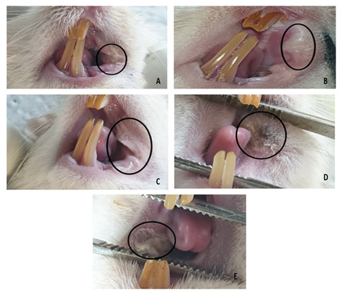
The left buccal mucosa of the rats was subjected to the intraperitoneal administration of 5-FU followed by mechanical injury, resulting in the development of visible ulcerative OM lesions. Random assignments were made to animals with a high macroscopic analysis score on the left side of their cheek pouch. Representative macroscopic findings are shown in Figure 2A–E. In group I (SM 2 mg/kg), only mild ulceration was observed, and the lesions healed rapidly. In groups II and III (SM 4 mg/kg and 6 mg/kg, respectively), no mucosal ulceration was detected, indicating a protective effect of SM (Figure 2A–C).
In contrast, the control groups (group IV, DMSO vehicle control, and group V, 5-FU only) exhibited severe clinical manifestations of OM, including erythema, extensive ulceration and abscess formation. Both groups showed marked mucosal ulceration following 5-FU injection in the absence of SM pre-treatment (Figure 2D,E).
Oral mucositis ulcer area
The dimensions of oral mucositis ulcers were measured using a digital caliper. Measurements were performed from day 3 to day 14 in the control groups and from day 11 to day 14 in the treatment groups. A highly significant reduction in ulcer area was observed in the SM-treated groups (I–III) compared with the control groups (p < 0.01). No significant differences in ulcer area were observed between the 2 control groups during the study period (Table 2).
Histopathological findings
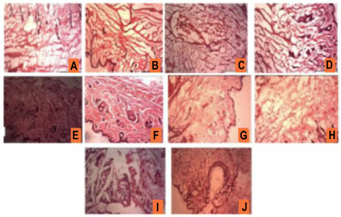
Histopathological examination demonstrated the architecture of the left (injured) and right (non-injured) buccal mucosae in all experimental groups (Figure 3A–J).
Group I: 2 mg/kg of silymarin
This group received the lowest dose of SM. Histopathological examination of the left buccal mucosa revealed vascular congestion and infiltration of acute inflammatory cells without evidence of ulceration (Figure 3A). The right buccal mucosa exhibited normal histological architecture (Figure 3B).
Group II: 4 mg/kg of silymarin
The left buccal mucosa showed vascular congestion with mild inflammatory cell infiltration (Figure 3C), whereas no histopathological abnormalities were observed in the right buccal mucosa (Figure 3D).
Group III: 6 mg/kg of silymarin
This group received the highest dose of SM. The left buccal mucosa demonstrated only mild inflammatory cell infiltration without ulcer formation (Figure 3E). The right buccal mucosa showed normal histological features with no evidence of inflammation or ulceration (Figure 3F).
Group IV: DMSO vehicle control
The left buccal mucosa showed marked infiltration of acute inflammatory cells, vascular congestion, ulceration, necrosis, and mild fibrosis of the subepithelial layer (Figure 3G). The right buccal mucosa demonstrated moderate inflammatory cell infiltration accompanied by mild fibrosis (Figure 3H).
Group V: 5-FU control
The left buccal mucosa exhibited extensive ulceration, necrosis, and dense inflammatory cell infiltration (Figure 3I). The right buccal mucosa also showed ulceration and necrosis, although inflammatory cell infiltration was less pronounced (Figure 3J).
Calprotectin and S100A12/EN-RAGE levels
Calprotectin
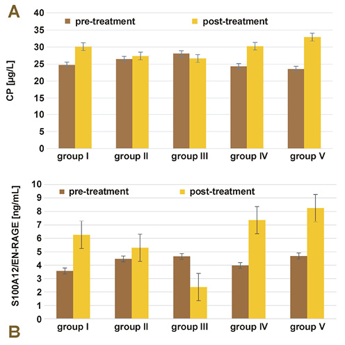
Groups I, II and III showed no significant changes in plasma CP levels compared with baseline. In contrast, plasma CP levels increased significantly in the control groups (groups IV and V) (p < 0.05), as shown in Figure 4A.
Dose-response analysis demonstrated that treatment with SM reduced plasma CP levels in a dose-dependent manner. The greatest reduction was observed in group III, followed by groups II and I. Analysis of variance demonstrated highly significant differences among the experimental groups (p < 0.01).
S100A12/EN-RAGE
Pre-treatment with SM at a dose of 6 mg/kg (group III) resulted in a highly significant reduction in plasma S100A12/EN-RAGE levels compared with baseline (p = 0.0001) and with the control groups (95% CI, p = 0.0002). There was an important growth in group I and a slight non-significant increase in group II compared to the baseline (Figure 4B).
Analysis of variance demonstrated highly significant differences among the experimental groups (p < 0.01). The strongest treatment effect was observed in group III, followed by groups II and I, indicating a dose-dependent effect of SM on plasma S100A12/EN-RAGE levels.
Correlation analysis
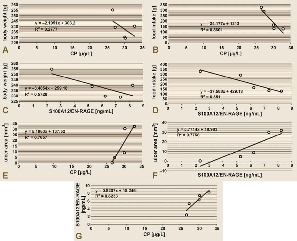
Pearson’s correlation coefficient (r) was used to evaluate the relationships between plasma biomarker CP and S100A12/EN-RAGE levels and the studied parameters, including body weight, food intake, ulcer area, and the correlation between the 2 biomarkers (Figure 5).
Correlation of calprotectin with body weight and food intake
There was a non-significant negative correlation between plasma CP levels and body weight (r = −0.527, p = 0.110) (Figure 5A). Similarly, plasma CP levels showed a non-significant negative correlation with food intake (r = −0.927, p = 0.060) (Figure 5B).
Correlation of S100A12/EN-RAGE with body weight and food intake
Plasma S100A12/EN-RAGE levels demonstrated a strong negative correlation with body weight (r = −0.756, p = 0.070), although the association was not statistically significant (Figure 5C).
Likewise, plasma S100A12/EN-RAGE levels showed a very strong negative correlation with food intake (r = −0.922, p = 0.060), without reaching statistical significance (Figure 5D).
Correlation of calprotectin and S100A12/EN-RAGE with oral mucositis ulcer area
Plasma CP levels showed a very strong positive correlation with OM ulcer area (r = 0.886, p < 0.0002) (Figure 5E). Similarly, plasma S100A12/EN-RAGE levels demonstrated a very strong positive correlation with ulcer area (r = 0.887, p < 0.0001) (Figure 5F).
Correlation between calprotectin and S100A12/EN-RAGE
Pearson’s correlation analysis demonstrated a very strong positive correlation between plasma CP and S100A12/EN-RAGE levels (r = 0.907, p < 0.0001) (Figure 5G).
Discussion
The present study is the first, to our knowledge, to evaluate the efficacy of different doses of SM as a mucoprotective agent against CIOM, while simultaneously assessing the plasma levels of inflammatory biomarkers CP and S100A12/EN-RAGE in an experimental animal model. Oral mucositis was induced using 5-FU. Previous studies have shown that 5-FU induces the production of pro-inflammatory cytokines, leading to basal epithelial cell damage, generation of ROS and inhibition of normal mucosal regeneration.27 The antioxidant and membrane-stabilizing properties of SM may therefore account for its protective effects observed in the present study.28
Chemotherapy-induced oral mucositis is one of the most common and clinically significant toxic adverse effects of anticancer therapy, resulting in extensive tissue damage.29 Previous studies have demonstrated that weight loss reaches its maximum between days 7 and 8 following chemotherapy, suggesting a link between the severity of mucositis and low immunological competence with weight loss.30 In the present study, body weight was measured daily in all groups throughout the experimental period. No significant changes in body weight were observed in the SM-treated groups, whereas significant weight loss was observed in the control groups. These results support the mucoprotective effect of SM.
Food intake was only slightly reduced in the SM-treated groups, whereas a significant reduction was observed in groups I, IV and V. These findings indicate the mucoprotective properties of SM, the efficacy of which was dose-dependent.
Regarding the development of OM, the severity of OM and ulcer size decreased progressively with increasing doses of SM. Rats treated with 2 mg/kg SM developed only mild ulcerative lesions, whereas no ulceration was observed in the groups receiving 4 mg/kg or 6 mg/kg, indicating a dose-dependent mucoprotective effect of SM.
Chemotherapeutic agents such as 5-FU interfere with DNA production primarily in rapidly proliferating cells, including oral epithelial cells. Consequently, mucosal injury is characterized by increased epithelial cell apoptosis and necrosis, accompanied by impaired epithelial regeneration.31 Consistent with these mechanisms, histopathological examination of SM-treated groups revealed only vascular congestion and mild inflammatory cell infiltration without ulcer formation. In contrast, the control groups exhibited severe inflammatory infiltration, vascular congestion, ulceration, necrosis, and mild subepithelial fibrosis.
Generation of ROS following treatment with chemotherapeutic agents such as 5-FU is considered a key even in the pathogenesis of OM, as demonstrated in numerous previous studies.32 After the administration of SM, attenuation of mucosal injury was observed. As a potent antioxidant, SM scavenges free radicals and reduces oxidative damage by modulating antioxidant defense systems and inflammatory mediators.32 Furthermore, SM is well-known for its antioxidant and chemoprotective effects on the liver.33
The antioxidant properties of SM promote tissue repair by stimulating RNA and protein synthesis. Silymarin also exhibits immunomodulatory and anti-inflammatory properties through inhibition of T-cell proliferation, suppression of NF-κB activation, and reduction of serum levels of inflammatory cytokines.34 The reduced inflammatory cell infiltration observed in the SM-treated groups is consistent with these biological activities and further supports the protective role of SM against chemotherapy-induced mucosal injury.
Dimethyl sulfoxide has been widely used as a pharmaceutical solvent, cryoprotectant and penetration enhancer, and has also been investigated for its potential therapeutic applications in oncology and tissue injury. In addition, it can be utilized in the palliative care and pain management sectors.35 In the current study, DMSO alone did not reduce the severity of OM, indicating that the observed protective effects were attributable to SM rather than the solvent vehicle.
Calprotectin is a neutrophil-derived inflammatory protein whose plasma concentration increases markedly in inflammatory and infectious conditions.36 The increase in the level of CP depends on the presence of inflammation. When leukocytes become stimulated as a direct result of pathology in different organs, the CP is released, leading to elevated concentrations in plasma and other biological fluids. Calprotectin has become an established biomarker of inflammation.37 In the present study, plasma CP levels significantly increased in the control groups due to the inflammation and mucositis but remained unchanged in the SM-treated groups. Furthermore, CP concentrations were significantly lower in the SM-treated groups than in the controls. These findings suggest that SM has protective effects on the mucous membrane, preventing inflammation and CIOM.
The association of S100A12 with the multi-ligand RAGE receptor and the sRAGE plays a vital role in pro-inflammatory pathways. In different inflammatory conditions, S100A12 is essential in local inflammatory responses, and serum S100A12 levels are elevated in chronic inflammatory diseases.38 Our findings are consistent with these observations. Plasma S100A12/EN-RAGE levels increase significantly in the control groups, whereas pre-treatment with 6 mg/kg SM resulted in a marked reduction compared with both baseline and the control groups. These results further support a dose-dependent anti-inflammatory effect of SM in experimental CIOM.
Correlation analysis demonstrated negative, although non-significant, associations between plasma CP and S100A12/EN-RAGE concentrations and both body weight and food intake. In contrast, both CP and S100A12/EN-RAGE levels showed very strong positive correlations with OM ulcer area. These findings suggest that both biomarkers reflect the severity of mucosal injury. An additional important finding was a highly significant correlation between plasma CP and S100A12/EN-RAGE levels.
Previous studies have shown that SM reduces inflammation and suppresses the production of pro-inflammatory cytokines.39 Bioactive constituents of S. marianum play a significant role in a wide range of pathological conditions, such as cancer.40 The findings of the present study are consistent with these reports and suggest that the mucoprotective effects of SM may be associated with attenuation of inflammation and oxidative stress, accompanied by reduced plasma CP and S100A12/EN-RAGE levels. These observations provide evidence for a potential additional mechanism underlying the protective effects of SM against CIOM.
Conclusions
Silymarin exerts dose-dependent mucoprotective effects against CIOM, which are associated with reduced CP and S100A12/EN-RAGE levels. Pre-treatment with SM attenuated inflammation and reduced the development of oral mucositis, as confirmed by both macroscopic and histopathological analyses. Furthermore, the very strong positive correlations between plasma CP and S100A12/EN-RAGE levels and OM ulcer area suggest that these biomarkers may have potential clinical value for assessing the severity of CIOM.
Ethics approval and consent to participate
The study protocol was approved by the Institutional Committee for Ethics in Animal Use at the Faculty of Pharmacy, Isra University, Amman, Jordan (registration No. 3-24/2019), and was conducted in accordance with the National Institutes of Health Guide for the Care and Use of Laboratory Animals.
Data availability
The datasets generated and/or analyzed during the current study are available from the corresponding author on reasonable request.
Consent for publication
Not applicable.
Use of AI and AI-assisted technologies
Not applicable.