Abstract
Background. The effective management of hemostasis is essential in surgical practice. Topical hemostatic agents become indispensable when conventional methods are insufficient or impractical. However, the wide range of commercially available products with similar compositional bases presents clinicians with numerous choices and potentially variable performance profiles. Notably, to the best of the authors’ knowledge, no previous study has comprehensively compared the biological functionality of these commercially available materials. Addressing this knowledge gap is essential for improving the precision and effectiveness of hemostatic interventions, ultimately contributing to better patient outcomes and advancing healthcare practice.
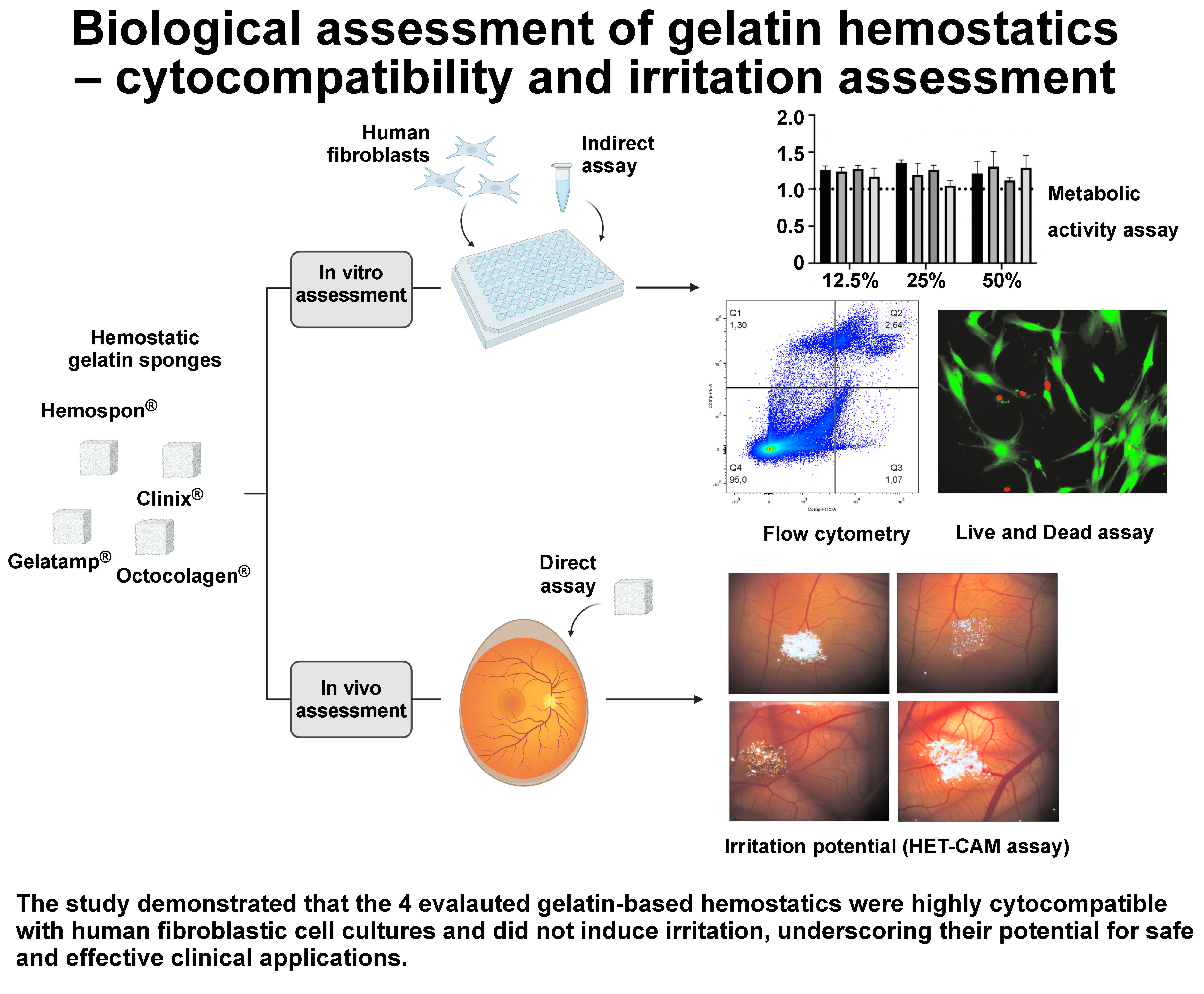
Objectives. This study aimed to conduct a comprehensive evaluation of the biological response to clinically available gelatin-based hemostatic agents, namely Hemospon®, Clinix®, Gelatamp®, and Octocolagen®. The evaluation encompassed in vitro studies to characterize the response of human fibroblasts upon exposure, together with an in vivo irritation assessment using the hen’s egg test – chorioallantoic membrane (HET-CAM) assay, thereby providing a comprehensive understanding of their biological effects.
Material and methods. Human fibroblasts were cultured and exposed to the leachables extracted from the hemostatic agents, prepared according to the ISO 10993-12:2021 guidelines, at concentrations of 50%, 25% and 12.5%. The cultures were characterized in terms of cellular viability (the live/dead assay and flow cytometry) and metabolic activity at multiple time points. In addition, irritation potential was evaluated using the HET-CAM assay.
Results. Data analysis revealed that the fibroblast cultures exposed to the leachables from the 4 hemostatic sponges proliferated actively throughout the culture period, with minimal evidence of cell death or significant impairment of metabolic activity. Furthermore, no irritant effects were observed in the HET-CAM assay.
Conclusions. Overall, despite minor variations in the biological response, the comprehensive analysis supports the overall biocompatibility of commercially available gelatin-based hemostatic sponges. These findings advance the current understanding of the biological safety of these materials, further reinforcing their clinical suitability and highlighting their potential to improve surgical healing outcomes.
Keywords: cytotoxicity, hemostatics, gelatin sponges, biological assays, chorioallantoic membrane
Introduction
The management of hemostasis remains a critical concern in surgical practice. The challenges associated with bleeding complications are further amplified in patients with altered hemostasis resulting from the underlying pathological conditions or ongoing pharmacological therapies. Beyond compromising surgical precision, such complications may impair wound healing and ultimately pose serious, potentially life-threatening risks.1 In this context, topical hemostatic agents assume a pivotal role. These agents are particularly valuable in situations where conventional methods, including pressure, electrocautery and suture ligatures, are insufficient or impractical.2 By enhancing and accelerating the coagulation process, topical hemostatic agents provide an effective adjunctive intervention that not only minimizes adverse effects, but also promotes rapid and efficient hemostasis, representing a significant advancement in the pursuit of optimal surgical outcomes.3
Topical hemostatic agents are broadly classified into active, non-active, and hybrid categories, each characterized by distinct mechanisms of action, efficacy, safety profiles, and formulations.4 Active hemostatic agents, such as those containing fibrinogen and thrombin, act directly within the coagulation cascade, and are therefore classified as adhesive hemostatics because of their ability to seal tissues.2, 5 Non-active hemostatic agents, including collagen, gelatin and regenerated oxidized cellulose, are regarded as passive hemostatic agents. These materials mechanically promote platelet adhesion, aggregation and activation, ultimately leading to the formation of a stable clot.2, 5 A major advantage of mechanical agents is their simplicity and immediate availability for use directly from the package, without the need for specialized storage conditions or pre-application preparation.6 This characteristic makes them particularly suitable for the management of minor bleeding events.7 Hybrid hemostatic agents combine the properties of both active and non-active materials; examples include formulations that integrate thrombin with mechanical hemostatic matrices. Commonly referred to as flowable hemostatics, these agents provide versatile options for hemostasis management.2, 8, 9
Within the category of non-active hemostatic agents, a variety of products have historically been employed, including bone wax and cellulose-, collagen-, and gelatin-based materials. Bone wax, primarily used to mechanically occlude vascular channels in bone, effectively controls bleeding, but does not actively participate in the hemostatic process. Moreover, its insoluble and non-resorbable nature is associated with several drawbacks, including an increased risk of inflammation and infection.10, 11, 12 Oxidized cellulose products, another important class of non-active agents, present limitations related to the low-pH microenvironment they generate, which may inactivate biologically active hemostatic agents such as thrombin. Consequently, their combined use with other agents is restricted, while their limited resorbability often necessitates post-surgical removal.13, 14 Microfibrillar collagen, although effective, demonstrates relatively high immunogenicity due to its xenogeneic origin, and is also more costly than its denatured derivative, gelatin, which is characterized by lower immunogenic potential.15, 16
Against this background, gelatin-based hemostatic agents have emerged as a preferred option because of their favorable biological and handling properties. These agents are derived from denatured xenogeneic collagen through processes that disrupt its native triple-helical structure into single strands by means of thermal denaturation or physical and chemical degradation.17 Gelatin-based materials exhibit remarkable hygroscopicity, enabling them to absorb large quantities of blood and other fluids, and to expand up to 200% of their original volume.7, 8 In addition, gelatin matrices are associated with minimal tissue reaction, undergo disintegration within approx. 1 week in the oral environment and are completely resorbed within 4–6 weeks, thereby offering a highly favorable profile for postoperative healing and recovery.18, 19
Despite the widespread use of gelatin-based hemostatic agents, a significant knowledge gap persists, particularly concerning their comparative efficacy. The large number of commercially available products sharing similar compositional foundations provides clinicians with a broad range of options that may nevertheless differ in performance characteristics.20 A clear understanding of the clinical effectiveness, advantages and limitations of hemostatic products derived from the same material is essential to support appropriate clinical selection and use, ultimately contributing to improved patient outcomes.21 Addressing this knowledge gap is therefore crucial for enhancing the precision and overall effectiveness of hemostatic interventions in surgical practice.
Therefore, the present study aimed to provide a comprehensive evaluation of the biological activity of clinically available gelatin-based hemostatic agents. Through in vitro investigations focused on the functional activity of human fibroblasts, together with an in vivo irritation assessment using the hen’s egg test – chorioallantoic membrane (HET-CAM) assay, this work seeks to generate valuable insights into the biological responses elicited by these materials. Such findings are fundamental for supporting informed clinical decision making, and may contribute significantly to the advancement and optimization of hemostatic agents in surgical practice.
Material and methods
In the present study, gelatin-based hemostatic sponges from 4 different manufacturers were evaluated, namely Hemospon® (Technew, Rio de Janeiro, Brazil), Clinix® (Biomaterials, Pisa, Italy), Gelatamp® (Roeke, Langenau, Germany), and Octocolagen® (Laboratorios Clarben, Madrid, Spain). The leachables obtained from these materials were employed for the in vitro assays, whereas the sponges themselves were directly applied in the in vivo irritation assay.
Preparation of leachables from hemostatic sponges
The leachables were prepared from the hemostatic sponges in accordance with the ISO 10993-12:2021 guidelines.22 Briefly, the sponges were immersed in alpha-Minimum Essential Medium (α-MEM; Gibco, Waltham, USA) and incubated for 24 h at 37°C under constant agitation (100 rpm). Following incubation, the leachables were collected and stored at −20°C until further use.
Establishment of cell cultures
Primary human gingival fibroblasts (AG09319; Coriell Institute for Medical Research, Camden, USA) were cultured in a growth medium consisting of α-MEM supplemented with 10% fetal bovine serum (FBS), 100 UI/mL penicillin, 100 UI/mL streptomycin, and 2.5 µg/mL amphotericin B (all from Gibco). The cell cultures were maintained at 37°C in a humidified atmosphere containing 5% CO2 until reaching approx. 70% confluence. The culture medium was renewed twice weekly. At the selected confluence, the cells were washed twice with phosphate-buffered saline (PBS, pH 7.4; Sigma-Aldrich, St. Louis, USA), and subsequently incubated with trypsin solution (0.05% trypsin in 0.25% ethylenediaminetetraacetic acid (EDTA); Sigma-Aldrich) for 5 min at 37°C to promote cell detachment. Trypsin was then neutralized, and the resulting cell suspension was centrifuged at 1,200 rpm for 10 min. The obtained cell pellet was resuspended in the growth medium, and the cells were seeded at a density of 104 cells/cm2 under the previously described culture conditions. After 24 h of incubation, the culture medium was replaced with the hemostatic sponge leachables diluted in α-MEM to final concentrations of 50%, 25% and 12.5%. The cells cultured in the growth medium alone, without hemostatic leachables, served as the negative control.
Analysis of cell viability by the live/dead assay
Cell viability was evaluated using the live/dead viability assay, following 24 h of exposure to the hemostatic sponge leachables. Briefly, cells were washed twice with PBS and incubated for 10 min at 37°C, protected from light, in a solution containing 2 μL/mL calcein AM (BioLegend, San Diego, USA) and 50 µL/mL propidium iodide (PI; BD Biosciences, Franklin Lakes, USA). Fluorescence images of viable and non-viable cells were immediately acquired using the CELENA® S digital imaging system (Logos Biosystems, Gunpo, South Korea) equipped with green (488 nm) and red (594 nm) filters. All experiments were performed in quintuplicate.
Five randomly selected images from each experimental condition were analyzed using ImageJ, v. 1.8 (National Institutes of Health – NIH) to quantitatively determine live and dead cell events. Channel separation was performed to isolate the fluorescence signals corresponding to viable (green) and non-viable (red) cells. Subsequently, image thresholding was applied using the MaxEntropy method, and the images were converted into a binary format. The “Analyze Particles” function was then used to detect, segment and quantify cellular events.
The percentage of cell death was calculated by dividing the number of events detected in the dead cell channel by the total number of events identified in both channels, and multiplying the result by 100.
Assessment of cell viability by flow cytometry
Cell viability was further evaluated by flow cytometry, following 24 h of exposure to the hemostatic sponge leachables. Briefly, the cells were stained using the Annexin V-FITC (fluorescein isothiocyanate) apoptosis detection kit (640914; BioLegend), according to the manufacturer’s instructions, and subsequently analyzed by flow cytometry using the FACSCalibur™ cytometer (BD Biosciences).
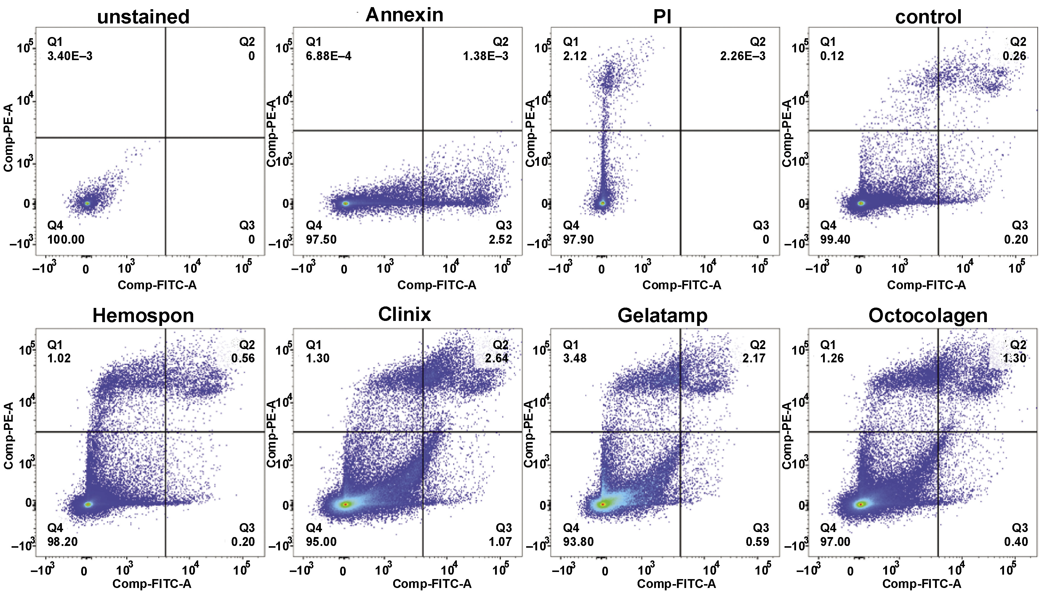
During early apoptosis, caspase activation and other proteolytic events lead to cytoskeletal cleavage and the loss of membrane rigidity, enabling the translocation of phosphatidylserine (PS) from the inner to the outer leaflet of the plasma membrane, where it is detected by Annexin V (Annexin V-FITC+/PI–; early apoptotic cells). In later stages, the loss of membrane integrity results in impaired exclusion of PI, corresponding to Annexin V-FITC+/PI– populations indicative of late apoptosis and/or necrosis. Viable cells remain unstained (Annexin V-FITC+/PI–).
Data acquisition and analysis were performed using FlowJo, v. 10.9 (BD Biosciences). All experiments were conducted in quintuplicate.
Evaluation of metabolic activity
Metabolic activity was quantitatively assessed using the MTT (3-(4,5-dimethylthiazol-2-yl)-2,5-diphenyltetrazolium bromide) assay on days 3 and 7 of culture, based on the reduction of MTT to a purple formazan product by metabolically active cells. Briefly, at each time point, the cells were incubated for 3 h at 37°C in a humidified atmosphere containing 5% CO2 in air, with 5 mg/mL MTT stock solution (Sigma-Aldrich). Following incubation, the resulting formazan crystals were solubilized in 100 μL of dimethyl sulfoxide (DMSO; Panreac, Barcelona, Spain), and absorbance was measured at 550 nm using the Synergy HT microplate reader (BioTek, Winooski, USA). All experiments were performed in quintuplicate.
In vivo assessment of the irritation potential – HET-CAM assay
The irritation potential of the hemostatic sponges was evaluated directly using the in vivo HET-CAM assay, in accordance with the Interagency Coordinating Committee on the Validation of Alternative Methods (ICCVAM) guidelines.23 Briefly, freshly laid fertilized chicken eggs were incubated at 37°C under 60% humidity in the Octagon Advance incubator (Brinsea, Sommerset, UK), with automatic rotation set at 1-hour intervals. On day 8 post-fertilization, a small window was created in the eggshell under aseptic conditions, and a fragment of each hemostatic sponge (1 mm3) was carefully placed onto the CAM of the chicken embryo. The chorioallantoic membrane was subsequently examined by visual inspection and documented using the Stemi 305 stereomicroscope equipped with the Axiocam 208 camera (Zeiss, Oberkochen, Germany), at contact times of 0.5, 2 and 5 min.
Irritation was assessed based on the presence of lysis, hemorrhage or coagulation. A scoring system according to the Luepke method,24 ranging from 0 (no reaction) to 21 (strong reaction), was used for irritation grading. A 0.9% NaCl solution served as the negative control, while 0.5 M NaOH was used as the positive control, in accordance with the ICCVAM recommendations. All experiments were performed in quintuplicate.
Statistical analysis
All quantitative data is presented as mean ± standard deviation (M ±SD), unless otherwise specified. Statistical analyses were performed using the GraphPad Prism 8 software (GraphPad, Boston, USA), employing the one-way analysis of variance (ANOVA) followed by Bonferroni’s multiple comparisons test. A p-value <0.05 was considered statistically significant.
Results
Analysis of cell viability by the live/dead assay
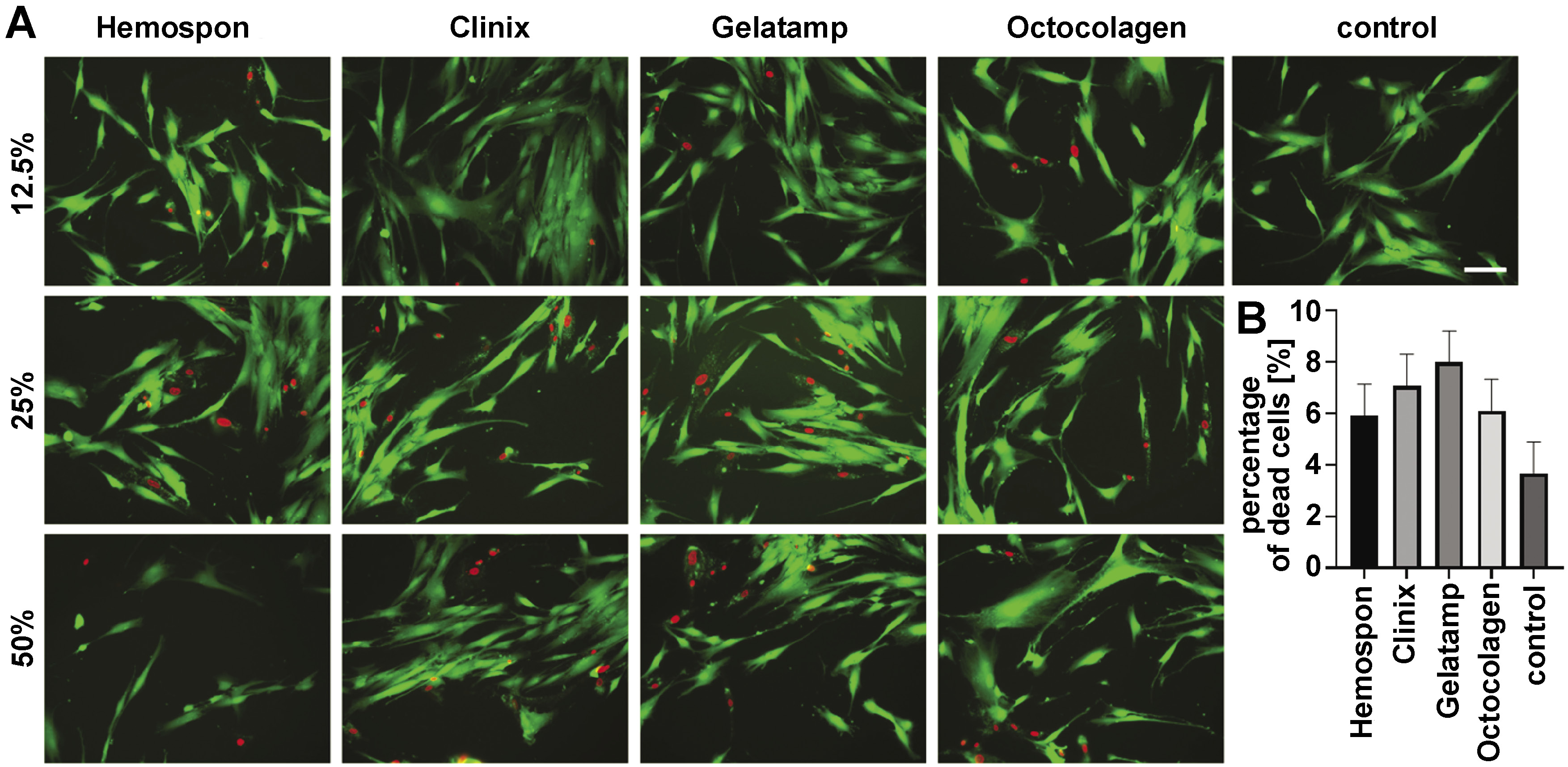
Cell viability following exposure to different concentrations (50%, 25% and 12.5%) of hemostatic sponge leachables was evaluated by live/dead double staining after a 24-hour incubation period. Under control conditions, the cultures exhibited a predominance of viable cells, as indicated by the extensive green fluorescence, with only sporadic dead cells observed throughout the culture. In comparison, the cultures exposed to the leachables demonstrated a higher frequency of cell death events. Nevertheless, despite this trend, all experimental groups maintained high levels of cell viability, comparable to those observed in the controls (Figure 1).
Analysis of cell viability by flow cytometry
Complementing the live/dead assay, cell viability was further evaluated by flow cytometry to distinguish viable cells from apoptotic and necrotic populations. The control cultures exhibited a high viability of approx. 99%. Similarly, the cultures exposed to the sponge leachables also maintained high viability levels, ranging from 94% to 98%, with no significant differences relative to the controls. Nevertheless, the cultures exposed to Clinix (95.00% viable cells, 1.07% early apoptotic cells, 2.64% late apoptotic cells, and 1.30% necrotic cells) and Gelatamp (93.80% viable cells, 0.59% early apoptotic cells, 2.17% late apoptotic cells, and 3.48% necrotic cells) demonstrated comparatively higher proportions of late apoptotic and necrotic cells than the cultures exposed to Hemospon (98.20% viable cells, 0.20% early apoptotic cells, 0.56% late apoptotic cells, and 1.02% necrotic cells) and Octocolagen (97.00% viable cells, 0.40% early apoptotic cells, 1.30% late apoptotic cells, and 1.26% necrotic cells) (Figure 2).
Analysis of metabolic activity
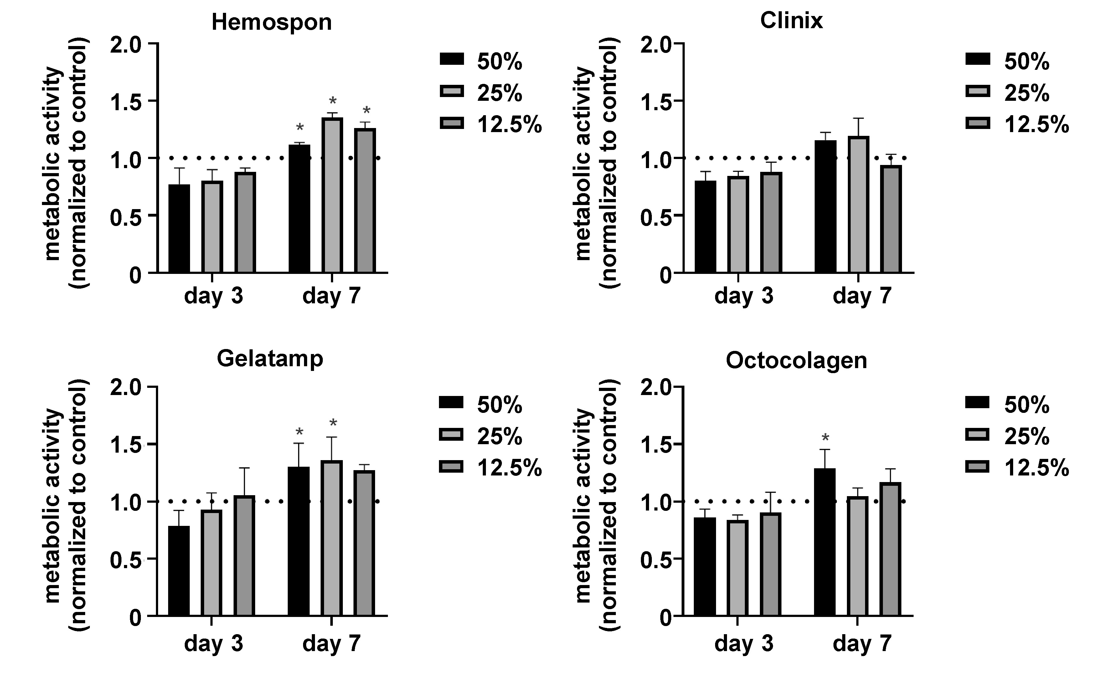
The metabolic activity of the cultures exposed to the hemostatic sponge leachables was assessed using the MTT assay. Under control conditions, the cells demonstrated a progressive increase in metabolic activity from day 3 to day 7 of culture. On day 3, the cultures exposed to the highest leachable concentration exhibited a tendency toward reduced metabolic activity across all tested materials, an effect that became less pronounced at lower concentrations. By day 7, the leachables from Hemospon induced significantly higher metabolic activity levels, a trend that was also observed for higher concentrations of the Gelatamp and Octocolagen leachables (Figure 3).
Analysis of the irritation potential – in vivo HET-CAM assay
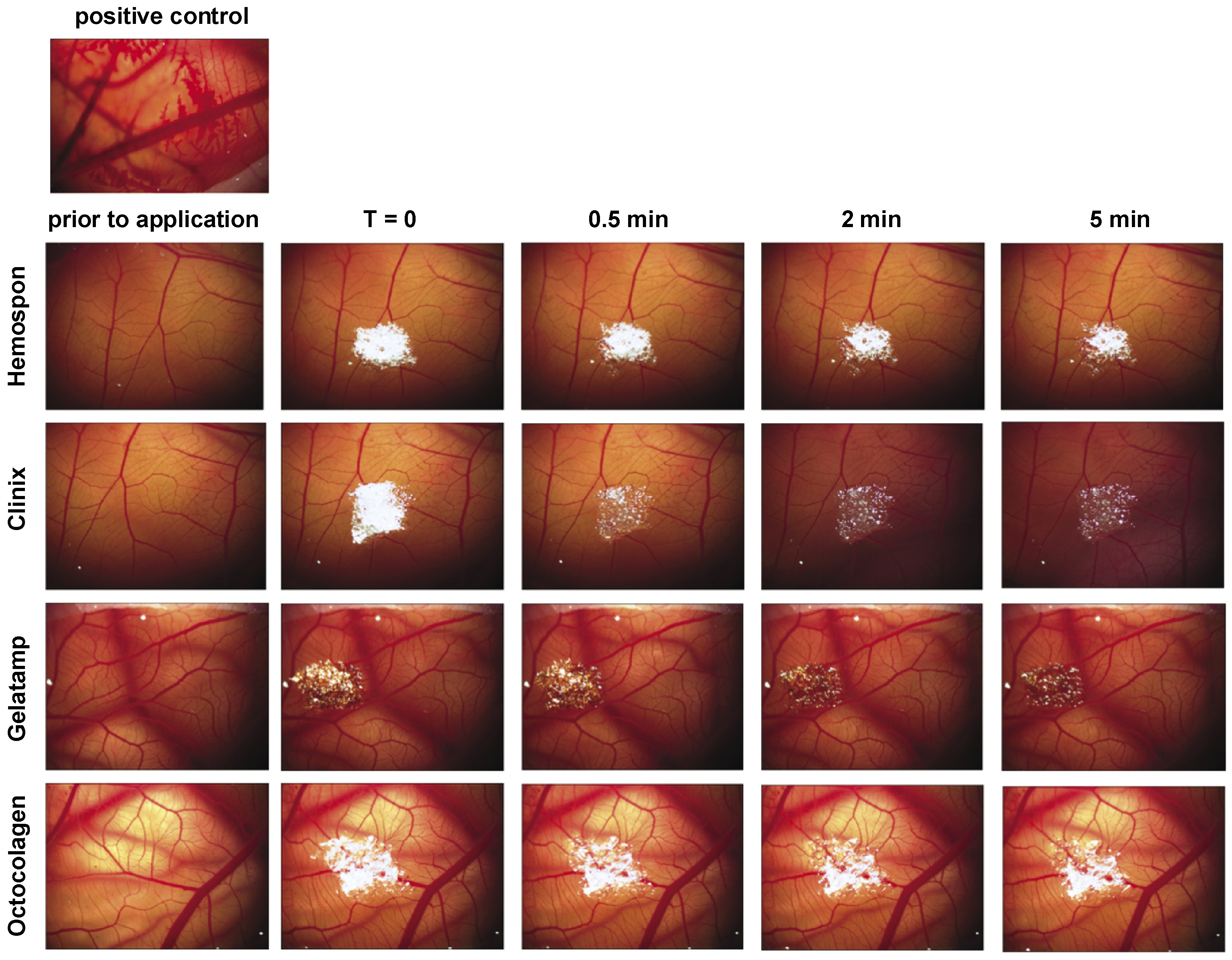
The in vivo HET-CAM assay was performed to evaluate the irritation potential of the hemostatic sponges, based on the occurrence of lysis, hemorrhage or coagulation. The test solutions were quantitatively graded according to the Luepke method (Table 1 and Table 2). The negative control (0.9% NaCl) produced no visible reaction throughout the 5-minute application period, and was therefore assigned a score of 0, corresponding to the absence of irritation. In contrast, the positive control (0.5 M NaOH) induced a severe hemorrhagic response, resulting in a score of 19, indicative of severe irritation. Regarding the hemostatic sponges, none of the tested materials induced visible signs of irritation within the duration of the assay. Consequently, all materials received a score of 0, similarly to the negative control (Table 3, Figure 4).
Discussion
Surgical bleeding can significantly affect both patient outcomes and healthcare costs, with inadequate hemorrhage control being closely associated with adverse outcomes and increased morbidity.20 In this context, gelatin-based hemostatic agents represent a valuable class among non-active hemostatic materials, helping to overcome clinical challenges while contributing to improved therapeutic outcomes and more cost-effective healthcare practices. Their high absorbency, minimal tissue reactivity and gradual resorption profile are particularly advantageous for achieving effective hemostasis and supporting postoperative healing.25 Despite their widespread clinical use, an important knowledge gap remains regarding the comparative biological performance of commonly used gelatin-based hemostatic agents. Accordingly, the present study aimed to biologically characterize 4 commercially available lyophilized porcine gelatin sponges – Hemospon, Clinix, Gelatamp, and Octocolagen – through cytocompatibility and irritation assays.
Human fibroblast cultures represent a relevant model for evaluating the biological response to materials intended for tissue contact. As the predominant cellular component of connective tissues, fibroblasts play a fundamental role in maintaining tissue structure and integrity, while also contributing significantly to tissue repair and regenerative processes during healing.26, 27 To assess fibroblast functionality, live/dead staining and flow cytometry analyses were performed at early culture time points, followed by metabolic activity evaluation at later stages of culture.
Viability assays demonstrated the absence of significant cytotoxic effects in the cultures exposed to the hemostatic sponge leachables when compared with the control condition at early culture time points. Although the exposed cultures exhibited a slightly greater number of dead cells relative to the control, these differences were not statistically significant, as confirmed by both the live/dead assay and flow cytometry. The evaluation of cell viability is a critical component of cytotoxicity assessment, with fluorescence-based microscopic and flow cytometric methods being widely recognized as reliable and effective approaches that demonstrate high correlation between datasets.28 These findings are consistent with those reported by Sezer et al., who evaluated different gelatin-based hemostatic agents using human umbilical vein endothelial cells (hUVECs), and observed no significant differences among groups with respect to cell death.29 Similarly, Awad et al. reported high viability levels (>95%) in human adipose-derived stem cells (hADSCs) exposed to a gelatin sponge.30 In addition, diluted extracts from a gelatin-based hemostatic agent were shown not to significantly affect the viability of rodent fibroblasts, following 24 h of exposure.31
Regarding the assessment of metabolic activity at later culture time points, the MTT assay revealed a consistent pattern across all 4 tested materials throughout the 7-day evaluation period. On day 3, the cultures exposed to more diluted extracts exhibited no significant differences relative to the control condition, whereas a tendency toward reduced MTT values was observed, particularly at higher extract concentrations. Nevertheless, these values remained above the 70% threshold established by ISO 10993-5:2009, below which a material or extract is considered cytotoxic.32 By day 7, the cultures exposed to all extract concentrations demonstrated metabolic activity levels comparable to or exceeding those of the control group. These findings further support the preservation of adequate cellular functionality in the presence of the sponge leachables. The initial reduction tendency observed in the MTT values may be associated with cumulative exposure within the in vitro culture environment, potentially resulting in concentrations exceeding those expected under clinical conditions, where continuous dilution and clearance by biological fluids would occur following application. At the later culture stage, however, a tendency toward increased metabolic activity became evident, corroborating previous findings reported by Sezer et al.29 and Lan et al.33 These authors evaluated hUVECs and murine fibroblasts, respectively, following exposure to gelatin-based materials, and reported favorable cytocompatibility without evidence of cytotoxicity.29, 33 This pattern is consistent with the well-documented ability of gelatin-based materials and their degradation products to stimulate cellular activity and support tissue healing processes.34, 35
Beyond the in vitro cytocompatibility assessment, irritation assessment – an essential component of the biocompatibility and biosafety evaluation of biomaterials and medical devices – was performed using the HET-CAM assay. This method has been widely recognized as a rapid, simple and reliable alternative approach for irritation assessment across different experimental settings.36, 37, 38 Accordingly, the hemostatic agents were evaluated following direct CAM exposure, with no evidence of hyperemia or irritative responses, including vessel lysis, hemorrhage or coagulation, throughout the duration of the assay. These findings are consistent with previous reports, such as the study by Rodrigues da Silva et al., who also employed the CAM assay to evaluate the irritation potential of a gelatin membrane, and observed no signs of hyperemia, hemorrhage or coagulation.39 Moreover, the extensive use of gelatin sponges in CAM-based studies, extending beyond irritation testing to applications involving angiogenesis assessment, further highlights their role as inert substrate materials capable of serving as platforms for the controlled delivery of cells or bioactive substances under investigation.40, 41, 42 Collectively, these observations reinforce the inert and non-irritant nature of gelatin-based sponges, and further support their suitability for a wide range of biomedical applications.
Conclusions
This comprehensive study characterized the biological response to 4 commercially available gelatin-based hemostatic materials, with particular emphasis on their effects on human fibroblast functionality and irritation potential. The findings demonstrated high cytocompatibility, consistent cellular responses and sustained metabolic activity, in agreement with the well-established capacity of gelatin-based materials to support tissue healing processes. The irritation assessment further confirmed the biocompatibility of these gelatin sponges, highlighting their inert nature and their inability to induce alterations in a well-established vascular tissue model. Although minor differences were observed among the tested materials regarding the biological response, the overall favorable biocompatibility profile reinforces the safety and effectiveness of commercially available gelatin-based hemostatic agents. Collectively, these findings support their suitability for clinical applications and their potential contribution to improved surgical outcomes.
Ethics approval and consent to participate
Not applicable.
Data availability
The datasets supporting the findings of the current study are available from the corresponding author on reasonable request.
Consent for publication
Not applicable.
Use of AI and AI-assisted technologies
Not applicable.