Abstract
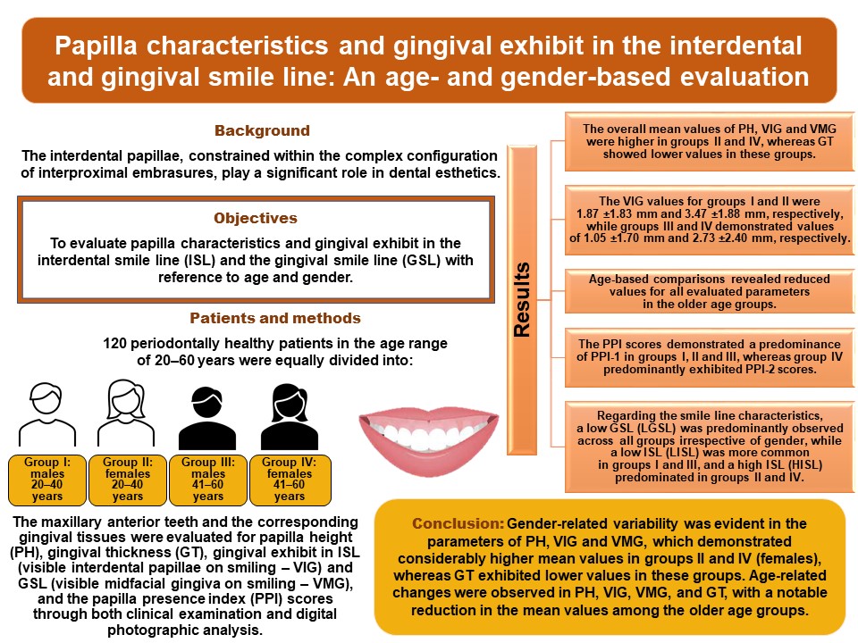
Background. The interdental papillae, constrained within the complex configuration of interproximal embrasures, play a significant role in dental esthetics.
Objectives. The aim of the present study was to evaluate papilla characteristics and gingival exhibit in the interdental smile line (ISL) and the gingival smile line (GSL) with reference to age and gender.
Material and methods. A total of 120 periodontally healthy patients aged 20–60 years were equally divided into 4 groups: group I – male patients aged 20–40 years; group II – female patients aged 20–40 years; group III – male patients aged 41–60 years; and group IV – female patients aged 41–60 years. The maxillary anterior teeth and the corresponding gingival tissues were evaluated for papilla height (PH), gingival thickness (GT), gingival exhibit in ISL (visible interdental papillae on smiling – VIG) and GSL (visible midfacial gingiva on smiling – VMG), and the papilla presence index (PPI) scores through both clinical examination and digital photographic analysis.
Results. The overall mean values of PH, VIG and VMG were higher in groups II and IV, whereas GT showed lower values in these groups. The VIG values for groups I and II were 1.87 ±1.83 mm and 3.47 ±1.88 mm, respectively, while groups III and IV demonstrated values of 1.05 ±1.70 mm and 2.73 ±2.40 mm, respectively. Age-based comparisons revealed reduced values for all evaluated parameters in the older age groups. The PPI scores demonstrated a predominance of PPI-1 in groups I, II and III, whereas group IV predominantly exhibited PPI-2 scores. Regarding the smile line characteristics, a low GSL (LGSL) was predominantly observed across all groups irrespective of gender, while a low ISL (LISL) was more common in groups I and III, and a high ISL (HISL) predominated in groups II and IV.
Conclusions. Gender-related variability was evident in the parameters of PH, VIG and VMG, which demonstrated considerably higher mean values in groups II and IV (females), whereas GT exhibited lower values in these groups. Age-related changes were observed in PH, VIG, VMG, and GT, with a notable reduction in the mean values among the older age groups.
Keywords: gingival thickness, esthetics, papilla height, gingival smile line, interdental smile line
Introduction
A smile transcends cultural and linguistic boundaries, and is universally recognized as a form of personal greeting. Individuals who smile are often perceived as happier, more pleasant and more trustworthy, reflecting an overall positive personality. The esthetic impact of an attractive smile extends beyond appearance, influencing an individual’s social, physical and psychological well-being.1, 2, 3
The increasing emphasis on smile esthetics has grown alongside greater public awareness, further amplified by media influence and digital editing platforms that allow patients to visualize and seek the desired esthetic outcomes with ease. In parallel, advancements in periodontal, orthodontic, restorative, and implant therapies have equipped clinicians with a wide range of treatment modalities to meet patients’ expectations for an attractive smile. Nevertheless, each patient presents unique characteristics and esthetic preferences, requiring clinicians to approach smile esthetics through an interdisciplinary perspective to achieve optimal effects. A multidisciplinary assessment and treatment approach facilitates comprehensive rehabilitation aimed at enhancing esthetics, particularly in cases with multifactorial underlying causes.4
The principal components of an attractive smile include the lip framework, the maxillary anterior teeth and gingival scaffold, all of which should be proportionate and harmonious with one another and with the surrounding facial structures.5, 6, 7 The parabolic contour of the gingiva is primarily determined by the position of the gingival margin, which exhibits a defined spatial orientation in both the anteroposterior and apicocoronal dimensions.8 The display of the teeth and gingival scaffold through the lip framework constitutes the “white” and “pink” components of the smile, both of which are essential elements of smile esthetics.
In addition, several parameters are known to influence smile characteristics, including the smile line, the smile arc, lip curvature, the gingival zenith position, gingival display, and tooth display at rest and during smiling. The interdental papillae, constrained within the complex anatomy of the interproximal embrasures, also play a significant role in esthetics.9, 10, 11 Although each parameter may be evaluated individually, all components must function in harmony to create the integrity required for an optimal final esthetic effect. Furthermore, these smile characteristics may vary among different populations, highlighting the importance of considering ethnicity as a relevant factor in the smile assessment.
For a more detailed assessment of smile esthetics, Hochman et al. proposed the classification of smiles into the interdental smile line (ISL) and the gingival smile line (GSL), based on the gingival tissue display of the interdental and midfacial gingiva during smiling.12 Preliminary studies evaluating tooth and gingival display in relation to ISL and GSL demonstrated that most participants exhibited a high ISL and a low GSL, suggesting a greater esthetic significance of the interdental papillae as compared to the midfacial gingiva.13
In addition, Cardaropoli et al. introduced the papilla presence index (PPI), a classification system based on the positional relationship among the interdental papilla, the cementoenamel junction (CEJ) and the adjacent teeth.14 However, the association between PPI and smile esthetics has not been extensively investigated, and the available literature on this topic remains limited.
Although substantial evidence exists regarding the factors influencing smile esthetics, there is still a paucity of literature exploring the interrelationship among these parameters. Understanding these associations may provide valuable clinical guidance for periodontal and restorative procedures in the maxillary esthetic zone. Given the close relationship between smile esthetics and gingival as well as interdental papillary display, the present study was designed to evaluate papilla presence and gingival exhibit in ISL and GSL with reference to age and gender.
Material and methods
A total of 120 periodontally healthy patients aged 20–60 years were recruited from the outpatient clinics of the Department of Periodontics and Implant Dentistry of Ranjeet Deshmukh Dental College and Research Centre, Nagpur, India. The study protocol and procedures were thoroughly explained to all participants, and written informed consent was obtained from each patient prior to enrollment. The study protocol adhered to the principles of the Declaration of Helsinki, and was reviewed and approved by the Institutional Ethics Committee. The study was also registered with the Clinical Trials Registry – India.
Sample size
A sample size of approx. 106 participants was estimated to provide a 90% confidence level, a 7.5% margin of error and an effect size of 0.31, based on the study conducted by Kolte et al., in which gingival exhibit and lip dimensions in the maxillary anterior region were evaluated and categorized according to gender.2 Considering these parameters, a final sample size of 120 participants was determined for the present study.
Eligibility criteria
Participants were enrolled in the clinical trial after fulfilling the predefined eligibility criteria. The inclusion criteria comprised: (a) fully erupted maxillary anterior teeth; (b) properly aligned teeth; (c) the absence of anatomical or pathological abnormalities15, 16; (d) clinically healthy periodontal tissues, as determined by the plaque index (PI)17 and the gingival index (GI)18 scores ranging from 0 to 1; and (e) the absence of dental restorations in the evaluated teeth. The exclusion criteria were as follows: (a) a history of periodontal surgical therapy; (b) previous orthodontic treatment; (c) the presence of systemic diseases, such as diabetes mellitus or hypertension; (d) the use of medications known to influence gingival tissues, including nifedipine, cyclosporine and phenytoin; and (e) facial asymmetry.
Grouping
A total of 120 periodontally healthy study participants who fulfilled the inclusion criteria were divided in to 4 groups: group I – 30 male patients aged 20–40 years; group II – 30 female patients aged 20–40 years; group III – 30 male patients aged 41–60 years; and group IV – 30 female patients aged 41–60 years.
Outcome variables
The primary outcome variables included the assessment of the PPI scores and gingival exhibit in ISL and GSL. The secondary outcome variables comprised the correlation of papilla height (PH) and gingival thickness (GT) with age and gender, as well as the association of gingival display with age, gender, ISL, and GSL within the study population.
Procedures
At the initial visit, all patients were evaluated for eligibility according to the study inclusion criteria, and standardized frontal digital photographs were obtained using a Canon camera equipped with a Canon EF 50 mm f/1.8 STM lens and built-in flash (Canon, Tokyo, Japan). The camera was positioned at a right angle to the patient to ensure image standardization. To maintain consistency, patients were seated at a distance of approx. 3 feet to avoid intrusion into personal space. Photographs were captured both at physiological rest and during smiling. The images were subsequently analyzed using Adobe Photoshop (Adobe, San Jose, USA) under a resolution of 1,680 × 1,050 pixels and an enlargement ratio of 1:1.2. Initial examinations and recording of study parameters were performed by 2 calibrated clinicians (APK and SPM), who underwent calibration prior to the commencement of the study. The following parameters were recorded:
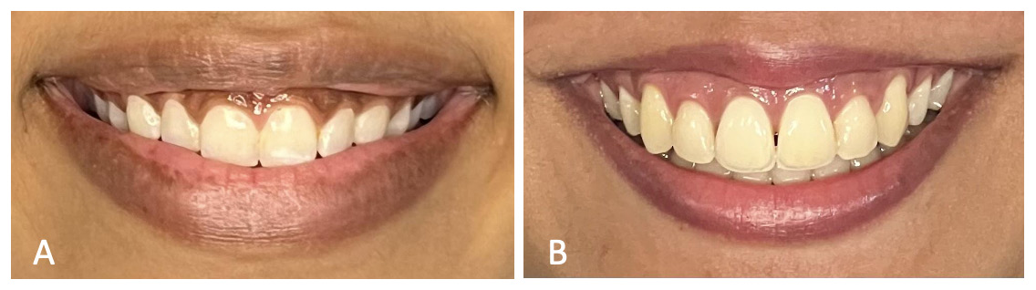
(1) ISL: The interdental smile line was assessed based on the visibility of the interdental papillae during smiling and was categorized into 3 groups: high ISL (HISL), when the interdental papillae of the maxillary anterior teeth were visible; low ISL (LISL), when no interdental papillary display was observed; and Cupid’s bow ISL (CB-ISL), when the central interdental papilla was not visible, but the papillae associated with the lateral incisors and/or more distal teeth were visible. All measurements and classifications were performed using standardized digital photographs (Figure 1).
(2) GSL: The gingival smile line was assessed based on the visibility of the midfacial gingiva during smiling and was categorized into 3 groups: high GSL (HGSL), when the midfacial gingiva of the maxillary anterior teeth was visible; low GSL (LGSL), when no midfacial gingival display was observed; and Cupid’s bow GSL (CB-GSL), when the midfacial gingiva of the central incisors was not visible, but gingival display was present in the lateral incisors and more distal teeth. All measurements and classifications were performed using standardized digital photographs (Figure 1).
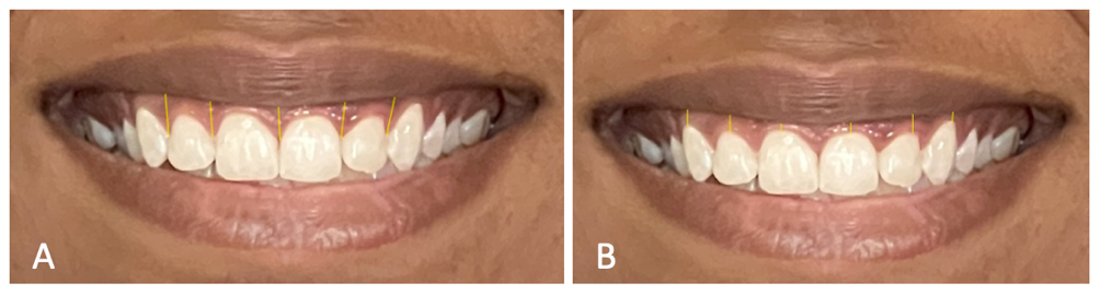
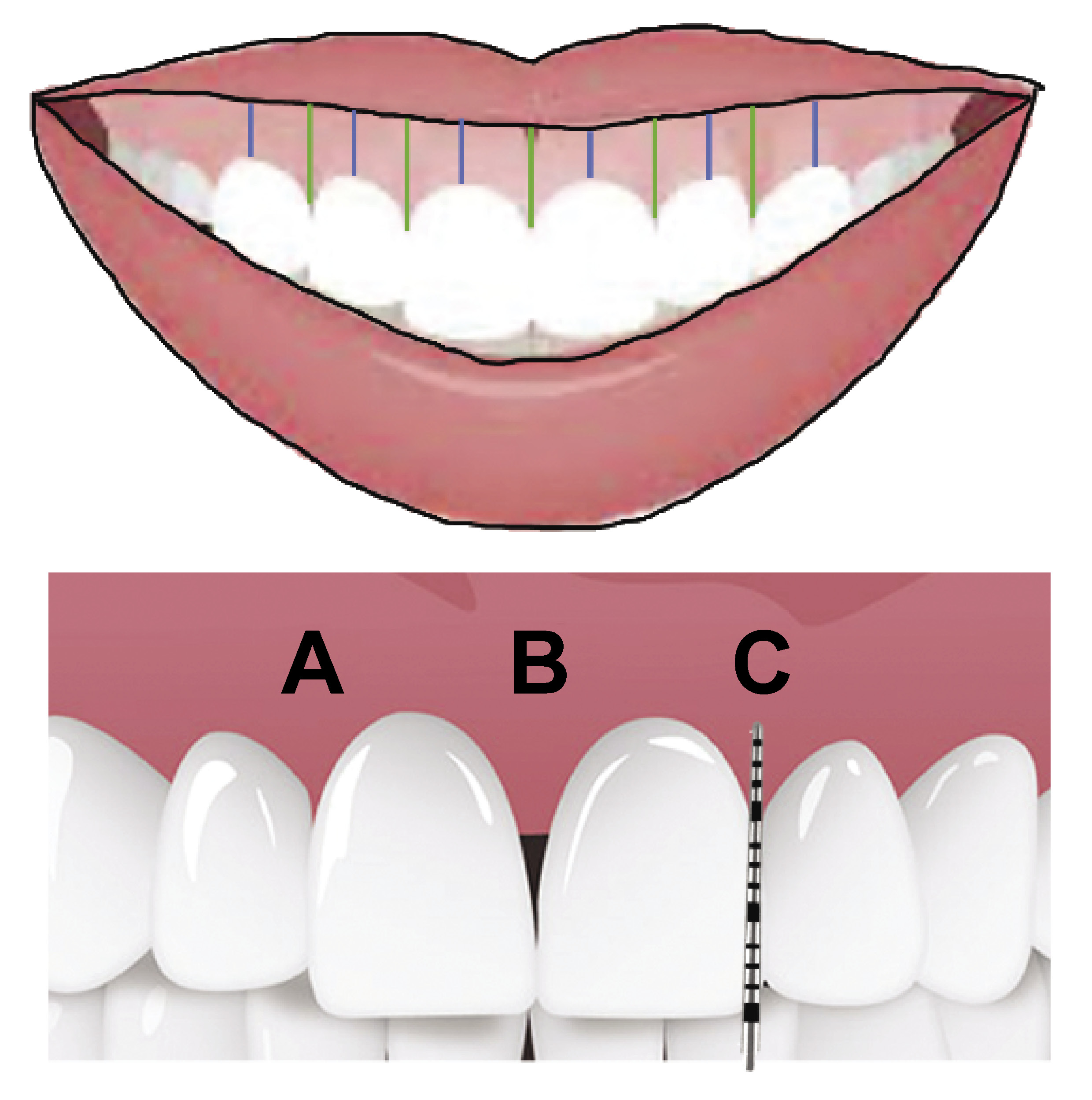
(3) Visible interdental papillae on smiling in ISL (VIG): Interdental papillary display on the right and left sides was recorded as the distance between the lower vermillion border and the tip of the interdental papilla. The amount of interdental papilla exhibited in the interdental smile line (ISL) was measured on frontal smiling photographs, from the tip of the interdental papillae of the maxillary anterior teeth to the lower vermillion border of the maxillary lip at the corresponding interdental positions on both the right and left sides. The mean value of these measurements was calculated for each patient. All measurements were performed using standardized digital photographs (Figure 2A, Figure 5).
(4) Visible midfacial gingiva on smiling in GSL (VMG): Midfacial gingival display on the right and left sides was recorded as the distance from the gingival zenith point of the respective teeth to the lower vermillion border of the maxillary lip at the midfacial positions. The mean value of these recordings was calculated for each patient. The measurement was done on photographs (Figure 2B, Figure 5).
(5) GT: Gingival thickness was evaluated for each individual tooth following the topical application of anesthetic gel, and measured at the midpoint between the gingival margin and the mucogingival junction (MGJ), using an endodontic spreader with a rubber stopper. The mean value obtained from all maxillary anterior teeth was recorded as the final score. All measurements were performed clinically.
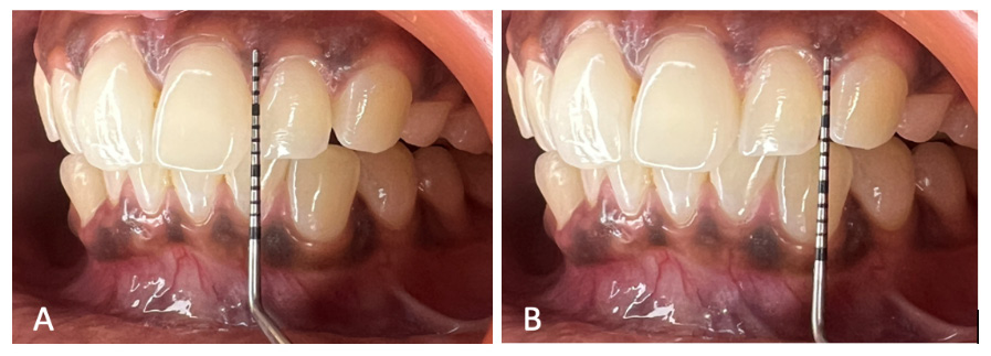
(6) PH: Papilla height was measured using a periodontal probe from an imaginary line drawn from the gingival zenith of the adjacent teeth to the tip of the papilla. All measurements were performed clinically. (Figure 3, Figure 5).
(7) PPI: The presence (PPI-1) or absence (PPI-2) of the papilla was determined based on the positional relationship between the interdental papilla, CEJ and the adjacent teeth, as described by Cardaropoli et al.13 All measurements were performed clinically. (Figure 4, Figure 5).
Statistical analysis
All measurements were continuous variables and were expressed as mean and standard deviation (M ±SD) for each group. Categorical parameters, such as PH, GT, VIG, and VMG were numerically represented in relation to age and gender, while the comparisons of the types of ISL and GSL, as well as the PPI scores across the study groups were performed using descriptive statistics, and the results were presented as percentages.
The paired sample t test was used to assess mean differences in PH, GT, VIG, and VMG with respect to age and gender, with two-sided p-values applied to determine statistical significance. The χ2 test was used to evaluate the association of ISL, GSL and PPI with the study groups, including different age and gender categories. Data analysis was performed using IBM SPSS Statistics for Windows, v. 29.0 (IBM Corp., Armonk, USA).
Results
The study included 120 periodontally healthy patients divided into 4 groups according to age and gender, providing a total of 720 maxillary anterior teeth and 600 interdental papillae for evaluation, along with the corresponding midfacial gingival displays. Both ISL and GSL served as reference parameters for the assessment of gingival display.
All clinical and photographic measurements were performed by 2 calibrated examiners, who demonstrated excellent inter-observer agreement for the evaluated parameters in both male and female participants.
Table 1 presents the mean values PH, GT, VIG, and VMG across all study groups, along with the intragroup comparisons. The mean PH values for groups I and II were 3.13 ±0.63 mm and 3.91 ± 0.48 mm, respectively, whereas groups III and IV demonstrated values of 2.96 ±0.60 mm and 3.55 ±0.48 mm, respectively. Comparisons based on gender within the corresponding age groups revealed statistically significant differences (p < 0.001). However, when comparisons were performed within the same gender across different age groups (groups I vs. III and groups II vs. IV), the former comparison was not significant, whereas the latter showed statistical significance (p = 0.004). Significant differences were also observed in the mean GT values among groups I, II, III, and IV. Similarly, comparisons between groups I and III and between groups II and IV demonstrated statistically significant differences (p = 0.039 and p = 0.002, respectively), indicating age-related changes in GT among both male and female participants. The mean VIG values for groups I, II, III, and IV were 1.87 ±1.83 mm, 3.47 ±1.88 mm, 1.05 ±1.70 mm, and 2.73 ±2.40 mm, respectively. The comparisons of this parameter revealed statistically significant differences (p = 0.001). Among male participants (groups I and III), the VIG values differed significantly between age groups, whereas no significant differences were observed among female participants (groups II and IV). For the VMG parameter, a significant difference was identified between groups I and II, with mean values of 0.31 ±0.64 mm and 0.81 ±1.01 mm, respectively. The remaining comparisons for this parameter did not demonstrate statistically significant differences, despite variations in the mean values.
Table 2 presents the ISL and GSL categories, and the PPI scores across all study groups, along with their respective comparisons. Regarding ISL, the predominant smile line categories for groups I, II, III, and IV were LISL, HISL, LISL, and HISL, with frequencies of 46.7%, 73.3%, 60.0%, and 66.7%, respectively. Intragroup comparison revealed a statistically significant difference between groups III and IV (p = 0.024). For GSL, the most predominant category in all 4 groups was LGSL, with frequencies of 76.7%, 46.7%, 93.3%, and 63.3% for groups I, II, III, and IV, respectively. In group I, the second most prevalent category was CB-GSL, accounting for 13.3%, whereas in group II, HGSL was the second most common category. The comparison of the GSL categories between groups II and IV demonstrated a statistically significant difference (p = 0.029). For PPI, PPI-1 was the most predominant score across all 4 groups, with a higher prevalence observed among male participants (groups I and III).
Discussion
The maxillary anterior segment, owing to its prominence and visibility, has gained considerable importance in smile esthetics, where the harmonious display of the teeth and gingival structures has the potential to enhance the overall attractiveness of the smile. Efforts to identify and evaluate intraoral and extraoral characteristics, such as the gingival zenith position,8 gingival display,19 tooth and gingival display,20 facial and dental proportions,21 maxillary lip dimensions,2 and the nasolabial angle,5 have been incorporated to varying extents into restorative and periodontal procedures aimed at improving esthetic outcomes.
The concepts of ISL and GSL, together with their role in defining smile characteristics and their relationship with peri- and intraoral features, warrant further investigation to establish harmonious relationships that contribute to the design of an esthetic smile. Furthermore, evaluating these parameters in relation to age and gender variability may provide clinically relevant information for procedures involving the maxillary anterior region. In addition, recent studies have utilized three-dimensional (3D) facial scans through linear and angular measurements, demonstrating no significant differences between 3D facial scanning and cone-beam computed tomography (CBCT) soft tissue volumetric assessment, thereby supporting its use as an alternative non-radiographic method.22
The observations of the present study indicate notable gender-related differences in the evaluated parameters. Papilla height, VIG in ISL, and VMG in GSL demonstrated considerably higher mean values in groups II and IV, whereas GT exhibited lower values in these groups. These findings may reflect the generally recognized biological differences between genders, with males typically exhibiting more robust anatomical structures than females. Papilla height, measured from an imaginary line drawn from the gingival zenith of the adjacent teeth to the tip of the papilla, is of particular relevance, as the present study is among the few investigations to clinically evaluate this parameter. The observed gender-related variability, favoring females in groups II and IV as compared to males in groups I and III, may be associated with triangular crown morphology and greater apicocoronal dimensions of the interproximal areas.9, 23 However, in both of these previous studies, PH was not directly measured and was assessed only in terms of papilla presence or absence. In addition, the PH values decreased with advancing age irrespective of gender, as observed in groups III and IV, and these findings are consistent with those reported by Chow et al.24
The display of teeth and gingival tissues during smiling, particularly the interdental and midfacial gingiva, has long attracted clinical interest, as these structures are considered essential components of an esthetic smile. In the present investigation, the parameters of VIG and VMG were quantitatively evaluated, and the findings demonstrated substantial differences between groups I and II as well as between groups III and IV, with female participants exhibiting greater gingival display both at the midfacial and interdental levels. The VIG values were considerably higher than the VMG values, which may be attributed to the more coronal position of the interdental papillae, a feature specifically represented in ISL. Although females in groups II and IV demonstrated greater VMG values than males in groups I and III within GSL, the extent of this display was markedly lower when compared with the gingival display observed in ISL. These observations are consistent with the findings reported by Kolte et al.,2 although the gingival display values described in their study were greater for both genders than those observed in the present investigation. This discrepancy may be attributed to differences in the measurement protocol, as Kolte et al. recorded measurements separately for each category of ISL and GSL, whereas the present study assessed gingival display cumulatively, irrespective of smile category. Another possible explanation for the greater VIG and VMG observed among females is the presence of a fuller smile pattern, in which the lip musculature undergoes greater stretching and reduction in fullness and length, thereby resulting in increased gingival display.25
An important strength of the present study is that all parameters were evaluated in 2 distinct age groups for both male and female participants, enabling the assessment of changes associated with the aging process. The evaluated parameters – PH, VIG and VMG – demonstrated notable age-related alterations, with overall reductions in their mean values observed with advancing age. The reduction in PH was particularly pronounced among females in groups II and IV as compared to males in groups I and III.
Gingival thickness is an important parameter in periodontal and implant therapy, as it serves as a valuable source of autogenous tissue for the management of gingival recession and dehiscence defects. Owing to its site-specific and individual variability, as well as its relevance in therapeutic procedures, GT has attracted considerable clinical interest. In the present study, GT was evaluated using the direct, predictive, and relatively simple measurement method described by Stein et al.,26 which identifies a reference point located midway between the gingival margin and MGJ, directly over the alveolar crest. The findings demonstrated substantially greater GT values in groups I and III as compared to groups II and IV. These observations are consistent with previous investigations,9, 27 and may be attributed to the generally more robust anatomical characteristics observed in males, as well as age-related epithelial thinning and reduced keratinization.
In the assessment of GSL, the most predominant category was LGSL, irrespective of gender. In contrast, for ISL, the predominant category was LISL in groups I and III, and HISL in groups II and IV, indicating a clear gender-related variability. These findings are particularly important, as they highlight the greater sensitivity of ISL in identifying interdental gingival display, which appears to be more prominently expressed than midfacial gingival display.
The present investigation is among the first studies to evaluate PPI, and the findings demonstrated a predominance of PPI-1 scores among the study participants. This observation may be attributed to the inclusion of only periodontally healthy individuals, most of whom exhibited interproximal areas completely filled by the interdental papillae, whereas only a limited number demonstrated PPI-2 scores. Comparisons between groups I and II and groups III and IV revealed a reduction in the proportion of participants with PPI-1 scores and a corresponding increase in PPI-2 scores in the older age groups. Similarly, comparisons between groups I and III and groups II and IV demonstrated greater differences among older female participants than among males, possibly reflecting the more delicate gingival tissues in females and their greater susceptibility to age-related alterations. In addition, the greater GT values observed in groups I and III may explain the predominance of PPI-1 scores in these groups, as thicker gingival tissues may be more resistant to structural changes.
Limitations
Several limitations of the present study should be acknowledged. First, as the clinical trial was conducted on a homogeneous population belonging to a specific racial and ethnic demographic, the findings may be more representative of the Asian population, and potential variations among other ethnic groups warrant further investigation to broaden the applicability of the results. Second, clinical measurements were performed using a periodontal probe, which may have introduced minor inaccuracies, particularly over convex and concave tooth surfaces. Third, factors such as mouth breathing and Angle’s classification, both of which may influence smile characteristics, were not included in the analysis. Finally, age-related maxillary remodeling and variations in lip and nasal support, which may also affect smile esthetics, were not evaluated in the present study.
Conclusions
The observations obtained from the present study may have important clinical implications and can be applied, in conjunction with specific treatment objectives, to assist clinicians in achieving an esthetically pleasing and functionally appropriate smile. Gender-related variability in intraoral characteristics was evident in the parameters of PH, VIG and VMG, which demonstrated considerably higher mean values in groups II and IV, whereas GT exhibited lower values in these groups. These findings also appeared to influence PPI scores, with PPI-1 predominating among participants in groups I and III. Age-related changes were observed across all evaluated parameters, including PH, GT, VIG, and VMG, with a notable reduction in the mean values associated with advancing age. The predominant presence of LGSL within the GSL categories, irrespective of gender, together with the predominance of LISL in groups I and III, and HISL in groups II and IV, not only demonstrated gender-related variability, but also highlighted the sensitivity of ISL in identifying interdental gingival display.
Trial registration
This clinical study was registered with the Clinical Trials Registry – India (CTRI/2023/06/053423).
Ethics approval and consent to participate
The trial was approved by the Institutional Ethics Committee at Ranjeet Deshmukh Dental College and Research Centre, Nagpur, India (IEC/VSPMDCRC/2/2023). Written informed consent was obtained from all participants.
Data availability
The datasets supporting the findings of the current study are available from the corresponding author on reasonable request.
Consent for publication
Not applicable.
Use of AI and AI-assisted technologies
Not applicable.