Dental and Medical Problems

Dent Med Probl
Impact Factor (IF 2024) – 3.9
Journal Citation Indicator (JCI 2024) - 1.36
Scopus CiteScore (2024) – 5.0
Index Copernicus Value (ICV 2023) – 181.00
MNiSW – 70 pts
ISSN 1644-387X (print)
ISSN 2300-9020 (online)
Periodicity – bimonthly


Download original text (EN)

Dental and Medical Problems

2025, vol. 62, nr 6, November-December, p. 1211–1221

doi: 10.17219/dmp/186354

Publication type: review

Language: English

License: Creative Commons Attribution 3.0 Unported (CC BY 3.0)

Download citation:

  • BIBTEX (JabRef, Mendeley)
  • RIS (Papers, Reference Manager, RefWorks, Zotero)

Cite as:


Rąpalska I, Chęciński M, Kaczmarzyk T. Impact of human papillomavirus (HPV) infection on the development of oral squamous cell carcinoma (OSCC): A systematic review. Dent Med Probl. 2025;62(6):1211–1221. doi:10.17219/dmp/186354

Impact of human papillomavirus (HPV) infection on the development of oral squamous cell carcinoma (OSCC): A systematic review

Iwona Rąpalska1,2,A,B,C,D,F, Maciej Chęciński2,3,4,B,C,D,F, Tomasz Kaczmarzyk1,A,C,D,E,F

1 Chair of Oral Surgery, Jagiellonian University Medical College, Krakow, Poland

2 Department of Oral Surgery, Preventive Medicine Center, Krakow, Poland

3 National Medical Institute of the Ministry of the Interior and Administration, Warsaw, Poland

4 Department of Maxillofacial Surgery, Hospital of the Ministry of the Interior and Administration, Kielce, Poland

Graphical abstract


Graphical abstracts

Highlights


  • Human papillomavirus (HPV) infection is frequently detected in patients with oral squamous cell carcinoma (OSCC), with prevalence rates reaching up to 74.5% in genetic testing studies.
  • Controlled studies show markedly higher odds of HPV-16 or HPV-18 detection in OSCC patients compared with non-OSCC controls (weighted mean OR ≈ 17).
  • Evidence linking OSCC with HPV types other than HPV-16 and HPV-18 remains limited and inconclusive.

Abstract

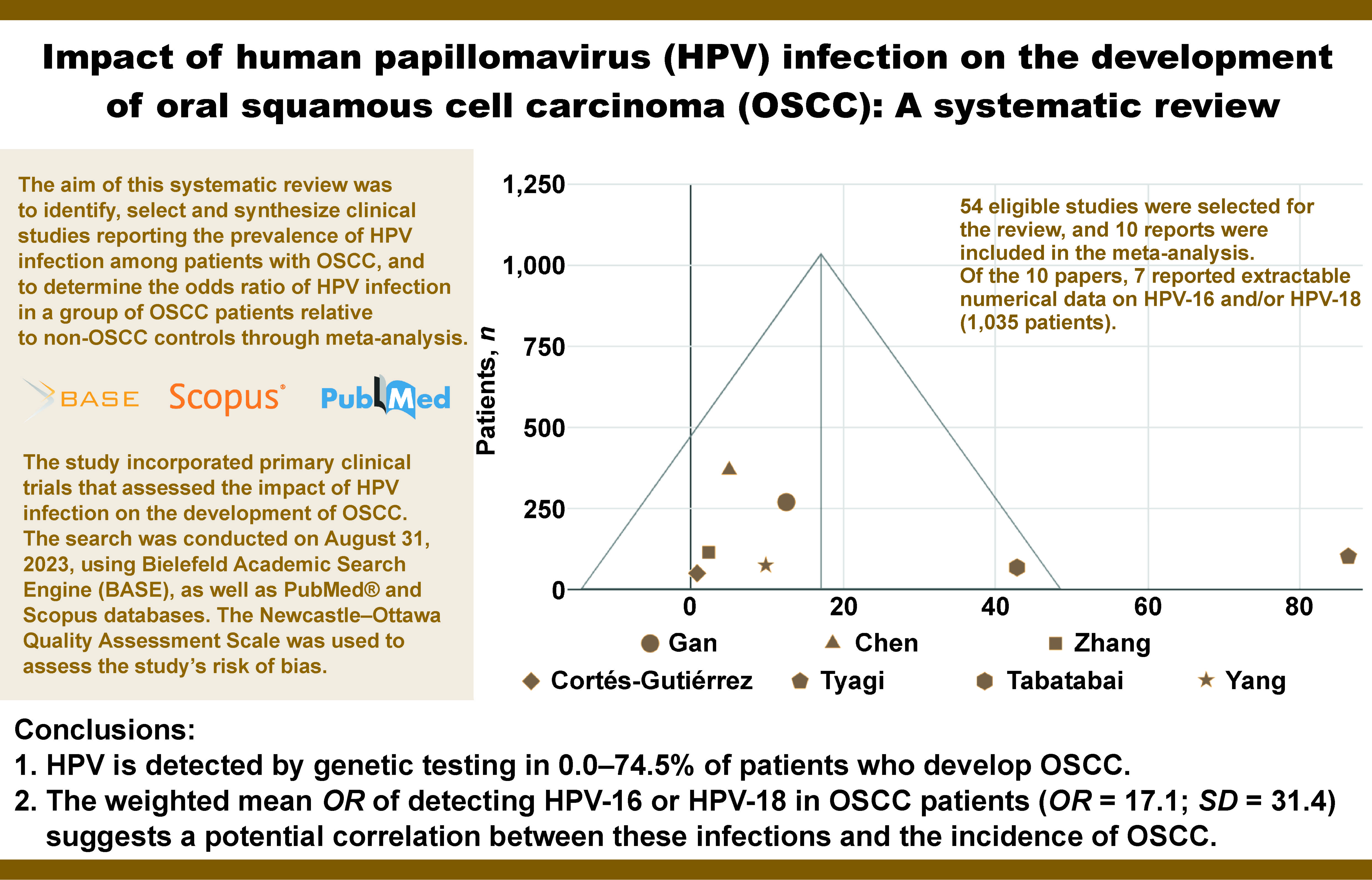
This systematic review aimed to identify, select and synthesize clinical studies reporting the prevalence of HPV infection among patients with OSCC, and to determine the odds ratio (OR) of HPV infection in a group of OSCC patients relative to non-OSCC controls through meta-analysis.

The study incorporated primary clinical trials that assessed the impact of HPV infection on the development of OSCC. The search was conducted on August 31, 2023, using Bielefeld Academic Search Engine (BASE), as well as PubMed® and Scopus databases. The Newcastle–Ottawa Quality Assessment Scale was used to assess the risk of bias of the included studies. The collected data was then synthesized in the form of tables and a funnel plot. A total of 54 eligible studies were selected for the review, and 10 reports were included in the meta-analysis. Of the 10 papers, 7 reported extractable numerical data on HPV-16 and/or HPV-18 (1,035 patients).

The limitations of the evidence included the following: inhomogeneity in terms of HPV type; small number of available controlled studies (not homogeneous in terms of virus type); small number of patients on whom controlled studies were conducted; and the risk of bias related to the selection of study and control groups (present in most studies qualified for the synthesis).

In conclusion, HPV is detected by genetic testing in 0.0–74.5% of patients who develop OSCC. The weighted mean OR of detecting HPV-16 or HPV-18 in OSCC patients (OR = 17.1; standard deviation (SD) = 31.4) suggests a potential correlation between these infections and the incidence of OSCC.

Keywords: HPV, OSCC, oral squamous cell carcinoma, systematic review, human papillomavirus

Introduction

Background

Head and neck cancers account for over 5% of all malignancies.1 This category includes cancers of the oral cavity, throat, larynx, paranasal sinuses, thyroid and salivary glands, as well as the surrounding soft and hard tissues.2, 3 Approximately 90% of cancers in this category are squamous cell carcinomas (SCCs).3

Oral squamous cell carcinoma (OSCC) is a term used to describe cancers with squamous cell differentiation developing within the oral mucosa and lips, excluding the skin of the mouth and the pharyngeal mucosa. Oropharyngeal SCC (OPSCC) refers to cancers located in the palatine tonsil, the root of the tongue, the glossotonsillar groove, and the mucous membrane of the lateral and posterior pharyngeal walls. In some publications,4 the term OPSCC is used imprecisely to describe both cancer of the oral cavity and cancer of the oropharynx. However, current evidence supports the conclusion that OSCC and OPSCC are distinct and unique, with differing etiopathogenesis, treatment and prognosis.3

The risk of OSCC increases significantly after the age of 50, and the condition is diagnosed 3 times more often in males.5 Despite advancements in technology, including self-learning systems, detecting oral cancer at an early stage remains challenging for clinicians.6 Therefore, the identification of OSCC risk factors seems particularly important. The influence of tobacco smoking and chronic alcohol consumption on the development of OSCC has been extensively documented.7 Additional significant risk factors include poor oral hygiene, chronic irritation of the mucous membrane due to faulty prosthetic restorations or dental fillings, candidiasis, and the presence of potentially malignant disorders such as leukoplakia, lichen planus or erythroplakia.8, 9

Rationale

In recent years, there has been a sharp increase in the incidence of OSCC among patients in younger age groups (approx. 20–40 years) who developed OSCC despite good oral hygiene and the absence of any of the aforementioned risk factors.5, 10 Current research on the impact of head and neck cancer on the quality of life confirms that available treatment leads to its permanent deterioration (e.g., it induces sexual issues, which are of particular importance to this age group).11 It has been speculated that in this group of patients, another initiator may be respon­sible for promoting the carcinogenesis process, and it is currently under debate whether high-risk human papillom­avirus infection (HR-HPV) plays a role in the devel­opment of OSCC.12 This hypothesis is related to the fact that, in relation to OPSCC, i.e., cancers developing in the immediate anatomical vicinity of the oral cavity, there is clear scientific evidence of the impact of HR-HPV infec­tion on the development of cancer.2 Recent studies have indicated that up to 70% of OPSCC cases are related to HR-HPV infection, primarily types 16 and 18.13 However, the relationship between HPV infection and OSCC is still unclear.14 The frequency of OSCC cases in which HR-HPV genetic material is detected, according to various authors, ranges between 2.6%15 and 74%.13 However, in most of the analyzed samples, HPV is detected in over 25% of OSCC cases.16

Revealing a direct relationship between viral infection and the occurrence of OSCC would have significant clinical implications. In the context of OPSCC, HPV(+) cancers are characterized by a less aggressive course and significantly better survival outcomes compared to HPV(−) cancers. Consequently, therapeutic interventions in this group of patients may be less invasive.17, 18

Aim

The aim of this systematic review was to identify, select and synthesize clinical studies reporting the prevalence of HPV infection among patients with OSCC, and to determine the odds ratio (OR) of HPV infection in a group of OSCC patients relative to non-OSCC controls through meta-analysis. The collection of this data is intended to indirectly assess whether the risk of OSCC is greater in HPV-positive individuals.

Material and methods

This systematic review is based on and arranged in accordance with the current version of the Preferred Reporting Items for Systematic reviews and Meta-Analyses (PRISMA) guidelines.19 The completed PRISMA checklist and the PRISMA checklist for abstracts constitute the supplementary material to this article (available on request from the corresponding author). This systematic review has been registered in the PROSPERO database (registration No. CRD42023483769).

Eligibility criteria

Primary clinical trials published in English that evaluated the impact of HPV infection on the development of OSCC were included. The detailed eligibility criteria are presented in Table 1, albeit the Control and Outcomes criteria only concerned the inclusion in the meta-analysis.

Information sources

A comprehensive search of medical databases was conducted using the Bielefeld Academic Search Engine (BASE), PubMed® and Scopus. All final searches were performed on August 31, 2023.

Search strategy

The search strategy, identical for each of the engines, was formulated based on the eligibility criteria and arranged in the form of the following query: (“oscc” OR “oral scc” OR “oral squamous cell carcinoma”) AND (“hpv” OR “papillomavirus” OR “papilloma virus”) AND (“correlation” OR “correlated” OR “effect” OR “affects” OR “impact” OR “influence” OR “link” OR “connection”) AND (“study” OR “trial” OR “primary”).

Article selection

The identified records were entered into the Rayyan automation tool (Rayyan Systems, Cambridge, USA).20 The duplicates were automatically removed, and the records indicated by the tool as potential duplicates were manually verified (IR and MC). A blind screening of abstracts and titles was performed by 2 researchers (IR and MC). The convergence of researchers’ ratings was expressed by Cohen’s kappa coefficient. In cases of non-compliance during the screening stage, the record underwent further processing. The full-text reports were evaluated by the same researchers (IR and MC). Discrepancies that emerged during the full-text evaluation phase were resolved through consensus.

Data collection

The data was extracted from the source articles by 2 independent authors (IR and MC). The present study exclusively utilized published data, specifically the content of articles and supplementary materials. The collection process did not involve the use of automation tools.

Extracted characteristics of the studies

The following items were extracted from the source studies: first author and year of publication; sex and aver­age age of patients; OSCC location; total number of patients in the OSCC group; number of patients in the OSCC group with genetically confirmed HPV; total number of patients in the non-OSCC group; number of patients in the non-OSCC group with genetically confirmed HPV; OR between HPV and OSCC.

Assessment of the risk of bias

The Newcastle–Ottawa Quality Assessment Scale was used to assess the risk of bias for studies included in the meta-analysis. The evaluation was performed by 2 inde­pendent researchers (IR and MC). Any discrepancies in assessment were resolved through consensus, and no automation tools were implemented in the process.

Effect measures

For studies with a control group, the OR was calculated using the MedCalc Statistical Software v. 22.018 (MedCalc Software Ltd, Ostend, Belgium).

Data synthesis

The collected data was synthesized in the form of tables and a funnel plot using Google Workspace (Google LLC, Mountain View, USA).

Reporting the assessment of bias

In instances of missing data, this fact was noted, yet the series was not discarded. No further reporting bias assessments were undertaken.

Results

Study selection

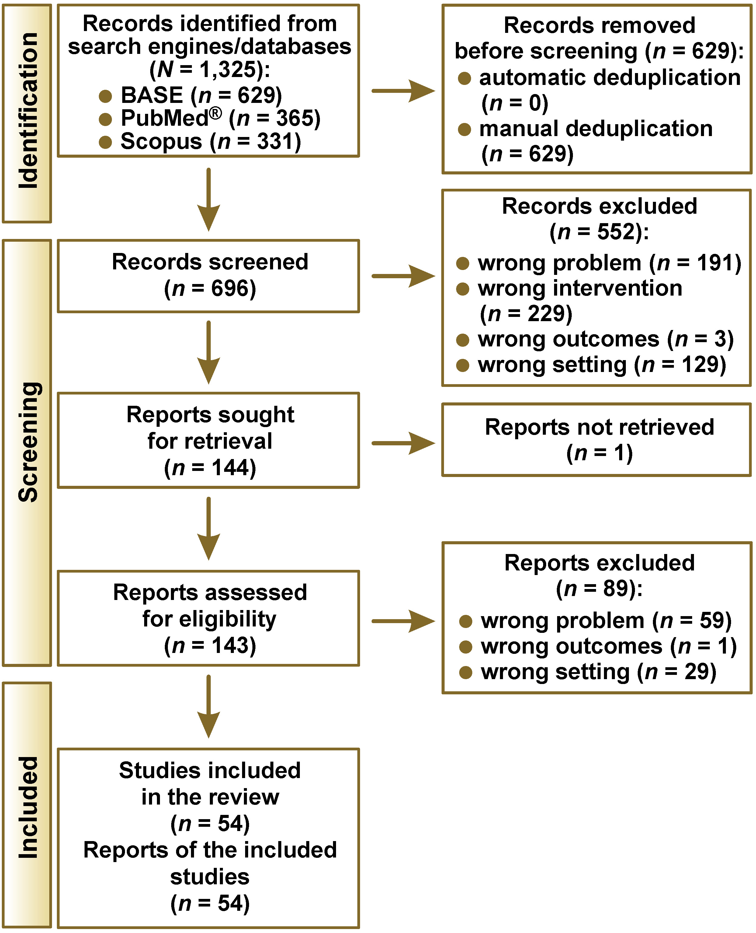
The number of records identified from each search engine/database is presented in Table 2. During the selec­tion process, 54 eligible studies were selected out of 1,325 identified records (Figure 1). The consistency index of the judges’ assessments at the screening stage was κ = 0.87, which represents almost perfect agreement. The study by Cao et al. was considered eligible based on the abstract, but its full text was not obtained due to the lack of digital archiving of articles from these years in the Chinese Journal of Dental Research.21

Study characteristics

Fifty-four studies included in the review are summarized in Table 3. Each study considered different locations of OSCC, making it impossible to create subgroups based on specific areas of the oral cavity. The collected material covered clinical trials since 1996, which resulted in an overview of over a quarter of a century of active research on the relationship between HPV infection and the occurrence of OSCC. The study samples did not exceed 254 patients, and most reports were based on material from fewer than 100 subjects. The preponderance of single-center studies made it challenging to extrapolate results to broader populations. The percentage of diagnosed HPV infections in OSCC patient groups fluctuated significantly (0.0–74.5%), which was partly related to the limited sample size and the heterogeneity of the identified virus types. The majority of the papers did not include a control sample and were limited to a single arm, precluding the possibility of synthesizing them to ensure a high level of evidence for the review. Therefore, studies incorporating a control group advanced to further stages of the review.

Risk of bias

The Newcastle–Ottawa Quality Assessment Scale scores for each of the studies included in the meta-analysis ranged from 6 to 9 (maximum) points (Table 4). Therefore, the overall risk of bias for the studies was low or raised some concerns. The ratings were decreased primarily due to the absence of statements regarding the inclusion of consecutively reporting patients and limited information on the control groups.

Results of individual studies

The results of individual studies are outlined in Table 5. Most studies had symmetrical sample sizes, though there were exceptions to this rule. The number of patients in the study groups ranged from 30 to 200, which enabled quantitative analysis. Depending on the study, the sex ratio was either similar or men predominated by up to 2.5 times. The percentage of HPV infections in the control samples did not exceed 55%. In numerous reports, the prevalence of HPV infection in the control sample was negligible or equal to 0. In each study sample, HPV was detected in at least 1 patient, and the percentage of infected individuals reached up to 74%. Interestingly, a study that encompassed a broader range of virus types observed a lower infection rate in comparison to the maximum recorded value.42 This could be due to the lack of identifica­tion of HPV-18, which is present in the majority of tests conducted by other research groups. Discrepancies can be observed in relation to the type of identified virus, with a clear dominance of HPV-16 and HPV-18. In studies with smaller sample sizes, the OR did not attain statistical significance. In cases with confirmed statistical signifi­cance, the OR ranged from 2.3 to 86.3, indicating a higher probability of identifying HPV in materials from patients diagnosed with OSCC.

Results of data synthesis

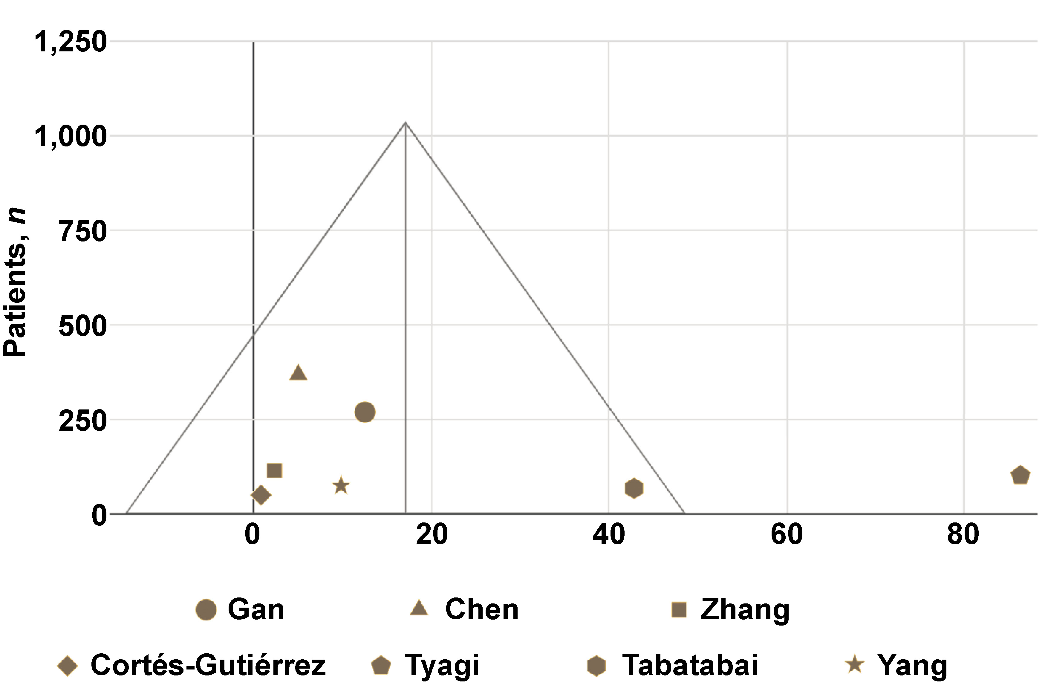
Of the 10 reports containing numerical data that enabled OR calculation, 8 assessed HPV-16 and/or HPV-18; however, only 7 studies provided extractable data specific to HPV-16 or HPV-18. The OR results for HPV-16 and HPV-18 were synthesized in a funnel plot (Figure 2). This synthesis was based on a total of 1,035  patients. The weighted mean OR was 17.1 (standard deviation (SD) = 31.4). The OR result reported in the study by Tyagi et al. was an outlier.

Discussion

General interpretation of the results

In the collected research material, the percentage of OSCC patients infected with HPV ranged from 0.0% to 74.5%. Significant discrepancies in the results have been confirmed in other reviews.65, 66 The discrepancies are likely attributable to the testing of different types of the virus within individual clinical trialws. Moreover, the method of testing for the HPV genome, even of the same type, is not uniform, which may be the reason for differences in the results presented by different teams of researchers.

Of the 54 eligible studies, only 10 included a control group. The ideal study design would involve a study group and a control group that are matched in terms of sex and age. In the available material, control groups were selected primarily from patients of the same institution (but not from the general population).

The results of the OR for the presence of the HPV genome in OSCC vs. non-OSCC patients indicate the pres­ence of a relationship between the occurrence of OSCC and HPV-16 or HPV-18 infection. With the exception of the study by Cortés-Gutiérrez et al.,15 which was conducted on a small group of patients, the OR for the HPV-16 and HPV-18 in OSCC vs. non-OSCC groups is greater than 1, indicating a higher probability of detecting these types of virus in patients with OSCC. Based on the patient-derived outcomes, it was confirmed that a positive result for HPV-16 and/or HPV-18 is associated with a significant risk of developing OSCC.

The collected research material is insufficient to draw similar conclusions regarding other types of the virus. The study by Yang et al. showed no statistically significant relationship between the incidence of OSCC and the presence of HPV-67 or HPV-68 infection.24

Biological risk factors for OSCC

The current state of knowledge suggests a correlation between HPV and the risk of OSCC, which is the subject of this paper. Additionally, an increased predisposition to OSCC is suspected in patients infected with Epstein–Barr virus (EBV).56 A similar relationship has been observed among individuals infected with bacteria that cause periodontitis, i.e., Porphyromonas gingivalis and Fusobacterium nucleatum. However, there is a lack of clinical evidence for this relationship, and assumptions are solely based on in vitro and animal studies.67 The previously ambiguous relationship between Candida spp. infection and the development of OSCC was confirmed in a systematic review from 2023.68 The demonstrated correlation supports the possibility of causality, which requires further research.

HPV diagnostics

The gold standard for the diagnosis of HPV infection is the identification of the genetic material of the virus by polymerase chain reaction (PCR). An alternative method involves the identification of cyclin-dependent kinase inhibitor 2A (p16) protein immunohistochemically.16 A meta-analysis of diagnostic methods for oral HPV showed high sensitivity but moderate specificity of immunohistochemical identification.69 It is, therefore, accepted that the detection of p16 is of high value as a screening test, but may not be sufficient for scientific purposes. The present systematic review was based solely on source studies, in which the presence of HPV genetic material in patient tissues was confirmed by PCR.

Benign lesions

The infection with HPV may also contribute to the development of benign lesions, commonly referred to as oral warts. These include, among others, focal epithelial hyperplasia (Heck’s disease), squamous cell papilloma of the oral cavity, common wart (verruca vulgaris), and condyloma acuminata of the oral cavity.70 For a progression to a malignant state, the presence of appropriate co-factors is necessary, including genetic predisposition, smoking and alcohol consumption.71

The significance of HPV infection in oral potentially malignant lesions, specifically in leukoplakia, remains unclear.72 A recent systematic review has estimated the overall HPV prevalence in leukoplakia at 6.66%, whereas the prevalence of HPV-16 at 2.95%.73 However, the current update from the World Health Organization (WHO) classification of head and neck tumors distinguishes the HPV-associated dysplasia as a separate lesion, with about 15% risk of malignant transformation.74, 75

Future research

A particular emphasis must be placed on the methodological problem that is present in all qualified studies. The authors of the source papers attempted to estimate the risk of OSCC among HPV(+) vs. HPV(−) patients by designing studies in which the presence of the viral genome was determined in OSCC and non-OSCC patients. In order to most appropriately investigate the incidence of OSCC among patients with HPV, a long-term follow-up of HPV(+) vs. HPV(−) cohorts in the context of OSCC occurrence is necessary. It is important to consider the possibility of HPV infection during observation, and to differentiate between the various types of the virus.

Limitations of the evidence

The limitations of the evidence included the following: inhomogeneity in terms of HPV type; small number of available controlled studies (not homogeneous in terms of virus type); small number of patients on whom controlled studies were conducted; and the risk of bias related to the selection of study and control groups (present in most studies qualified for the synthesis).

Limitations of the review process

The main limitation of the review process included the use of a query in English, thereby excluding reports that lacked at least a title or abstract in this language. Moreover, the search engine and databases used do not ensure the identification of articles published in locally indexed journals.

Conclusions

Human papillomavirus is detected through genetic testing in 0.0–74.5% of patients with OSCC. The weighted average OR for detecting HPV-16 or HPV-18 in OSCC patients is 17.1, suggesting that these viral variants may contribute to the development of OSCC.

Ethics approval and consent to participate

Not applicable.

Data availability

The datasets generated and/or analyzed during the current study are available from the corresponding author on reasonable request.

Consent for publication

Not applicable.

Use of AI and AI-assisted technologies

Not applicable.

Tables


Table 1. Eligibility criteria for study inclusion and exclusion

PICOS

Criteria for inclusion

Criteria for exclusion

Population, Problem

OSCC diagnosis

animal studies

Intervention

detection of the HPV

methods not detecting the genome

Control*

non-OSCC group

none

Outcomes*

OR

none

Timeframe

unlimited

preprints

Setting

primary studies on groups of 10 and more

none

* applied to the meta-analysis only; OSCC – oral squamous cell carcinoma; HPV – human papillomavirus; OR – odds ratio.
Table 2. Number of records identified through database search

Search engine/database

Coverage, n

Identified records, n

BASE

340,488,332

629

PubMed®

>36,000,000

365

Scopus

>87,000,000

331

Total

1,325

BASE – Bielefeld Academic Search Engine.
Table 3. Characteristics of studies included in the review

Study

Country

Presence of a control group (n)

OSCC group, n

Males/females in the OSCC group (control group),
n

Mean age in the OSCC group (control group)
[years]

HPV infections in the OSCC group
[%]

HPV type

Mneimneh et al.
202110

USA

no

150

89/61

34.0

0.0

16

Saleh et al.
202322

USA

no

114

56/58

70.8

12.0

16

Al-Dabbagh et al.
202223

UK

no

124

85/39

60.0

0.0

16

Yang et al.
201924

China

yes (30)

30

14/16
(14/16)

58.0
(50.0)

3.3

67, 68

Loeschke et al.
201625

Germany

no

91

59/32

55.7

7.7

16

Popović et al.
201026

Serbia

no

60

47/13

N/S

10.0

16

Zhao et al.
200917

China

no

52

35/17

N/S

40.4

6, 16, 18

Oliveira et al.
200827

Brazil

no

87

73/14

N/S

19.5

16, 18

de Freitas Cordeiro-Silva et al.
20125

Brazil

no

45

35/10

58.0

6.0

16

Singh et al.
201512

India

no

250

200/50

N/S

9.2

16, 18

Adnan Ali et al.
201828

Pakistan

no

140

82/58

N/S

67.9

16, 18

Gan et al.
201429

China

yes (68)

200

143/57
(27/41)

N/S
(N/S)

27.5

16, 18

Rivero and Nunes
200630

Brazil

no

40

32/8

57.0

0.0

16, 18

Sivakumar et al.
202131

India

no

26

23/3

58.2

14.0

16

Duray et al.
201232

Belgium

no

147

N/S

N/S

70.1

16, 45, 53, 58, 59, 66, 67

Grewal et al.
201833

India

no

47

36/11

46.7

74.5

16, 18

Schwartz et al.
200118

USA

no

254

162/92

54.2

15.1

16

Rushatamukayanunt et al.
201434

Japan

yes (40)

40

N/S
(N/S)

31.5
(61.0)

5.0

16, 18

Chen et al.
20167

China

yes (189)

178

110/68
(117/72)

58.9
(56.6)

14.0

16, 18

Polz-Gruszka et al.
201535

Poland

no

154

131/23

56.8

30.4

6, 11, 16, 18, 26, 31, 33, 35, 39, 40, 43, 44, 45, 51, 52, 53, 54, 56, 58, 59, 66, 68, 69, 70, 71, 73, 74, 82

Jalouli et al.
201036

India

no

62

50/12

58.2

24.0

16, 18

Chen et al.
201237

Taiwan

no

65

52/13

53.0

36.9

16

Zhang et al.
200413

China

yes (40)

73

48/25
(24/16)

N/S
(N/S)

74.0

16, 18

Premoli-De-Percoco and Ramirez
200138

Venezuela

no

50

0/50

48.2

60.0

6, 11, 16, 18

Antunović et al.
202239

Montenegro

no

60

47/13

62.0

23.3

16

Chen et al.
201640

China

no

40

29/11

N/S

0.0

16, 18

Campisi et al.
200641

Italy

no

63

28/35

68.9

38.1

16, 18

Saini et al.
201142

Malaysia

yes (105)

105

51/54
(58/47)

56.2
(42.0)

51.4

6, 11, 16, 26, 31, 33, 35, 45, 51, 53, 54, 58

Termine et al.
201243

Italy

no

83

43/40

64.0

15.7

16, 39, 51

Nagpal et al.
200244

India

no

110

68/42

N/S

33.6

16, 18

Laco et al.
20121

Czech Republic

no

48

N/S

N/S

15.0

16

Lee et al.
201214

Taiwan

no

173

N/S

N/S

22.0

16, 18

Penhallow et al.
19989

UK

no

28

N/S

65.6

50.0

6,16

Nemes et al.
200645

Hungary

no

79

67/12

55.8

41.8

16

Correnti et al.
200446

Venezuela

no

16

7/9

54.0

50.0

16, 18

Premoli-De-Percoco et al.
199847

Venezuela

no

50

0/50

55.0

70.0

6, 11, 16, 18

González-Moles et al.
199648

Spain

no

37

26/11

60.0

19.1

18

Cortés-Gutiérrez et al.
202115

Mexico

yes (10)

38

N/S
(N/S)

43.0
(45.5)

2.6

16

Shima et al.
200049

Japan

no

46

32/14

N/S

73.9

16, 18

Ali et al.
200850

Pakistan

no

140

N/S

N/S

68.0

16, 18

Tyagi et al.
201951

Nepal

yes (50)

50

29/21
(N/S)

48.6
(N/S)

46.0

16, 18

Sharma and Prakash
202352

India

no

15

7/8

50.8

26.7

N/S

Soares et al.
200353

Brazil

no

27

N/S

N/S

40.7

6, 11, 16, 18

Tabatabai et al.
201554

Iran

yes (27)

39

22/17
(18/9)

64.2
(63.6)

43.6

16, 18

Pandey et al.
201855

India

no

24

21/3

53.1

20.8

16, 18

de Lima et al.
202256

Brazil

no

100

64/36

N/S

31.0

6, 11, 16, 18, 31, 33, 35, 39, 45, 51, 52

Ibieta et al.
200557

Mexico

no

51

37/14

N/S

42.0

16, 18

Ali et al.
201458

Pakistan

no

140

N/S

60.3

68.0

16, 18

Yang et al.
200459

Taiwan

yes (36)

37

N/S
(N/S)

N/S
(N/S)

10.8

16, 18

Panzarella et al.
202160

Italy

no

40

23/17

66.5

10.0

16, 31, 51, 66, 67, 68

Ono et al.
201461

Japan

no

93

59/34

N/S

10.7

6, 11, 16, 18, 22, 26, 31, 33, 35, 39, 40, 42, 45, 51, 52, 53, 54, 55, 56, 58, 59, 61, 62, 64, 66, 67, 68, 69, 70, 71, 72, 73, 81, 83, 84

Ali et al.
201462

Pakistan

no

140

N/S

N/S

68.0

16, 18

Kozomara et al.
200563

Montenegro

no

50

42/8

55.4

64.0

16, 18, 31, 33

Ali and Awan
201664

Pakistan

no

140

N/S

N/S

67.9

16, 18

N/S – not specified.
Table 4. Assessment of the risk of bias for studies included in the meta-analysis

Study

Newcastle–Ottawa Quality Assessment Scale

Selection

Comparability

Exposure

Sum (0–9)

Yang et al.
201924

1-1-1-1

2

1-1-1

9

Gan et al.
201429

1-0-1-1

2

1-1-1

8

Rushatamukayanut et al.
201434

1-0-0-0

2

1-1-1

6

Chen et al.
20167

1-0-0-1

2

1-0-1

6

Zhang et al.
200413

1-0-1-0

2

1-1-1

7

Saini et al.
201142

1-0-1-1

2

1-1-1

8

Cortés-Gutiérrez et al.
202115

1-0-1-1

2

1-1-1

8

Tyagi et al.
201951

1-0-1-1

2

1-1-1

8

Tabatabai et al.
201554

1-0-0-0

2

1-1-1

6

Yang et al.
200459

1-0-0-1

2

1-1-1

7

Table 5. Results of individual studies included in the meta-analysis

Study

Non-OSCC group (male/female), n;
mean age [years]

HPV infections in the non-OSCC group, n (%)

OSCC group (male/female), n;
mean age [years]

HPV infections in the OSCC group, n (%)

OR and significance level

HPV type

Yang et al.
201924

30 (14/16)
50.0

1 (3.3)

30 (14/16)
58.0

1 (3.3)

1.0
p = 1.00

67, 68

Gan et al.
201429

68 (27/41)
N/S

2 (2.9)

200 (143/57)
N/S

55 (27.5)

12.5
p < 0.05

16, 18

Rushatamukayanut et al.
201434

40 (N/S)
61.0

1 (2.5)

40 (N/S)
31.5

2 (5.0)

2.1
p = 0.56

N/S

Chen et al.
20167

189 (117/72)
56.6

6 (3.2)

178 (110/68)
58.9

25 (14.0)

5.0
p < 0.05

16, 18

Zhang et al.
200413

40 (24/16)
N/S

22 (55.0)

73 (48/25)
N/S

54 (74.0)

2.3
p < 0.05

16, 18

Saini et al.
201142

105 (58/47)
42.0

26 (24.8)

105 (51/54)
56.2

54 (51.4)

3.2
p < 0.05

6, 11, 16, 26, 31, 33, 35, 45, 51, 53, 54, 58

Cortés-Gutiérrez et al.
202115

10 (N/S)
45.5

0 (0.0)

38 (N/S)
43.0

1 (2.6)

0.8
p = 0.92

16

Tyagi et al.
201951

50 (N/S)
N/S

0 (0.0)

50 (29/21)
48.6

23 (46.0)

86.3
p < 0.05

16, 18

Tabatabai et al.
201554

27 (18/9)
63.6

0 (0.0)

39 (22/17)
64.2

17 (43.6)

42.8
p < 0.05

16, 18

Yang et al.
200459

36 (N/S)
N/S

0 (0.0)

37 (N/S)
N/S

4 (10.8)

9.8
p = 0.13

16, 18

Figures


Fig. 1. Preferred Reporting Items for Systematic reviews and Meta-Analyses (PRISMA) flowchart of the study
BASE – Bielefeld Academic Search Engine.
Fig. 2. Funnel plot displaying odds ratios (x-axis) derived from reports on human papillomavirus (HPV)-16 and/or HPV-18

References (75)

  1. Laco J, Nekvindova J, Novakova V, et al. Biologic importance and prognostic significance of selected clinicopathological parameters in patients with oral and oropharyngeal squamous cell carcinoma, with emphasis on smoking, protein p16(INK4a) expression, and HPV status. Neoplasma. 2012;59(4):398–408. doi:10.4149/neo_2012_052
  2. Ang KK, Harris J, Wheeler R, et al. Human papillomavirus and survival of patients with oropharyngeal cancer. N Engl J Med. 2010;363(1):24–35. doi:10.1056/NEJMoa0912217
  3. Chi AC, Day TA, Neville BW. Oral cavity and oropharyngeal squamous cell carcinoma—an update. CA Cancer J Clin. 2015;65(5):401–421. doi:10.3322/caac.21293
  4. de Menezes SAF, Miranda YMS, da Silva YM, et al. Prevalence and genotyping of HPV in oral squamous cell carcinoma in Northern Brazil. Pathogens. 2022;11(10):1106. doi:10.3390/pathogens11101106
  5. de Freitas Cordeiro-Silva M, Stur E, Agostini LP, et al. Promoter hypermethylation in primary squamous cell carcinoma of the oral cavity and oropharynx: A study of a Brazilian cohort. Mol Biol Rep. 2012;39(12):10111–10119. doi:10.1007/s11033-012-1885-4
  6. Beristain-Colorado MP, Castro-Gutiérrez MEM, Torres-Rosas R, et al. Application of neural networks for the detection of oral cancer: A systematic review. Dent Med Probl. 2023;61(1):121–128. doi:10.17219/dmp/159871
  7. Chen F, Yan L, Liu F, et al. Oral human papillomavirus infection, sexual behaviors and risk of oral squamous cell carcinoma in southeast of China: A case–control study. J Clin Virol. 2016;85:7–12. doi:10.1016/j.jcv.2016.10.011
  8. Spirito F, Caponio VCA, Lo Muzio E, et al. Oral lichen planus in children: An Italian case series. Pediatr Dermatol. 2023;40(3):489–493. doi:10.1111/pde.15318
  9. Penhallow J, Steingrimsdottir H, Elamin F, et al. p53 alterations and HPV infections are common in oral SCC: p53 gene mutations correlate with the absence of HPV 16-E6 DNA. Int J Oncol. 1998;12(1):59–68. doi:10.3892/ijo.12.1.59
  10. Mneimneh WS, Xu B, Ghossein C, et al. Clinicopathologic characteristics of young patients with oral squamous cell carcinoma. Head Neck Pathol. 2021;15(4):1099–1108. doi:10.1007/s12105-021-01320-w
  11. Regeer J, Enzlin P, Prekatsounaki S, Van Der Cruyssen F, Politis C, Dormaar JT. Sexuality and intimacy after head and neck cancer treatment: An explorative prospective pilot study. Dent Med Probl. 2022;59(3):323–332. doi:10.17219/dmp/148156
  12. Singh V, Husain N, Akhtar N, et al. Do human papilloma viruses play any role in oral squamous cell carcinoma in North Indians? Asian Pac J Cancer Prev. 2015;16(16):7077–7084. doi:10.7314/apjcp.2015.16.16.7077
  13. Zhang ZY, Sdek P, Cao J, Chen WT. Human papillomavirus type 16 and 18 DNA in oral squamous cell carcinoma and normal mucosa. Int J Oral Maxillofac Surg. 2004;33(1):71–74. doi:10.1054/ijom.2002.0443
  14. Lee LA, Huang CG, Liao CT, et al. Human papillomavirus-16 infection in advanced oral cavity cancer patients is related to an increased risk of distant metastases and poor survival. PloS One. 2012;7(7):e40767. doi:10.1371/journal.pone.0040767
  15. Cortés-Gutiérrez EI, Garza Molina JG, Dávila-Rodríguez MI, Zapata Benavides P, Faz Eguía JM, Cerda-Flores RM. DBD-FISH, an effective marker for detecting genotoxicity in buccal mucosa exfoliated cells of patients with oral cancer. Toxicol Mech Methods. 2021;31(5):343–348. doi:10.1080/15376516.2020.1862379
  16. Smitha T, Mohan CV, Hemavathy S. Prevalence of human papillomavirus16 DNA and p16INK4a protein in oral squamous cell carcinoma: A systematic review and meta-analysis. J Oral Maxillofac Pathol. 2017;21(1):76–81. doi:10.4103/jomfp.JOMFP_248_16
  17. Zhao D, Xu QG, Chen XM, Fan MW. Human papillomavirus as an independent predictor in oral squamous cell cancer. Int J Oral Sci. 2009;1(3):119–125. doi:10.4248/IJOS.09015
  18. Schwartz SR, Yueh B, McDougall JK, Daling JR, Schwartz SM. Human papillomavirus infection and survival in oral squamous cell cancer: A population-based study. Otolaryngol Head Neck Surg. 2001;125(1):1–9. doi:10.1067/mhn.2001.116979
  19. Page MJ, McKenzie JE, Bossuyt PM, et al. The PRISMA 2020 statement: An updated guideline for reporting systematic reviews. BMJ. 2021;372:n71. doi:10.1136/bmj.n71
  20. Ouzzani M, Hammady H, Fedorowicz Z, Elmagarmid A. Rayyan – a web and mobile app for systematic reviews. Syst Rev. 2016;5(1):210. doi:10.1186/s13643-016-0384-4
  21. Cao J, Zhang ZY, Patima, Zhang YX, Chen WT. Human papillomavirus infection and p53 alteration in oral squamous cell carcinoma. Chin J Dent Res. 2000;3(3):44–49. PMID:11314535.
  22. Saleh W, Cha S, Banasser A, et al. Localization and characterization of human papillomavirus-16 in oral squamous cell carcinoma. Oral Dis. 2023;29(2):436–444. doi:10.1111/odi.13920
  23. Al-Dabbagh R, Al-Hazmi N, Alhazzazi TY, Barrett AW, Speight PM. Human papillomavirus and head and neck squamous cell carcinoma in a UK population: Is there an association? Indian J Cancer. 2022;59(1):65–72. doi:10.4103/ijc.IJC_599_19
  24. Yang LQ, Xiao X, Li CX, et al. Human papillomavirus genotypes and p16 expression in oral leukoplakia and squamous cell carcinoma. Int J Clin Exp Pathol. 2019;12(3):1022–1028. PMID:31933914.
  25. Loeschke S, Ohlmann AK, Bräsen JH, Holst R, Warnke PH. Prognostic value of HMGA2, P16, and HPV in oral squamous cell carcinomas. J Craniomaxillofac Surg. 2016;44(9):1422–1429. doi:10.1016/j.jcms.2016.06.009
  26. Popović B, Jekić B, Novaković I, et al. Cancer genes alterations and HPV infection in oral squamous cell carcinoma. Int J Oral Maxillofac Surg. 2010;39(9):909–915. doi:10.1016/j.ijom.2010.05.007
  27. Oliveira LR, Ribeiro-Silva A, Ramalho LNZ, Simões AL, Zucoloto S. HPV infection in Brazilian oral squamous cell carcinomapatients and its correlation with clinicopathological outcomes. Mol Med Rep. 2008;1(1):123–129. PMID:21479388.
  28. Adnan Ali SM, Awan MS, Atif S, Ali N, Mirza Y. Correlation of human papillomavirus infection and clinical parameters with five-year survival in oral squamous cell carcinoma. J Laryngol Otol. 2018;132(7):628–635. doi:10.1017/S0022215118000361
  29. Gan LL, Zhang H, Guo JH, Fan MW. Prevalence of human papillomavirus infection in oral squamous cell carcinoma: A case–control study in Wuhan, China. Asian Pac J Cancer Prev. 2014;15(14):5861–5865. doi:10.7314/apjcp.2014.15.14.5861
  30. Rivero ERC, Nunes FD. HPV in oral squamous cell carcinomas of a Brazilian population: Amplification by PCR. Braz Oral Res. 2006;20(1):21–24. doi:10.1590/s1806-83242006000100005
  31. Sivakumar N, Narwal A, Kamboj M, Devi A, Kumar S, Bhardwaj R. Molecular and immunohistochemical cognizance of HPV16 in oral leukoplakia, oral squamous cell carcinoma and oropharyngeal squamous cell carcinoma. Head Neck Pathol. 2021;15(3):882–892. doi:10.1007/s12105-021-01309-5
  32. Duray A, Descamps G, Decaestecker C, et al. Human papillomavirus DNA strongly correlates with a poorer prognosis in oral cavity carcinoma. Laryngoscope. 2012;122(7):1558–1565. doi:10.1002/lary.23298
  33. Grewal RK, Sircar K, Bhat KG, Grewal DS, Tyagi KK, David S. Detection of human papilloma virus-E6/E7 proteins of high-risk human papilloma virus in saliva and lesional tissue of oral squamous cell carcinoma patients using nested multiplex polymerase chain reaction: A comparative study. J Oral Maxillofac Pathol. 2018;22(3):318–324. doi:10.4103/jomfp.JOMFP_15_18
  34. Rushatamukayanunt P, Morita KI, Matsukawa S, et al. Lack of association between high-risk human papillomaviruses and oral squamous cell carcinoma in young Japanese patients. Asian Pac J Cancer Prev. 2014;15(10):4135–4141. doi:10.7314/apjcp.2014.15.10.4135
  35. Polz-Gruszka D, Morshed K, Stec A, Polz-Dacewicz M. Prevalence of human papillomavirus (HPV) and Epstein–Barr virus (EBV) in oral and oropharyngeal squamous cell carcinoma in south-eastern Poland. Infect Agent Cancer. 2015;10:37. doi:10.1186/s13027-015-0031-z
  36. Jalouli J, Ibrahim SO, Mehrotra R, et al. Prevalence of viral (HPV, EBV, HSV) infections in oral submucous fibrosis and oral cancer from India. Acta Otolaryngol. 2010;130(11):1306–1311. doi:10.3109/00016481003782041
  37. Chen SF, Yu FS, Chang YC, Fu E, Nieh S, Lin YS. Role of human papillomavirus infection in carcinogenesis of oral squamous cell carcinoma with evidences of prognostic association. J Oral Pathol Med. 2012;41(1):9–15. doi:10.1111/j.1600-0714.2011.01046.x
  38. Premoli-De-Percoco G, Ramirez JL. High risk human papillomavirus in oral squamous carcinoma: Evidence of risk factors in a Venezuelan rural population. Preliminary report. J Oral Pathol Med. 2001;30(6):355–361. doi:10.1034/j.1600-0714.2001.300605.x
  39. Antunović M, Lopičić M, Vučković L, Raonić J, Mugoša S. Prevalence and clinical implications of the HPV16 infection in oral cancer in Montenegro – Evidence to support the immunization program. Acta Microbiol Immunol Hung. 2022;69(3):241–246. doi:10.1556/030.2022.01794
  40. Chen XJ, Sun K, Jiang WW. Absence of high-risk HPV 16 and 18 in Chinese patients with oral squamous cell carcinoma and oral potentially malignant disorders. Virol J. 2016;13:81. doi:10.1186/s12985-016-0526-2
  41. Campisi G, Giovannelli L, Calvino F, et al. HPV infection in relation to OSCC histological grading and TNM stage. Evaluation by traditional statistics and fuzzy logic model. Oral Oncol. 2006;42(6):638–645. doi:10.1016/j.oraloncology.2005.11.007
  42. Saini R, Tang TH, Zain RB, et al. Significant association of high-risk human papillomavirus (HPV) but not of p53 polymorphisms with oral squamous cell carcinomas in Malaysia. J Cancer Res Clin Oncol. 2011;137(2):311–320. doi:10.1007/s00432-010-0886-8
  43. Termine N, Giovannelli L, Rodolico V, Matranga D, Pannone G, Campisi G. Biopsy vs. brushing: Comparison of two sampling methods for the detection of HPV-DNA in squamous cell carcinoma of the oral cavity. Oral Oncol. 2012;48(9):870–875. doi:10.1016/j.oraloncology.2012.03.002
  44. Nagpal JK, Patnaik S, Das BR. Prevalence of high-risk human papilloma virus types and its association with P53 codon 72 polymorphism in tobacco addicted oral squamous cell carcinoma (OSCC) patients of Eastern India. Int J Cancer. 2002;97(5):649–653. doi:10.1002/ijc.10112
  45. Nemes JA, Deli L, Nemes Z, Márton IJ. Expression of p16(INK4A), p53, and Rb proteins are independent from the presence of human papillomavirus genes in oral squamous cell carcinoma. Oral Surg Oral Med Oral Pathol Oral Radiol Endod. 2006;102(3):344–352. doi:10.1016/j.tripleo.2005.10.069
  46. Correnti M, Rivera H, Cavazza ME. Detection of human papillomaviruses of high oncogenic potential in oral squamous cell carcinoma in a Venezuelan population. Oral Dis. 2004;10(3):163–166. doi:10.1046/j.1601-0825.2003.00989.x
  47. Premoli-De-Percoco G, Ramírez JL, Galindo I. Correlation between HPV types associated with oral squamous cell carcinoma and cervicovaginal cytology: An in situ hybridization study. Oral Surg Oral Med Oral Pathol Oral Radiol Endod. 1998;86(1):77–81. doi:10.1016/s1079-2104(98)90153-6
  48. González-Moles MA, Rodríguez-Archilla A, Ruiz-Avila I, González-Moles S, Marfil-Alvarez R. Increase of proliferating cell nuclear antigen (PCNA) expression in HPV-18 positive oral squamous cell carcinomas. Acta Stomatol Belg. 1996;93(3):113–118. PMID:9487740.
  49. Shima K, Kobayashi I, Saito I, et al. Incidence of human papillomavirus 16 and 18 infection and p53 mutation in patients with oral squamous cell carcinoma in Japan. Br J Oral Maxillofac Surg. 2000;38(5):445–450. doi:10.1054/bjom.2000.0162
  50. Ali SMA, Awan MS, Ghaffar S, et al. Human papillomavirus infection in oral squamous cell carcinomas: Correlation with histologic variables and survival outcome in a high risk population. Oral Surg. 2008;1(2):96–105. doi:10.1111/j.1752-248X.2008.00018.x
  51. Tyagi KK, Pradhan M, Grewal RK, Sherchan P, Pasha KSA. Assessment of role of human papilloma virus in patients with oral squamous cell carcinoma. JCMS Nepal. 2019;15(2):125–127. doi:10.3126/jcmsn.v15i2.22707
  52. Sharma S, Prakash S. Association of oral squamous cell carcinoma and human papilloma virus status in chronic periodontitis patients: A cross-sectional study. J Clin Diagn Res. 2023;17(1):ZC48–ZC51. doi:10.7860/JCDR/2023/58178.17419
  53. Soares CP, Benatti Neto C, Fregonezi PAG, et al. Computer-assisted analysis of p53 and PCNA expression in oral lesions infected with human papillomavirus. Anal Quant Cytol Histol. 2003;25(1):19–24. PMID:12630078.
  54. Tabatabai SH, Nabieyan M, Sheikhha MH, et al. Detection of human papillomavirus 16 and 18 types in oral squamous cell carcinoma patients in Yazd, Iran: A case–control study. Arch Adv Biosci. 2015;6(1). doi:10.22037/jps.v6i1.8046
  55. Pandey M, Kannepali KK, Dixit R, Kumar M. Effect of neoadjuvant chemotherapy and its correlation with HPV status, EGFR, Her-2-neu, and GADD45 expression in oral squamous cell carcinoma. World J Surg Oncol. 2018;16(1):20. doi:10.1186/s12957-018-1308-7
  56. de Lima MAP, Cavalcante RB, da Silva CGL, et al. Evaluation of HPV and EBV in OSCC and the expression of p53, p16, E-cadherin, COX-2, MYC, and MLH1. Oral Dis. 2022;28(4):1104–1122. doi:10.1111/odi.13814
  57. Ibieta BR, Lizano M, Fras-Mendivil M, et al. Human papilloma virus in oral squamous cell carcinoma in a Mexican population. Oral Surg Oral Med Oral Pathol Oral Radiol Endod. 2005;99(3):311–315. doi:10.1016/j.tripleo.2004.04.010
  58. Ali AM, Awan MS, Pervez S. Human papillomavirus infection and p53 expression in oral squamous cell carcinoma. Otolaryngol Head Neck Surg. 2014;151(S1):170. doi:10.1177/0194599814541629a102
  59. Yang YY, Koh LW, Tsai JH, et al. Involvement of viral and chemical factors with oral cancer in Taiwan. Jpn J Clin Oncol. 2004;34(4):176–183. doi:10.1093/jjco/hyh037
  60. Panzarella V, Campisi G, Giardina Y, et al. Low frequency of human papillomavirus in strictly site-coded oral squamous cell carcinomas, using the latest NHI/SEER-ICD systems: A pilot observational study and critical review. Cancers (Basel). 2021;13(18):4595. doi:10.3390/cancers13184595
  61. Ono K, Sugahara K, Nomura T, Takano N, Shibahara T, Katakura A. Multiple HPV subtypes infection in Japanese oral squamous cell carcinoma. J Oral Maxillofac Surg Med Pathol. 2014;26(2):128–132. doi:10.1016/j.ajoms.2013.01.001
  62. Ali SMA, Awan S, Pervez S. P0023 human papillomavirus and p53 mutation in oral cavity cancers of Pakistani patients: Correlation with histological variables and disease outcome. Eur J Cancer. 2014;50(Suppl 4):e15–e16. doi:10.1016/j.ejca.2014.03.067
  63. Kozomara R, Jović N, Magić Z, Branković-Magić M, Minić V. p53 mutations and human papillomavirus infection in oral squamous cell carcinomas: Correlation with overall survival. J Craniomaxillofac Surg. 2005;33(5):342–348. doi:10.1016/j.jcms.2005.05.004
  64. Ali A, Awan S. Prevalence of human papillomavirus infection in Pakistani patients with oral cancer. Eur J Cancer. 2016;60(Suppl 1):e14. https://www.ejcancer.com/article/S0959-8049(16)32011-1/abstract. Accessed February 27, 2024.
  65. Aghaeipour F, Salehiniya H, Abbaszadeh H. Prevalence of human papillomavirus (HPV) in oral mucosal lesions in Iran: A systematic review and meta-analysis. J Med Virol. 2021;93(11):6089–6099. doi:10.1002/jmv.27161
  66. Melo BAC, Vilar LG, de Oliveira NR, et al. Human papillomavirus infection and oral squamous cell carcinoma – a systematic review. Braz J Otorhinolaryngol. 2021;87(3):346–352. doi:10.1016/j.bjorl.2020.10.017
  67. Perera M, Al-Hebshi NN, Speicher DJ, Perera I, Johnson NW. Emerging role of bacteria in oral carcinogenesis: A review with special reference to perio-pathogenic bacteria. J Oral Microbiol. 2016;8:32762. doi:10.3402/jom.v8.32762
  68. Tasso CO, Ferrisse TM, de Oliveira AB, Ribas BR, Jorge JH. Candida species as potential risk factors for oral squamous cell carcinoma: Systematic review and meta-analysis. Cancer Epidemiol. 2023;86:102451. doi:10.1016/j.canep.2023.102451
  69. Prigge ES, Arbyn M, von Knebel Doeberitz M, Reuschenbach M. Diagnostic accuracy of p16INK4a immunohistochemistry in oropharyngeal squamous cell carcinomas: A systematic review and meta-analysis. Int J Cancer. 2017;140(5):1186–1198. doi:10.1002/ijc.30516
  70. Feller L, Khammissa RAG, Wood NH, Marnewick JC, Meyerov R, Lemmer J. HPV-associated oral warts. SADJ. 2011;66(2):82–85. PMID:21608502.
  71. Syrjänen S. Oral manifestations of human papillomavirus infections. Eur J Oral Sci. 2018;126 Suppl 1(Suppl 1):49–66. doi:10.1111/eos.12538
  72. Della Vella F, Pannone G, Patano A, et al. Detection of HPV in oral leukoplakia by brushing and biopsy: Prospective study in an Italian cohort. Clin Oral Investig. 2020;24(5):1845–1851. doi:10.1007/s00784-019-03048-y
  73. Radzki D, Kusiak A, Ordyniec-Kwaśnica I, Bondarczuk A. Human papillomavirus and leukoplakia of the oral cavity: A systematic review. Postepy Dermatol Alergol. 2022;39(3):594–600. doi:10.5114/ada.2021.107269
  74. Lerman MA, Almazrooa S, Lindeman N, Hall D, Villa A, Woo SB. HPV-16 in a distinct subset of oral epithelial dysplasia. Mod Pathol. 2017;30(12):1646–1654. doi:10.1038/modpathol.2017.71
  75. Muller S, Tilakaratne WM. Update from the 5th Edition of the World Health Organization Classification of Head and Neck Tumors: Tumours of the Oral Cavity and Mobile Tongue. Head Neck Pathol. 2022;16(1):54–62. doi:10.1007/s12105-021-01402-9