Abstract
Background. The oral and maxillofacial region comprises a wide spectrum of pathological conditions, among which odontogenic lesions (OLs) are particularly prevalent. Ongoing academic research in this field focuses on advancing diagnostic modalities, improving therapeutic strategies and elucidating disease pathogenesis, thereby promoting interdisciplinary collaboration across medicine, pathology and dentistry.
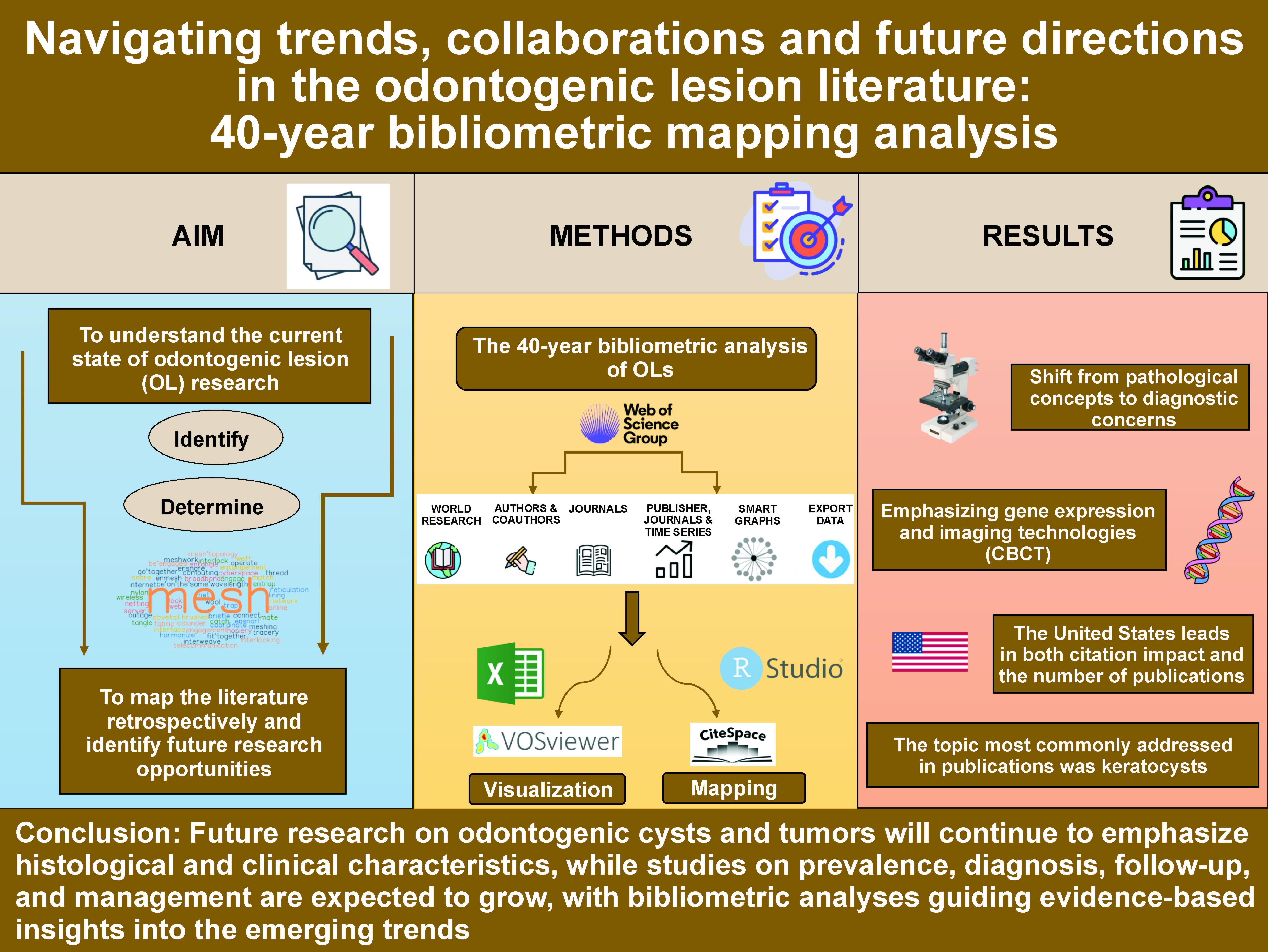
Objectives. Within this context, the present bibliometric analysis aimed to evaluate the current landscape of OL research, retrospectively map the field, and identify the emerging trends and future research directions.
Material and methods. Guided by the principles of the Leiden Manifesto, this comprehensive bibliometric study analyzed 4,298 publications published between 1980 and 2023, and retrieved from the Web of Science Core Collection. The analysis examined international collaboration patterns, keyword evolution, institutional affiliations, co-citation networks, and thematic clustering. CiteSpace, RStudio Bibliometrics and VOSviewer were employed to explore bibliometric relationships and generate visualizations.
Results. The 40-year bibliometric mapping analysis of OL research revealed a diverse and evolving scientific landscape. A total of 4,298 publications encompassing 40,209 references demonstrated extensive international collaboration, represented by 111 nodes and 385 collaborative links. India emerged as the most prolific contributor (670 publications), while the USA led in citation impact (12,169 citations). Co-citation analysis identified highly influential works, notably updates to the World Health Organization (WHO) classifications. Thematic clustering highlighted major research domains, including odontogenic keratocysts, ameloblastomas and calcifying odontogenic lesions. Trend analysis further indicated a shift from traditional pathological concepts toward advanced diagnostic approaches, with growing emphasis on gene expression profiling and imaging technologies, such as cone-beam computed tomography (CBCT).
Conclusions. In the present study, the historical development of OL literature was evaluated using bibliometric analysis, identifying the most influential publications, major thematic domains and research hotspots. The emerging trends in OL research continue to shape the field and drive advances in both scientific understanding and clinical practice.
Keywords: oral pathology, bibliometric analysis, odontogenic cysts, odontogenic tumor, medical bibliography
Introduction
The oral and maxillofacial region encompasses a wide spectrum of pathological conditions of diverse etiology.1 Dental caries, oral lesions and odontogenic lesions (OLs) are among the most common disorders affecting this region.2, 3 Dental caries is a multifactorial disease initiated by microbiological shifts within the complex oral biofilm, and influenced by factors such as salivary flow and composition, fluoride exposure, dietary sugar intake, and preventive behaviors.2, 4 Oral lesions are defined as abnormal or pathological alterations of the tissues within the oral cavity and the associated structures, presenting as changes in color, texture or surface appearance. These lesions may arise from a variety of causes, including infection, inflammation, trauma, or systemic disease.3, 5
Within this spectrum, odontogenic lesions (OLs) are particularly prevalent.6 According to the World Health Organization (WHO), OLs are pathological conditions associated with tooth development or tissues of dental origin, and commonly occur within the jaw bones.7 Odontogenic tissues develop through time-dependent, tightly regulated interactions between epithelial and mesenchymal components, giving rise to a wide spectrum of morphological patterns in the lesions derived from these tissues.8 Although most OLs are benign, they exhibit considerable clinical and histopathological diversity, and represent an important entity in clinical practice.7 In the oral and maxillofacial region, OLs typically arise from the abnormalities occurring during embryonic tooth development. These conditions may manifest in various forms, including jaw bone masses, dental dysplasia, or other dental anomalies.9 The stimuli initiating odontogenic epithelial growth and the subsequent lesion development remain poorly understood.10 Clinically, OLs most often present as painless masses; however, depending on their size and anatomical location, they may exert pressure on the adjacent structures, leading to functional impairment or esthetic concerns.11
Odontogenic lesions play a significant role in dentistry, pathology, oral surgery, and radiology.12 At the academic level, ongoing research continues to expand knowledge on OLs through the investigation of novel diagnostic modalities, therapeutic strategies and the underlying pathogenetic mechanisms. Such studies enhance the understanding of these lesions, and contribute to improved clinical management and patient care. Moreover, academic research on OLs promotes interaction and collaboration among the disciplines of medicine, pathology and dentistry. A multidisciplinary approach to the evaluation and management of these lesions enables more comprehensive patient care, and fosters continuous advancement in this field. In this context, a thorough and critically appraised review of the available scientific literature is essential for informed clinical decision making.
In 1977, Dr. E. Garfield pioneered bibliometric studies by identifying highly cited publications to investigate the factors underlying their citation impact and influence within a given field.13 Bibliometric analysis is a quantitative method used to evaluate the productivity, impact and evolving trends of a scientific discipline by analyzing the characteristics of selected publications within specific databases, including citation counts, co-citation patterns, and the year of publication.14 The cumulative nature of scientific knowledge, global advances in health systems, and the growing number and diversity of academic journals have contributed to a substantial increase in the published research. Despite this growth, there remains a lack of quantitative and objective evaluations identifying the publication patterns related to OLs within the field of oral and maxillofacial surgery. A comprehensive bibliometric analysis in this area would enable researchers to identify research hotspots, assess influential contributions, and gain insight into current perspectives and the emerging trends.
The aim of this study was to analyze and visualize the existing body of literature on OLs in order to retrospectively map the field and identify potential directions for future research.
Material and methods
This study was conducted in accordance with the principles set forth in the Leiden Manifesto. Bibliometric analysis is exempt from the institutional ethics committee review, as it relies solely on publicly available electronic sources and does not involve the generation of novel data or the use of private patient information.
Articles were retrieved from the Web of Science Core Collection (WoS-CC) database on the same day (December 2, 2023) to avoid bias due to daily database updates. The literature was filtered for the period between 1980 and 2023. Medical Subject Headings (MeSH) were used to select the following search terms: “ALL=(“odontogenic’’ and (“lesion’’ or “cyst’’ or “tumor’’ or “pathology’’ or “malignity’’ ) not (“nonodontogenic’’)) and Dentistry Oral Surgery Medicine or Pathology or Surgery or General Internal Medicine (Research Fields) and Soft Tissue, Bone & Nerve Cancers or Dentistry & Oral Medicine or Cosmetic Surgery or Molecular & Cell Biology – Cancer & Development (Citation Topics Meso) and Article, Review Article or Book (Document Types)’’. No language restrictions were applied. Articles unrelated to oral and maxillofacial surgery were excluded (this exclusion was done through the Research Fields and Citation Topics Meso filters).
A list of articles was compiled in a Microsoft Excel spreadsheet (Microsoft Corporation, Redmond, USA), and the following information was recorded: journal name; citation rank; citation density (citations per year); first author’s name; year of publication; first author’s institution and country of origin; study type; study design; research areas; keywords; author keywords; and indexing information. The finalized dataset was imported into CiteSpace (v. 6.2.R6; Drexel University, Philadelphia, USA), RStudio (v. 4.3.2; Posit, Boston, USA) and VOSviewer (v. 1.6.20; Leiden University, the Netherlands) for statistical analysis and the generation of maps and graphs. CiteSpace and VOSviewer were used to examine relationships among authors, institutions, countries, and keywords, while co-citation analysis was conducted to identify common research foci and the emerging hotspots. Descriptive statistics were used to summarize study characteristics. The analysis excluded the examinations of author self-citations and H-index values, as these metrics are database-specific and not consistently available across sources.
Results
Overall results
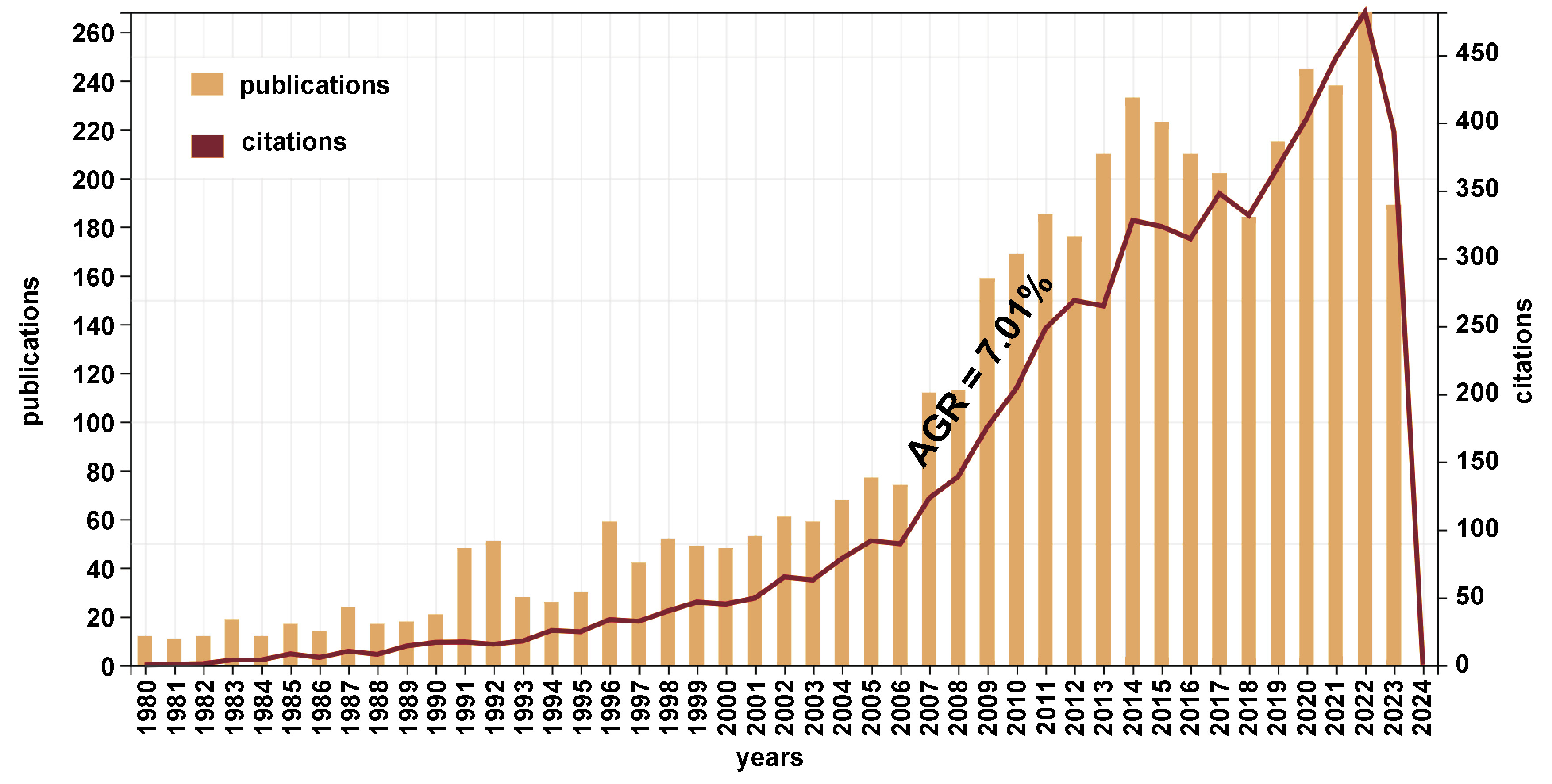
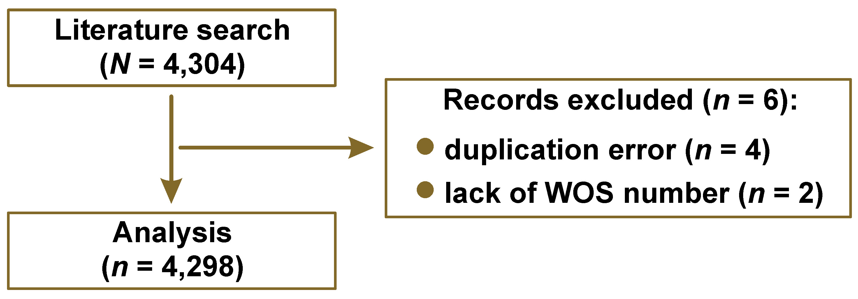
A total of 4,298 publications and 40,209 references were retrieved from the WoS-CC database to investigate the basic publication characteristics, hotspots and the boundaries of this research area (Table 1). A total of 373 sources (journals, books, etc.) were identified. The average number of citations per article was 13.73. The average logistic annual growth rate (AGR) with regard to the number of publications was 7.01% (Figure 1). The search retrieved a total of 4,304 articles, of which 6 were excluded. A total of 4,298 articles were included in the analysis (Figure 2).
Collaboration across countries
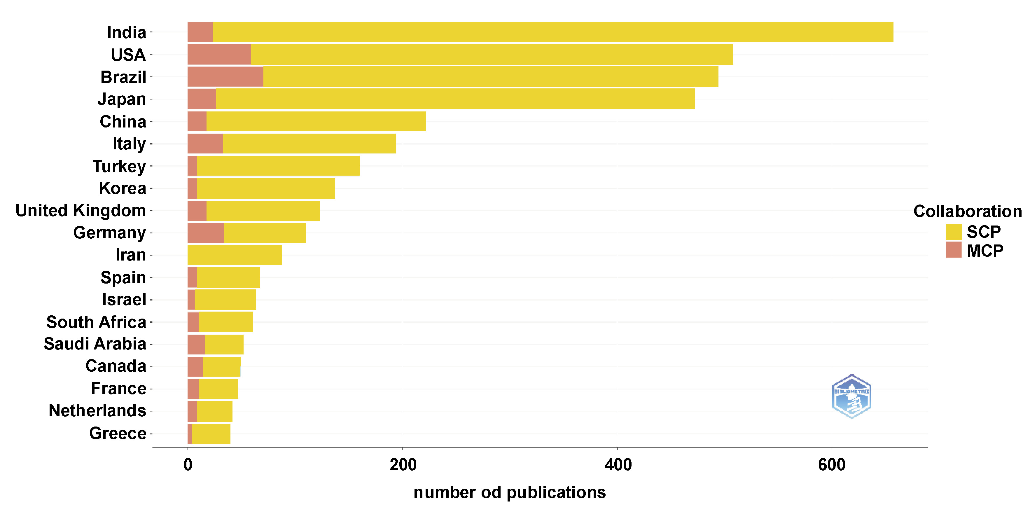
In terms of cooperation across countries, the nodes and links were created using CiteSpace. A total of 111 nodes and 385 links were identified (Figure 3). The size of the nodes indicates the frequency of co-citation, and the links between the nodes indicate connections between co-citations. Nodes of different colors represent specific years. India was the top publishing country with 670 publications, followed by the USA (609), Brazil (536), Japan (462), China, and Italy. The top 5 countries in terms of centrality (the purple circle), which reflects the extent to which a country functions as a bridging node within international collaboration networks rather than the publication volume alone, were the USA (0.59), India (0.24), the UK (0.15), Denmark (0.13), and Germany (0.12). The MCP index (multiple-country publications) illustrates the number of documents with at least one co-author from a different country for each respective country, thus measuring the extent of international collaboration within that country (Figure 4). In the visualization based on the nationality of authors in the dataset, India (2,057), Brazil (1,996) and the USA (1,714) emerged as the leading nations. The color intensity deepens as the number of publicatIons increases (Figure 5).
Keyword analysis
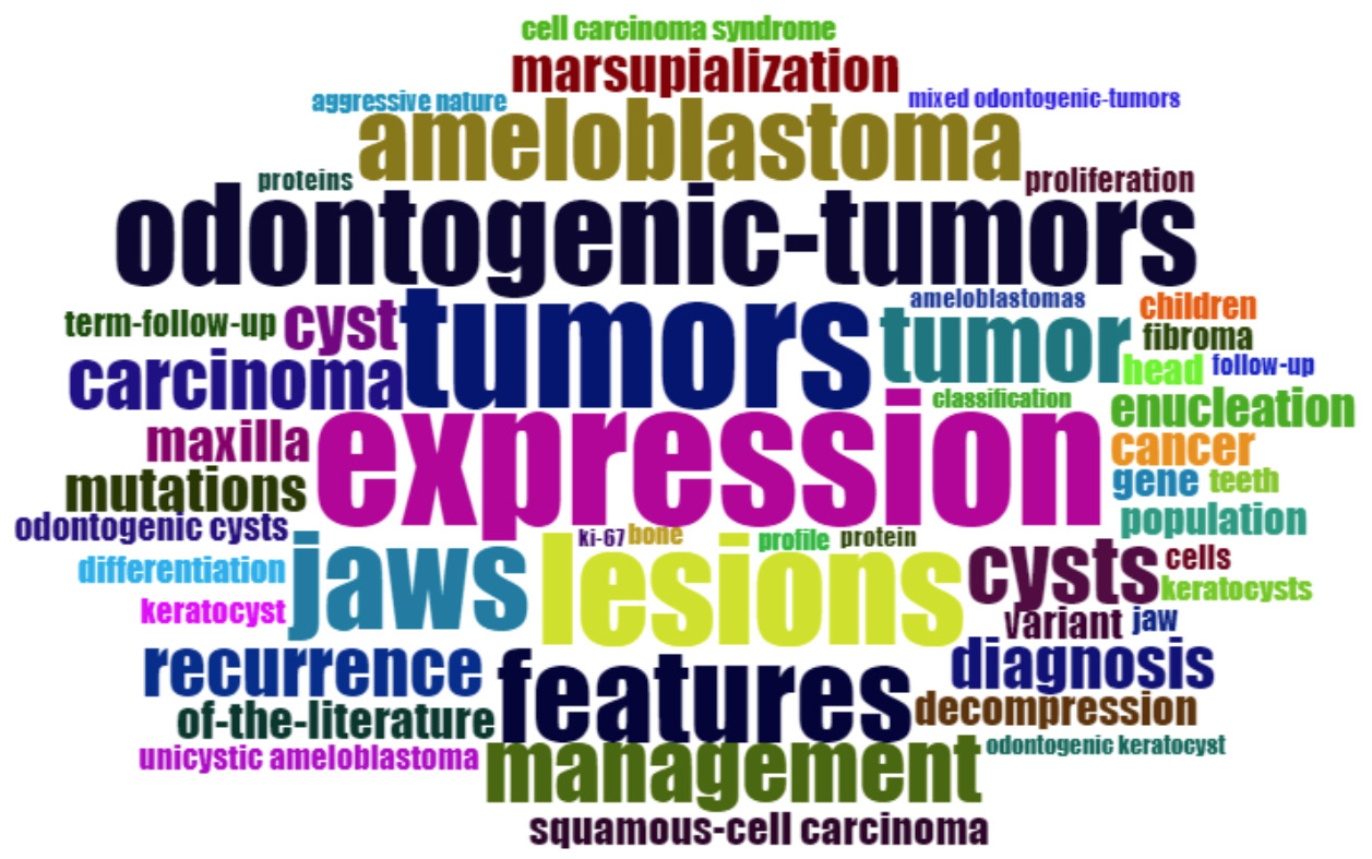
In Figure 6, the WordCloud illustrates a network of keywords derived from the cited literature. Among the KeyWord Plus terms extracted from the titles of the articles, a clear distribution can be observed around the most prominent keyword “expression’’ (338). Other frequently occurring terms include “tumor”, “lesion”, “odontogenic tumors”, and “ameloblastoma”, each appearing 199 times or more. In the visualization, the font size reflects the frequency of each keyword, while more central placement indicates greater relevance within the overall research theme.
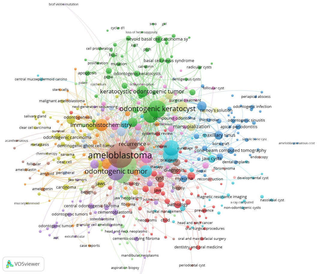
The construction of a keyword co-occurrence network, as shown in Figure 7, enables the exploration of the conceptual structure of the research domain. This analysis examined relationships among author-assigned keywords based on the frequency and repetition of their co-occurrences. When a minimum co-occurrence threshold of 5 was applied, the KeyWord Plus co-occurrence network comprised 417 keywords. In the network, the links between the nodes indicate that keywords were frequently associated within the same publications. Three major clusters are evident, centered on the keywords “ameloblastoma”(total link strength of 1,015), “odontogenic tumors” (743) and “odontogenic keratocyst” (546).
Analysis of affiliations
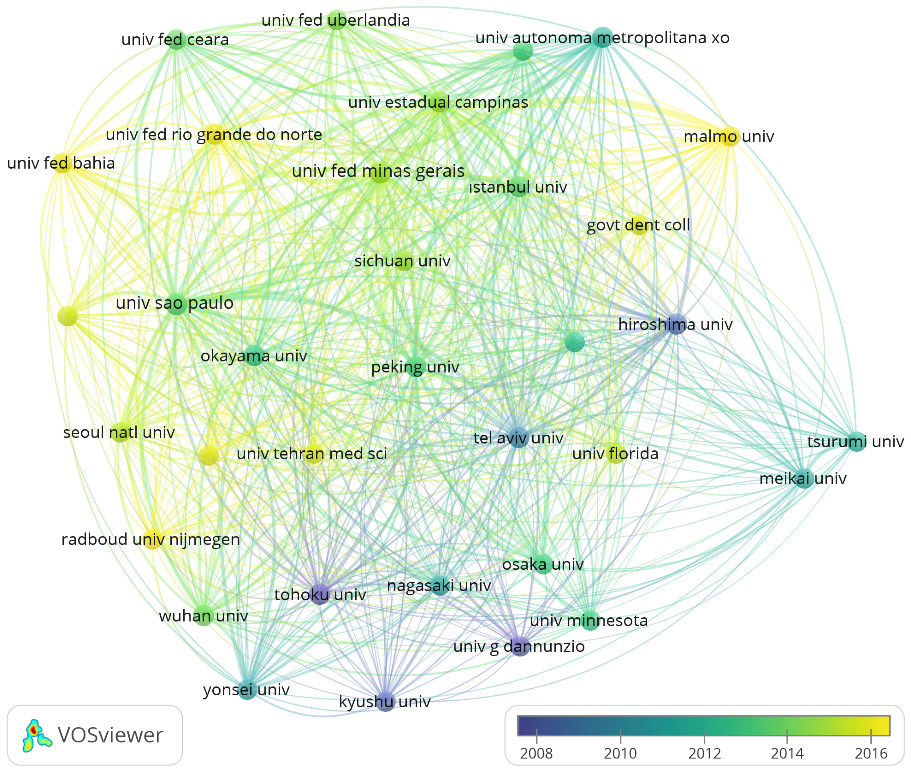
A total of 34 institutions that published more than 20 documents and received at least 20 citations were analyzed using VOSviewer (8). The 4 institutions with the highest total link strength were the University of Minas Gerais, Belo Horizonte, Brazil (total link strength of 25,142), the University of São Paulo, Brazil (17,824), the State University of Campinas, Brazil (16,168), and Peking University, China (11,205).
Analysis of co-citation
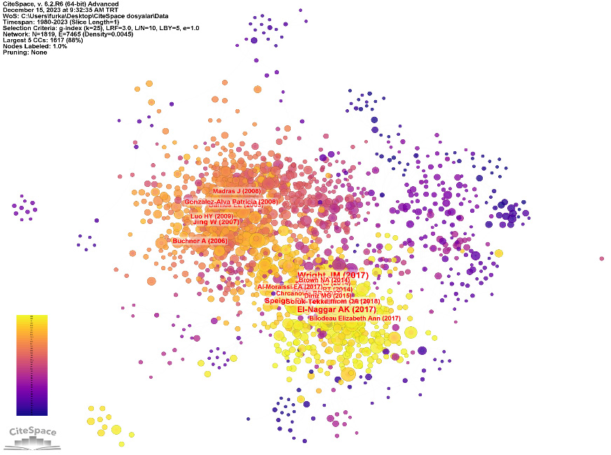
Figure 9 illustrates the co-citation network of the publications related to OLs. In this network, the nodes represent the cited references, and the links between the nodes indicate co-citation relationships. Co-citation analysis identified a total of 1,270 nodes and 11,617 links. The top 10 articles in terms of citation count, co-citation frequency and citation burst strength are summarized in Table 2. A study by Reichart et al.15 was the most cited publication, receiving 816 citations. An article by Wright and Vered25 had the largest node radius, indicating the highest co-citation frequency, with 148 co-citations. A study by Slootweg and El-Naggar18 exhibited the strongest citation burst (strength = 89.15), with a duration of 5 years.
Analysis of clusters
When the studies were automatically clustered, several major thematic groups emerged, including cluster #0 “odontogenic keratocyst”, cluster #1 “odontogenic myxoma”, cluster #2 “calcifying epithelium”, cluster #3 “calcifying odontogenic lesion”, cluster #4 “ameloblastoma”, and cluster #5 “apical periodontal cyst”. The timeline visualization of the network, which incorporates publication characteristics such as cluster distribution and citation bursts, is presented in Figure 10.
Analysis of topic trends
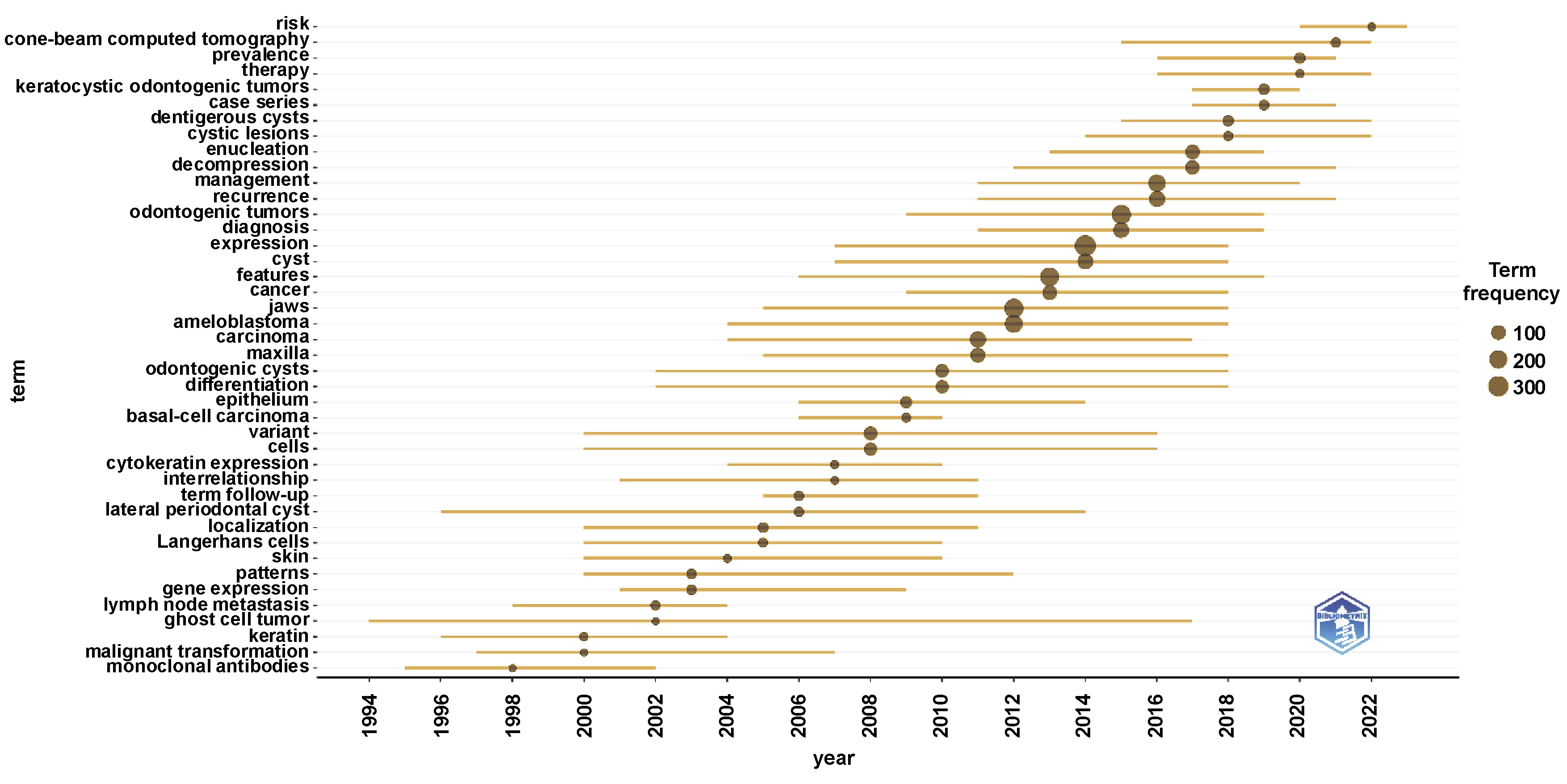
Figure 11 illustrates the temporal trends of OL research topics over the period 1994–2021. Topic trends were identified based on term frequency, with larger point sizes indicating higher frequencies. In the early 2000s, research predominantly focused on terms such as “gene expression” (25), “keratin” (14) and “monoclonal antibodies” (7). In contrast, more recent studies have increasingly emphasized topics such as “risk” (13), “cone-beam computed tomography” (23) and “prevalence” (36).
Discussion
The Leiden Manifesto is widely regarded as an important guide in bibliometrics.36 Comprising 10 principles, it aims to discourage the use of persuasive or overly conclusive statements in bibliometric analyses, and instead promotes the transparency and contextualized interpretation of data.36 Bibliometric analysis has emerged as a widely used approach for evaluating the impact and productivity of articles, journals, authors, institutions, and countries, as well as for mapping collaborative networks. By systematically analyzing the scientific literature within a specific field, bibliometric methods enable the identification of research trends, the evolution of topics over time and potential future research directions.14
In dentistry, evaluating lesions and critically reviewing the associated scientific literature are essential for informed clinical decision making and improved patient care.37 Accordingly, the present study provides a comprehensive analysis of global publication trends related to OLs and tracks the development of OL research over the past 4 decades. Visualization techniques and bibliometric mapping were employed to enhance the interpretability of the findings. The results offer insights into recent advances in the field, including patterns of global research collaboration, trends in clinical research and the emerging areas of interest.
The number of publications on OLs in dental journals increased markedly during the 2010s, reaching a peak in 2022, followed by a decline in 2023. Given that the preparation, submission and peer-review processes of academic publications require substantial time, the COVID-19 pandemic may have contributed to the observed decrease in publications from 2022 onward. This influence may reflect disruptions in research priorities, workflows and productivity among investigators studying OLs during the pandemic period (2019–2021).38 From the patient perspective, the postponement of elective dental treatment procedures and the disruption of routine check-ups during the same period likely resulted in fewer diagnosed and treated OL cases. Łazarz-Półkoszek et al. reported that, despite efforts to establish optimal sanitary conditions, patients remained anxious about attending dental appointments during the COVID-19 pandemic.39 Reduced patient motivation and attendance may have further delayed the diagnosis and management of OLs, particularly in asymptomatic cases. We anticipate that the normalization of dental care services in the post-pandemic period will lead to a relative increase in the detection and reporting of OL cases, which is expected to be reflected in future publications. Moreover, the observed AGR of 7.01% in the literature supports the view that OLs will remain an important and increasingly discussed topic in the coming years (Figure 1).
India had the highest number of publications (670) but ranked 6th in total citations (2,794). In contrast, the USA ranked 2nd in the publication count (609), yet achieved the highest total number of citations (12,169). Consistent with numerous bibliometric studies in oral and maxillofacial surgery, the USA continues to lead in both citation impact and research output, a trend that is expected to persist in the future (Figure 3).14, 40, 41
International collaboration varied considerably across countries and authors. Although India produced the largest number of publications, it collaborated with only 31 countries. By comparison, Germany, despite publishing substantially fewer articles (122), demonstrated broader international engagement, collaborating with 38 countries. One possible explanation for the relatively lower citation impact of Indian publications is their frequent appearance in locally indexed journals, which may limit international visibility and collaboration. Strengthening cross-border research partnerships, particularly for high-output countries such as India and Japan, could enhance the research quality, clinical applicability and global impact of future OL studies (Figure 4).
Cooperation analysis was employed to evaluate collaborative interactions among institutions. In the network visualization, the node size represents the number of publications produced by each institution, while the link thickness reflects the intensity of collaboration. Overall, the level of cooperation among countries and institutions was sufficient to sustain a robust scientific network. Notably, several Brazilian institutions are highlighted in yellow and light green, indicating active and ongoing contributions to the OL literature. Given that Brazil ranks 3rd in national research productivity (536) and hosts multiple institutions with strong engagement in OL research, it may be inferred that Brazil has the potential to emerge as a leading country in this field in the coming years.
The WordCloud is a useful visualization tool for identifying key terms that define a research topic and for guiding future studies. In this type of visualization, the font size, color coding and the spatial arrangement of words provide insights into keyword frequency, relative importance and associations with other topics. In keyword co-occurrence analysis, larger nodes or words typically indicate higher dominance of a keyword, smaller distances between 2 nodes suggest stronger relationships and thicker links reflect more frequent co-occurrence of 2 keywords.
A wide variety of keywords were identified, ranging from lesion types, such as “keratocysts”, “ameloblastomas” and “carcinomas”, to surgical procedures, including “endoscopy” and “aspiration”, as well as molecular and genetic factors, such as cell cycle-related proteins (“Bcl-2” (B-cell lymphoma 2), “Cyclin D1”) and cancer-associated mutant genes (e.g., “BRAF V600E”). A particularly prominent keyword in recent years is “next-generation sequencing”. Studies utilizing this technology to detect pathogenic variants in ameloblastoma, particularly somatic mutations such as FGFR2 and SMO, may serve as important references for future research.42 Three keywords – “odontogenic sinusitis”, “maxillary sinusitis” and “cone-beam computed tomography” – were highlighted due to their recent rise in frequency. The results indicate that researchers are now paying more attention to evaluating OLs in the maxillary sinus with cone-beam computed tomography (CBCT), which is widely regarded as the gold standard for sinus imaging.43
The primary objective of co-occurrence analysis is to map the conceptual structure of a research field by examining how words co-occur within a bibliographic collection. Keywords play a critical role in disseminating research to a broader audience and in influencing how findings are recognized within the scientific community.44 However, overreliance on generic keywords can weaken research visibility, and potentially lead to the misinterpretation of results.45 Literature reviews recommend a balanced approach that incorporates both generic and specific keywords to prevent overlooking relevant evidence while minimizing misleading conclusions.45 In the present study, the high density of generic terms such as “mandible”, “maxilla” or “jaw” may overshadow more specific keywords that could guide future research. The authors therefore suggest limiting the excessive use of generic terms to enhance clarity and relevance in bibliometric analyses.
Co-citation coupling is a technique used to assess subject similarity between 2 documents. When both documents appear in the reference list of a third document, they are considered co-cited, indicating a relational link between them. The strength of this relationship increases as the number of shared citations across different sources grows. Co-citation frequency reflects how often 2 documents are cited together, and serves as an indicator of their conceptual or thematic similarity.46
In this study, the most cited, most co-cited and strongest citation-burst articles related to OLs were evaluated. When analyzing Table 2, the publications with the greatest citation burst strength were the article by Slootweg and El-Naggar entitled “World Health Organization 4th edition of Head and Neck Tumour Classification: Insight into the consequential modifications”18 and the article by Wright and Vered entitled “Update from the 4th edition of the World Health Organization Classification of Head and Neck Tumours: Odontogenic and maxillofacial bone tumors”.25 In addition to being the 2 studies with the strongest citation burst in the last 5 years, they were also the most co-cited publications. This can be explained by the fact that these 2 publications reviewed the 4th edition of the World Health Organization (WHO) classification of head and neck tumors published in 2017. The significance of employing a precise and up-to-date classification system is particularly evident in situations where differential diagnosis is challenging. In this regard, Gupta et al. demonstrated that differences in the reported frequency distribution of gingival lesions across 2 distinct classification systems were attributable to heterogeneous terminology rather than actual geographical variations.47
As shown in Figure 10, the publications exhibiting citation bursts (the red rings surrounding larger circular areas) first appeared in the early 1980s, with the number of such circular areas continuing to increase over time. This sustained pattern over the past 4 decades suggests that research on OLs has remained of ongoing scientific interest. The analysis of the timeline view and the clustering of topics further reveals a shift in the thematic focus, with earlier studies commonly addressing topics such as apical periodontal cysts, ameloblastic carcinoma and glandular odontogenic cysts, whereas more recent research has increasingly emphasized themes such as ameloblastoma and maxillary sinus involvement. Publications describing a specific tumor, e.g. those by Gorlin19 and Pindborg et al.,33 experienced a citation burst between 1980 and 2000. For example, the calcifying odontogenic cyst (Gorlin’s cyst) was first described by Gorlin in 1980 as an intraosseous, solid, non-neoplastic cystic lesion.19 In the 2017 WHO classification, Slootweg and El-Naggar categorized this entity under the category “Developmental Odontogenic Cysts”, and this publication subsequently demonstrated a notable citation burst.18 Given the strength of this citation burst, this work is expected to gain increasing prominence in the coming years and to serve as a key reference for future studies on calcifying odontogenic cysts.
The trending topics related to OLs were analyzed as well. As expected, the most frequently occurring terms were “tumors” (325) and “lesions” (275). In the early 2000s, research predominantly focused on pathological and molecular concepts, with a frequent use of terms such as “gene expression” (25), “keratin” (14) and “monoclonal antibodies” (7). In contrast, more recent years have seen increased emphasis on diagnostic and epidemiological concepts, reflected by the prominence of terms such as “risk” (13), “cone-beam computed tomography” (23) and “prevalence” (36). This shift highlights a transition in OL research from primarily pathological characterization toward diagnostic evaluation and population-based assessment. Cone-beam computed tomography enables the three-dimensional (3D) evaluation of the maxillofacial region while delivering a relatively low radiation dose.48 Introduced into dental practice in the late 1990s, this technology has substantially advanced diagnostic and treatment planning capabilities in dentistry.49 Shweel et al. reported that CBCT was the most appropriate radiological modality for the preoperative assessment of odontogenic tumors.50 These advantages likely explain why CBCT has emerged as a prominent topic in the OL literature.
The high frequency of keywords such as “prevalence” and “risk” further indicates that assessing the epidemiology of OLs and evaluating their potential clinical risks are current priorities in the literature. It is therefore anticipated that research focusing on the prevalence, treatment, risk assessment, and imaging of OLs will continue to attract significant scholarly attention in the coming years.
Limitations
This study has several limitations. First, the data source was restricted to the literature indexed in the Web of Science (WoS) database; therefore, relevant publications from other databases may not have been included. Second, no formal assessment of bias was conducted when evaluating the included publications, and the study findings were presented without the critical appraisal of methodological quality. These limitations should be considered when interpreting the results.
Conclusions
In the near future, studies focusing on the histological and clinical characteristics of various odontogenic cysts and tumors are expected to remain highly relevant, while research on prevalence, diagnostic approaches, follow-up protocols, and management strategies is likely to gain parallel importance. Conducting similar bibliometric analyses in the future will enable evidence-based monitoring of research progress, and provide valuable insights into the emerging trends and future directions in the literature.
Ethics approval and consent to participate
This study was conducted in accordance with the principles set forth in the Leiden Manifesto. Bibliometric analysis is exempt from the institutional ethics committee review, as it relies solely on publicly available electronic sources and does not involve the generation of novel data or the use of private patient information.
Data availability
The datasets supporting the findings of the current study are available from the corresponding author on reasonable request
Consent for publication
Not applicable.
Use of AI and AI-assisted technologies
Not applicable.