Abstract
Background. Since impacted canines are a frequent eruptive anomaly, it is imperative to study their etiological aspects. A possible cause of impaction is the presence of odontogenic lesions close to the impacted canine.
Objectives. The aim of this study was to evaluate the frequency of odontogenic lesions in patients with impacted canines and their association with the characteristics of impaction.
Material and methods. This cross-sectional study was carried out with the scans of 93 impacted maxillary canines obtained from cone-beam computed tomography (CBCT) studies performed in 3 radiological centers. The selection criteria regarded male and female patients older than 12 years, and were based on the CBCT scans of impacted canines, showing unilateral or bilateral maxillary impaction, with or without odontogenic lesions. The position of the impacted canines, the sector of impaction, the presence or absence of a dentigerous cyst, an odontoma or follicular enlargement, as well as the total diameter of the lesion, were evaluated. The χ2 test and logistic regression analysis were performed, and the level of statistical significance was set at p < 0.05.
Results. The frequency of odontogenic lesions near the canine impaction area was generally low (7.5% for a dentigerous cyst, 6.5% for follicular enlargement and 3.2% for a mesiodens). However, there was a significant association between the presence of a dentigerous cyst and buccal or mid-alveolar impacted canines (p = 0.032). The alpha and beta angles influenced the possibility of the occurrence of dentigerous cysts, with the alpha angle increasing the risk (B = 1.22; p = 0.041) and the beta angle decreasing the chance of developing a dentigerous cyst (18%) (p = 0.024).
Conclusions. The presence of odontogenic lesions in impacted maxillary canine cases is low, and involves mainly dentigerous cysts and follicular enlargement in buccal or mid-alveolar impacted canines. The alpha and beta angles may influence the development of dentigerous cysts.
Keywords: cone-beam computed tomography, impacted maxillary canines, odontogenic lesions
Introduction
Eruptive processes can occasionally present anomalies due to obstruction in the trajectory of eruption. Dental impactions are primarily asymptomatic, and in the orthodontic diagnosis, they are initially identified by means of panoramic radiographs.1, 2, 3 The maxillary canine is the 2nd most frequently impacted tooth (0.8–2.8%), and its position can divert into the palatal or buccal direction, inside or outside the arch, with the palatal position being the most prevalent with a ratio of 2:1, although some authors report a ratio of 3:1.2, 3, 4, 5 Even greater significant differences were reported with regard to canine eruption anomalies between age-matched individuals with and without Down syndrome.6
Impacted canines can be associated with odontogenic lesions that cause a mechanical obstacle preventing the normal eruption of the tooth; on the other hand, impaction damages the surrounding tissues and structures, and, in turn, lead to the occurrence of lesions.3, 4 These associations are determined using cone-beam computed tomography (CBCT), showing that the prevalence of canine impaction associated with some pathology ranges between 1.0% and 9.9% as compared to other teeth, and is surpassed only by third molars.3, 4, 5 Likewise, previous studies assessed the thickness of the dental follicle of the impacted maxillary canine, and found that in 22% of cases, the dental follicle thickness exceeded 3 mm,5 but no significant correlation was found between the dental follicle width and sex, the impaction side and localization.7 Although panoramic and periapical radiographs are frequently used for the initial diagnosis, odontogenic tumors may require more detailed imaging observation. Therefore, greater visual capacity is required for this type of finding to avoid tissue overlapping and to perform a three-dimensional (3D) analysis.8
It is essential to know the prevalence of odontomas, supernumerary teeth, root dilacerations, lesions of traumatic origin, deciduous teeth without root resorption, congenital deformities, and cysts in association with impacted maxillary canines.9, 10, 11 Three-dimensional images can help determine the anatomical variations presented by impacted teeth or the areas invaded by the abovementioned lesions, and provide a more precise diagnosis.12, 13 It is worth mentioning that dentigerous cysts are the most frequent lesions, with a prevalence of 9.9%, and they affect the adjacent structures, including bones and roots.14, 15
For the treatment planning of impacted maxillary canines, it is crucial to assess the presence of associated injuries and know the characteristics of the surrounding structures, as well as their condition before any procedure.16, 17 It also facilitates the determination of appropriate treatment alternatives for each case. Although some studies report data on the frequency of odontogenic lesions in patients with impacted maxillary canines, few carry out 3D evaluations or have an adequate sample size for extrapolation.18, 19, 20, 21 In this way, it would be possible to determine exactly what percentage of maxillary canine impactions could be the consequence of an odontogenic lesion and to know the real impact of lesions on the etiology of this condition. Thus, the aim of the present study was to evaluate the frequency of odontogenic lesions in patients with impacted maxillary canines, as well as the association between odontogenic lesions and impaction characteristics, using CBCT.
Materials and methods
This cross-sectional study was approved by the institutional Ethics and Research Committee at the School of Dentistry of the Scientific University of the South (Universidad Científica del Sur), Lima, Peru, with approval number 704-2021-POS70. The study was carried out in accordance with the Declaration of Helsinki.
The study sample included the CBCT scans of 93 impacted maxillary canines (30 male patients aged 16.92 ±4.24 years and 63 female patients aged 16.70 ±4.58 years), obtained from 3 radiological centers in 3 Latin American countries (Mexico, Colombia and Peru). The CBCT images were performed for reasons external to the present study. The inclusion criteria comprised CBCT scans of patients with impacted maxillary canines, patients over 12 years of age (at this age, the eruption of the maxillary canine must have occurred), of both sexes, with unilateral or bilateral maxillary impaction, in any sector of impaction according to the Ericson and Kurol classification,22 with and without associated pathology, such as a compound odontoma, a dentigerous cyst or a follicular cyst. The CBCT scans of patients with previous orthodontic treatment, syndromes or craniofacial anomalies, dental agenesis, ankylosis, or localized infection were excluded.
The sample size calculation was performed using a formula, with a confidence level of 95%, precision of 5%, and an estimated proportion of 6.5% for the frequency of odontogenic lesions associated with impacted maxillary canines (data obtained from a pilot study). Thus, a minimum of 93 impacted maxillary canines were necessary.
Collection of CBCT scans
Three different radiological centers located in Mexico, Colombia and Peru were requested for CBCT images with a field of view (FOV) of 8×8 cm and 10×10 cm. The images were analyzed using the CS 3D Imaging (Carestream Health, Inc., Rochester, USA), Blue Sky Plan (https://www.blueskyplan.com) and Xelis Dental-3DViewer (INFINITT Europe, Frankfurt am Main, Germany) software, chosen for their compatibility with the image output formats of the CBCT machines used (Vatech Co., Ltd., Yongin, South Korea, and Carestream Health, Inc.).
Training and calibration
A researcher was trained and calibrated by 3 orthodontists with more than 10 years of experience to carry out all the measurements of the qualitative variables. The weighted Cohen’s kappa test was applied to determine the intra- and inter-evaluator calibration values, until values greater than 0.9 were achieved in all measurements.
Measurement of impacted maxillary canines
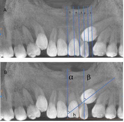
The position of the impacted maxillary canine (palatal, buccal or mid-alveolar), as well as the location of the impaction, whether unilateral or bilateral, were established. In addition, the sector of impaction was registered as follows according to the Erikson and Kurol classification:22 sector 1 – between the mesial side of the first premolar to the distal side of the lateral incisor; sector 2 – between the distal side of the lateral incisor and its median axis; sector 3 – between the median axis of the lateral incisor and the distal side of the central incisor; sector 4 – between the distal of the central incisor and its median axis; and sector 5 – between the median axis of the central incisor and the dental midline (Figure 1A).22, 23, 24, 25
The height of impaction in millimeters in relation to the occlusal plane, as well as the angulation of the impacted maxillary canine in relation to the mid-sagittal plane (the alpha angle) and to the medium axis of the lateral incisor (the beta angle), were also measured (Figure 1B).22
Measurement of odontogenic lesions
The presence or absence of odontogenic lesions – dentigerous cysts, follicular cysts and compound odontomas – was evaluated. A dentigerous cyst was considered as the presence of the epithelium formed around the crown of the impacted tooth, showing a hypodense image with a well-defined sclerotic border around the crown of the tooth and a diameter larger than 5 mm. A follicular cyst was defined as the epithelial growth of the eruption membrane of the canine, not larger than 3 mm in diameter. Lastly, a compound odontoma was determined to be a set of miniature rudimentary dental structures in the epithelial tissue, obstructing or associated with the maxillary canine, with small, hyperdense masses similar to the teeth observed in CBCT. Sizes of 4 mm and 5 mm were not taken into account in order to ensure well-defined criteria for diagnosing a follicular cyst or follicular enlargement, and to avoid borderline cases.
Statistical analysis
The IBM SPSS Statistics for Windows software, v. 24.0 (IBM Corp., Armonk, USA) was employed for statistical analysis. The associations between the study variables was analyzed using the χ2 test. Also, the multivariate analysis was performed with the logistic regression test (p < 0.05).
Results
The characteristics of maxillary canine impaction showed that palatally impacted canines were mainly found in impaction sectors 3 and 5, while buccally impacted canines were mainly identified in sectors 1 and 3 (p = 0.001) (Table 1). The frequency of odontogenic lesions near the canine impaction area was generally low, showing the presence of a dentigerous cyst in 7.5%, follicular enlargement in 6.5% and a mesiodens in 3.2% of the sample (Table 2). There were no associations between the type of impaction (palatal, buccal or mid-alveolar) and follicular enlargement or other lesions (p > 0.05). However, there was a significant association between a dentigerous cyst and maxillary canine impaction, with this lesion found in approx. 15% of buccal and mid-alveolar cases (p = 0.032) (Table 3). When evaluating the influence of the predictor variables (sex, impaction type, impaction sector, alpha and beta angles, and impaction height) on the occurrence of odontogenic lesions, only the alpha and the beta angles had an impact with regard to dentigerous cysts, showing that for each degree of increase in the alpha angle, the risk of a dentigerous cyst increased 1.22 times (p = 0.041). On the contrary, for each degree of increase in the beta angle, the possibility of developing a dentigerous cyst decreased by 18% (p = 0.024) (Table 4).
Discussion
Taking into account the high prevalence of impacted maxillary canines observed during the initial radiographic diagnosis of patients over 12 years of age, we considered the determination of the occurrence of odontogenic lesions as one of the possible causes of maxillary canine impaction to be a priority, since the presence of odontogenic lesions can originate mechanical barriers to the normal eruption of the maxillary canine.26, 27, 28, 29 Dentigerous cysts, compound odontomas and the enlargement of the follicular sac were the lesions evaluated in our study, as they are most commonly associated with the occurrence of an impacted maxillary canine in the literature.29, 30 Additionally, in order to better understand and treat this challenging condition, the present study evaluated the severity of canine impaction, the alpha and beta angles, and the impaction sector as intervening variables.31
This study evaluated the CBCT scans of patients older than 12 years with impacted maxillary canines, given that the eruption of maxillary canines must have already occurred at this age. Likewise, the impaction could be in any sector according to the Ericson and Kurol classification,22 which, although based on panoramic radiographs, has been applied in various studies with regard to panoramic images derived from tomography, reinforcing its reliability.23, 24, 25 It is also important to highlight that the researchers were trained and calibrated to carry out all the measurements of the variables evaluated.
One of the consequences of untreated canine impaction can be the root resorption of the neighboring teeth, which may be severe. For this reason, it is essential to carry out a radiographic evaluation of the eruption of this tooth at the age of 9–11 years as a diagnostic test or for treatment planning if necessary. The results of this study show that the most frequent impaction sectors were 3 and 5 for palatally impacted canines, and sectors 1 and 3 for buccally impacted canines (p = 0.001). Orthodontists should take this into consideration, even though odontogenic lesions associated with canine impaction mainly involve buccally impacted maxillary canines. In addition, the frequency of the lesions related to canine impaction – 7.5% for a dentigerous cyst, 6.5% for follicular enlargement and 3.5% for a mesiodens – should be regarded by orthodontists as a possible etiological factor for this condition despite the low prevalence values.
The results of the present study are useful for clinical practice, since there is a lack of evidence in the scientific literature on the exact percentage of maxillary canine impaction due to the occurrence of cystic lesions. We observed that dentigerous cysts were present in approx. 8% of cases, and were exclusively found in patients with buccal or mid-alveolar canine impaction, demonstrating that the presence of these odontogenic lesions is scarce in the case of palatally impacted canines. Likewise, the alpha and beta angles were found to affect the appearance of an impacted canine. It was observed that for each degree of increase in the alpha angle, the risk of the presence of a dentigerous cyst increased 1.22 times (p = 0.041). In contrast, for each degree of increase in the beta angle, the likelihood of presenting a dentigerous cyst decreased by 18% (p = 0.024). Orthodontists should consider both angular results in the early planning of the treatment of such cases. In this regard, it is essential to note that the early diagnosis and treatment of maxillary canine impaction reduce treatment complexity and possible complications. Besides, the development of additional complications, such as the root resorption of the neighboring teeth in buccal or mid-alveolar impacted maxillary canines, must also be considered.32, 33, 34
The scientific literature provides information on the measurement methods, differential radiographic and tomographic diagnosis, the characteristics and classification of odontogenic lesions, diagnostic imaging interpretation, and even genetic information that can guide and enrich clinical practice.35, 36, 37, 38 In this regard, the results of the present study provide data that can help develop metric and positional analysis methods for volumetric images, and CBCT storage guidelines for imaging findings, among other aspects.
Finally, according to the present results, the probability of maxillary canine impaction being dependent on the presence of an odontogenic lesion is very low. Although these lesions may be circumstantial in cases of buccal and mid-alveolar impaction, there is no absolute association between these lesions and impaction, and more studies regarding this research line should be done in different racial groups.
Conclusions
The presence of odontogenic lesions as a possible factor for the appearance of impacted maxillary canines is low, and involves mainly dentigerous cysts and follicular enlargement, which are primarily observed in buccal or mid-alveolar impacted canines. In addition, the alpha and beta angles could influence the development of dentigerous cysts, with the alpha angle increasing and the beta angle decreasing the risk of the lesion.
Ethics approval and consent to participate
The present study was approved by the institutional Ethics and Research Committee at the School of Dentistry of the Scientific University of the South (Universidad Científica del Sur), Lima, Peru (No. of approval: 704-2021-POS70). The study was carried out in accordance with the Declaration of Helsinki.
Data availability
The datasets generated and/or analyzed during the current study are available from the corresponding author on reasonable request.
Consent for publication
Not applicable.
Use of AI and AI-assisted technologies
Not applicable.