


Abstract
Background. Parsley has been traditionally used as a food additive and herbal medicament. The flavonoid apigenin and its glycosides constitute the most abundant phenolic compounds found in parsley. They exhibit numerous pharmacological effects, including antioxidant, anti-inflammatory, antitoxic, and anticancer properties.
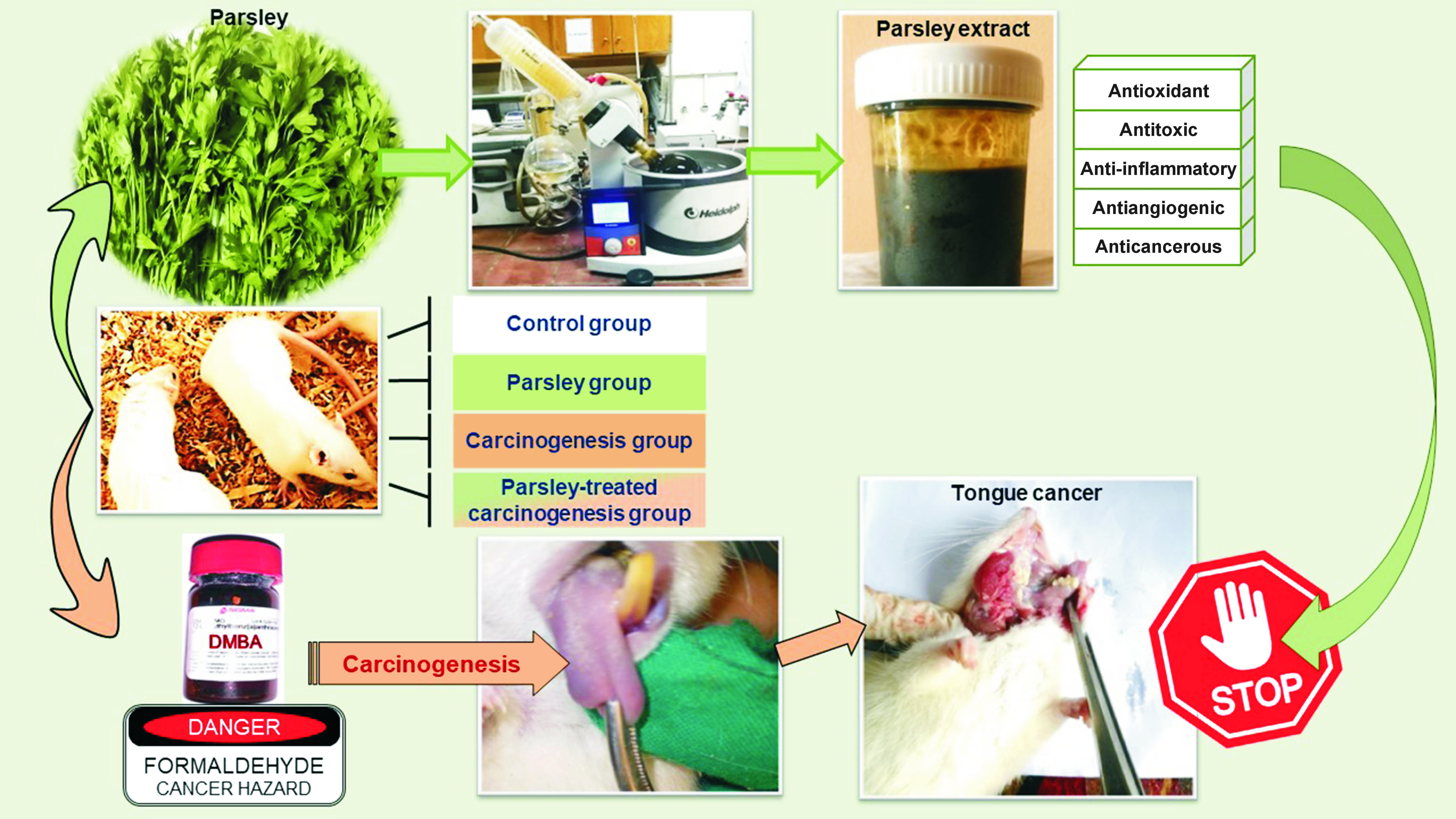
Objectives. The aim of the study was to evaluate the potential chemopreventive effect of orally administered parsley extract on tongue cancer induced by 7,12-dimethylbenz[a]anthracene (DMBA) and formaldehyde in rats.
Material and methods. A total of 36 adult male albino rats were randomly allocated into 3 equal groups: the parsley group was administered 2 g/kg body weight parsley extract through oral gavage 3 times per week; the carcinogenesis group received a topical application of 0.5% DMBA in acetone and formaldehyde to the tongues; and the parsley-treated carcinogenesis group was administered parsley extract combined with a topical application of DMBA and formaldehyde. Additionally, a group of 5 rats served as a negative control group. After 8 weeks, the tongues of the rats were dissected and subjected to histopathological, immunohistochemical, histomorphometric, and quantitative real-time polymerase chain reaction (qRT-PCR) analyses.
Results. Histopathologically, the tongues from the carcinogenesis group revealed several signs of hyperplasia, dysplasia and the invasion of dysplastic cells into the underlying connective tissue. The tongues of the parsley-treated carcinogenesis group exhibited a reduction in dysplastic changes and almost regained their normal architecture, as observed in both the control and parsley groups. The immunohistochemical analysis of the area percentage of caspase-3 immunoexpression revealed a significant increase in the parsley-treated carcinogenesis group compared to the carcinogenesis group, which approached the results observed in both the control and parsley groups. The qRT-PCR results of tumor necrosis factor-alpha (TNF-α) expression displayed a significantly decreased expression in the parsley-treated carcinogenesis group as compared to the carcinogenesis group. These findings were comparable to those observed in the control and parsley groups.
Conclusions. In a rat model, oral administration of parsley extract has been shown to impede the initiation of several cellular carcinogenic changes in tongue tissues.
Keywords: apigenin, plant extracts, Petroselinum, anthracene, tongue neoplasms
Introduction
Cancer is one of the leading causes of death worldwide. Among the most prevalent forms of the disease is oral squamous cell carcinoma (OSCC). Oral carcinoma includes cancer of the lip, floor of the mouth, buccal surface, hard palate, alveolar surfaces, salivary glands, and tongue.1, 2
Tongue cancer accounts for approx. 40–60% of all deaths caused by oral cancer. Tongue squamous cell carcinoma (TSCC) is reported to be a life-threatening condition due to the complexity of the tongue, which is characterized by rich vascular and lymphatic networks. The musculature of the tongue fosters aggressive behavior, manifesting as local invasion and regional lymph node metastasis.3, 4
The downregulation or high inactivation of caspase-3 expression is typically observed in cancer cells, making them resistant to local microenvironmental stresses as well as various treatments.5 Caspase-3 and cleaved caspase-3 are considered biomarkers in the diagnosis of TSCC. The co-expression of cleaved caspase-3 and/or caspase-3 has shown tumor-suppressing properties in patients with TSCC.6
In carcinogenesis, tumor necrosis factor-alpha (TNF-α) is essential for the epithelial–mesenchymal transition of tumor cells, loss of intercellular adhesion, increased motility of tumor cells, and direct invasiveness or vascular metastasis. These effects are the result of changes in the tumor microenvironment that enhance tumor cell proliferation, migration, invasiveness, and metastasis.7
The primary goal of tongue cancer treatment is to eradicate the primary tumor lesion in the tongue by surgical excision or cryosurgery, control any other neck diseases (nodal metastasis) through chemotherapy or radiotherapy, and preserve tongue functions to the greatest extent possible.8 Various treatment modalities are associated with serious side effects such as pain, necrosis and loss of function. Therefore, there is an urgent need for chemoprevention, defined as the administration of drugs or natural agents with the aim of preventing cancer.9
Cancer chemoprevention refers to the application of agents designed to hinder, delay, or even reverse carcinogenesis before the initiation, progression and invasion of cancerous tissue. This process usually involves a prolonged administration of natural or synthetic chemopreventive compounds.9
Parsley (Petroselinum crispum, family: Apiaceae) is a bright green, biennial plant that thrives in temperate climates. The leaves and stems, whether fresh or dried, as well as their seeds, have been introduced into the food industry, creams, soaps, and perfume manufacturing.10 The flavonoid apigenin is the main polyphenolic compound of parsley, which has antioxidant, anti-inflammatory, antiproliferative, antiulcer, and immunosuppressive properties. It has been shown to inhibit the induction, proliferation and migration of various human cancer cell lines.11 Thus, the present study aimed to investigate the potential protective effect of parsley in preventing or hindering the progression of carcinogenesis in induced tongue cancer in a rat model. The evaluation of this effect was conducted through histopathological analysis, immunohistochemical assessment of caspase-3, and by quantitative real-time polymerase chain reaction (qRT-PCR) analysis to determine TNF-α gene expression.
Material and methods
Ethics statement
The study was approved by the Institutional Animal Care and Use Committee (IACUC), Faculty of Science, Cairo University, Egypt (approval No. CU-III-F-15-19).
Preparation of parsley extract
Fresh leaves of parsley (P. crispum) were air-dried at room temperature and ground into fine powder. The powdered leaves were then soaked in ethanol at room temperature (27°C) and extracted for 48 h with occasional shaking to obtain parsley extract. The extract was filtered and evaporated under reduced pressure (60°C) until it was completely dry. Subsequently, it was dissolved in distilled water prior to its administration to rats.12
High-performance liquid chromatography analysis
The quantification of the active compound (apiin) was conducted by transferring a 7.5-mg sample of the alcoholic extract to a 10-mL measuring flask. The volume was then adjusted to the designated mark with methanol and sonicated for a few seconds. Subsequently, a sample of this solution was filtered through a 0.45-μm membrane filter prior to the high-performance liquid chromatography (HPLC) analysis.13
Sample size
The sample size was calculated using the G*power software, v. 3.0 (https://www.psychologie.hhu.de/arbeitsgruppen/allgemeine-psychologie-und-arbeitspsychologie/gpower). The estimated sample size was 36, with 12 rats allocated to each experimental group. The primary outcome (hindering or preventing carcinogenic changes) was based on the study by Kasem et al.,14 the results of which indicated that the administration of Mentha piperita extract, which has a similar antioxidant effect as parsley, resulted in a reduction of dysplastic carcinogenic changes induced by DMBA or formaldehyde by 61% and inhibited tumor incidence by 100%. A large effect size of approx. 0.6 was anticipated. The statistical power was set at 80% and the α error probability was set at 0.05.
Animals
Thirty-six adult male albino Wistar rats (Rattus norvegicus), with an average age of 4–6 months and an average weight of 150–200 g, were used in this study. The subjects were randomly allocated into 3 equal groups. The animals were housed in specially designed stainless steel cages with a controlled environment (temperature: 25 ±2° and 12-hour dark/light cycles) under good ventilation. The rats were fed a standardized balanced laboratory diet of regular rat chow and distilled water ad libitum. The diet was formulated to meet the nutrient needs of rodents for the duration of the experiment. An additional 5 rats were used as a negative control group.
Induction of tongue carcinogenesis
Formaldehyde and DMBA were used in carcinogenesis. These carcinogens were purchased from Sigma-Aldrich Chemical Pte. Ltd. (Cairo, Egypt). The dorsal and ventral surfaces of each rat’s tongue were painted with 0.5% DMBA in acetone, 3 days per week, with the use of a size #3 camel hair brush. After 9 days, a 10% formaldehyde solution in water was applied in conjunction with DMBA for an additional 3 days per week over the course of 8 weeks.14
Experimental design
The 36 rats were divided into the following groups:
– parsley group: 12 healthy rats supplied with a dose of 2 g/kg body weight of parsley extract via oral gavage 3 days per week for a period of 8 weeks15;
– carcinogenesis group: 12 rats with induced tongue carcinogenesis. This group received no treatment;
– parsley-treated carcinogenesis group: 12 rats provided with parsley extract as in the parsley group, combined with DMBA and formaldehyde as in the carcinogenesis group.
Postmortem specimen processing
Eight weeks after the beginning of the experiment, all rats were sacrificed via intraperitoneal injection of a mixture of ketamine (300–360 mg/kg) and xylazine (30–40 mg/kg), as outlined in the American Veterinary Medical Association (AVMA) guidelines.16 Whole tongues were dissected from all subjects. Subsequently, the specimens were fixed in 10% neutral buffered formalin for 24 h, dehydrated in ascending grades of ethyl alcohol, cleared in xylene, and embedded in paraffin. Sections measuring 4–5 µm were obtained and subjected to the histopathological, immunohistochemical, histomorphometric, and qRT-PCR analyses.
Histopathological examination
The hematoxylin and eosin (H&E) stains were used in accordance with the standard technique to assess the histopathological changes observed in the tongues of the subjects.17
Immunohistochemical examination
Paraffin sections obtained from each group were mounted over positively charged slides. The caspase-3 antibody was used as the immunohistochemical marker. The immunostained sections were then examined with the use of a light microscope (Leica DMLB 2; Leica Microsystems GmbH, Wetzlar, Germany). The positive reaction manifested as brown membranous, cytoplasmic, and/or nuclear staining.18
Histomorphometric analysis of caspase-3 immunoexpression
The immunohistochemically stained sections were evaluated using Leica QWin 500 image processing and analyzing software (Leica Microsystems GmbH). The image analyzer was calibrated automatically to convert the measurement units [px] produced by the program into micrometers [μm]. Immunostaining was measured as area and area percentage in a standard measuring frame in 5 fields for each specimen using ×400 magnification by light microscopy transferred to the screen. The areas exhibiting brown 3,3’-diaminobenzidine (DAB) immunostaining were selected for evaluation. These areas were masked by a red binary color to be measured by the computer system. The mean and standard deviation (M ±SD) values were obtained for each group.
qRT-PCR analysis for the evaluation of TNF-α gene expression
The tissue homogenate was processed for RNA extraction using the Promega spin or vacuum (SV) Total RNA Isolation System (Thermo Fisher Scientific, Waltham, USA), followed by reverse transcriptase for complementary DNA (cDNA) synthesis and qRT-PCR. The qRT-PCR amplification and analysis were performed using a StepOne™ System software v. 3.1 (Applied Biosystems, Foster City, USA).19, 20 Primer sequences of the studied genes are listed in Table 1.
Statistical analysis
The data obtained from the histomorphometric analysis as well as the qRT-PCR analysis was presented as M ±SD. The data was coded and evaluated using the IBM SPSS Statistics for Windows software, v. 21.0 (IBM Corp., Armonk, USA). The Kolmogorov–Smirnov test revealed a normal distribution of the data. Comparisons between the studied groups for normally distributed numeric variables were conducted using analysis of variance (ANOVA). A Tukey’s post hoc test was employed for multiple pairwise comparisons. The value of p < 0.05 was considered statistically significant.
Results
HPLC quantification of apiin
The quantity of apiin, a diglycoside of the flavone apigenin, was found to be 6.41 g% (w/w) in the alcoholic extract of parsley, as determined by the standard calibration curve of apiin, constructed based on the HPLC analysis (Figure 1).
Histopathological results
The histopathological examination of the tongues of the rats in the control group revealed normal tongue architecture, characterized by keratinized stratified squamous epithelium and filiform papillae covering the dorsal surface (Figure 2A). The ventral surface displayed normal stratified squamous epithelium with a thin keratin layer and short, saw-like rete pegs. The underlying connective tissue showed no evidence of infiltration of the inflammatory cells (Figure 3A). The specimens in the parsley group did not exhibit marked histopathological differences in either the dorsal (Figure 2B) or ventral (Figure 3B) surfaces as compared to those of the control group.
The dorsal and ventral surfaces of the rats’ tongues in the carcinogenesis group exhibited hyperplastic stratified squamous epithelium with hyperkeratinization, basal cell hyperplasia and dysplastic cellular changes. These changes included abnormal mitosis with atypical mitotic figures, alteration of the nuclear–cytoplasmic (N/C) ratio, nuclear hyperchromatism, nuclear atypia, and prominent nucleoli. Numerous vacuolated cells and individual cell keratinization (dyskeratosis) were easily identified across different epithelial layers. The connective tissue also exhibited areas of subepithelial inflammatory cell infiltration as well as dilated blood vessels (Figure 2C,Figure 3C). The ventral surface demonstrated the rupture of the basement membrane, with the microinvasion of the dysplastic, hyperchromatic basal cells into the subepithelial connective tissue (Figure 3C).
The histological examination of the parsley-treated carcinogenesis group revealed normal histology of both the dorsal and ventral surfaces of the rats’ tongues, similar to those of the control group. However, 3 specimens were still showing signs of epithelial hyperplasia and hyperkeratinization (Figure 2D,Figure 3D).
Immunohistochemical results
The immunohistochemical examination of the tissue sections from rats’ tongues in the control, parsley and parsley-treated carcinogenesis groups revealed moderate to strong membranous, cytoplasmic, and nuclear caspase-3 immunoreactivity in the entire epithelial thickness of both dorsal (Figure 4A,Figure 4B,Figure 4D) and ventral surfaces (Figure 5A,Figure 5B,Figure 5D). However, the carcinogenesis group revealed negative to weak cytoplasmic and nuclear caspase-3 immunoreactivity in the entire epithelial thickness of the dorsal surface (Figure 4C). Additionally, the ventral surface showed a negative to weak cytoplasmic and nuclear caspase-3 immunoreactivity in the basal and granular cell layers. In contrast, the spinous cell layer exhibited moderate cytoplasmic and nuclear caspase-3 immunoreactivity (Figure 5C).
Histomorphometric results
The analysis of variance revealed a significant difference among all groups (p = 0.000). The highest mean value of caspase-3 immunoexpression was recorded in the control group and the lowest value was observed in the carcinogenesis group. The Tukey’s post hoc test for pairwise comparisons revealed a statistically significant decrease in the area percentage of caspase-3 immunoexpression in the carcinogenesis group in comparison to the control, parsley and parsley-treated carcinogenesis groups (p = 0.000). Additionally, a statistically significant decrease in the area percentage of caspase-3 immunoexpression was observed in the parsley-treated carcinogenesis group compared to the control group (p = 0.006). No statistically significant differences were identified in caspase-3 immunoexpression between the parsley and control groups (p = 0.280), nor between the parsley and parsley-treated carcinogenesis groups (p = 0.204) (Table 2).
Results of the qRT-PCR analysis
The analysis of variance revealed a significant difference among all groups (p = 0.000). The highest mean value of TNF-α gene expression was documented in the carcinogenesis group, while the lowest value was reported in the parsley group. The Tukey’s post hoc test for pairwise comparisons revealed a statistically significant increase in TNF-α mRNA gene expression in the carcinogenesis and parsley-treated carcinogenesis groups compared to the control and parsley groups (p = 0.000). In addition, a statistically significant decrease in TNF-α gene expression was observed in the parsley-treated carcinogenesis group compared to the carcinogenesis group (p = 0.000). However, no statistically significant difference in TNF-α gene expression was observed between the parsley and control groups (p = 0.999) (Table 2).
Discussion
Cancer chemoprevention is defined as the use of pharmaceutical agents, including diet-derived nutritional compounds, to delay, disrupt, or reverse the process of tumorigenesis, especially at the promotion stage.21
Parsley, a natural herb, has been identified as a valuable candidate for anticancer treatment due to its anti-inflammatory, antioxidant, antiproliferative, antitumor, and apoptotic properties.22
The current study was based on the two-stage carcinogenesis model. The DMBA was applied topically as a chemical initiator mutagen, followed by formaldehyde, which was used as a chemical promotor of previously transformed cells. The continuous application of formaldehyde has been demonstrated to accelerate carcinogenesis.14
In this work, ethanol was used as a solvent for obtaining the alcoholic extract from parsley leaves. Using ethanol provides the highest extraction value of polyphenolic compounds, especially apigenin, due to their enhanced solubility in the substance compared to water.23
Regarding the histopathological findings of the present investigation, the tongue specimens of the control group exhibited normal tissue and cellular architecture of the keratinized stratified squamous epithelium with normal underlying connective tissue. These findings were also observed in the tongue specimens from the parsley group. The observations were corroborated by Silvan and Manoharan, who reported similar results upon administration of the flavonoid apigenin.24
On the other hand, tongue specimens from the carcinogenesis group showed variable degrees of epithelial dysplastic changes as well as subepithelial inflammatory cell infiltration. These results align with the studies by Abd El Hameed et al.25 and Said,26 who used DMBA to induce carcinogenesis in albino rats’ lips and hamsters’ buccal pouches, respectively.
The observed changes in the histological architecture of the rats’ tongue mucosa can be explained based on the study by El Yaagoubi et al., who characterized DMBA as an effective carcinogen that induced intracellular accumulation of reactive oxygen and nitrogen species (ROS, RNS).27 This process resulted in DNA damage and mutations in several proto-oncogenes and tumor suppressor genes. Additionally, an elevated expression of proinflammatory mediators responsible for cell proliferation, maturation, angiogenesis, and metastasis was observed.27
In the present study, histopathological changes in the carcinogenesis group were observed in the ventral, rather than the dorsal, surfaces of the rats’ tongues. Similar results were demonstrated by Vendruscolo et al.28 and Thomson,29 who reported that the ventral surface exhibited a thin covering epithelium with elevated proliferative cell activity in comparison to the thicker epithelium of the dorsal surface. The elevated proliferative cell activity prolongs the synthesis (S) phase DNA during the cell division cycle, resulting in chromosomal instability and many DNA damages. These chromosomal aberrations are considered endogenous predisposing factors for malignant transformation, especially under the influence of chemical carcinogens.
In our study, the parsley-treated carcinogenesis group showed a significant reduction in dysplastic changes. This anticancer effect may be attributed to the active compound apiin present in parsley extract, as quantified by the HPLC analysis. This finding aligns with the study by Zhou et al., who reported that the apigenin content of parsley can be considered a potent chemopreventive agent in human keratinocytes.30
In addition, El-fiky et al. examined rats’ lung tissues and demonstrated that the concurrent oral administration of parsley and nicotine significantly ameliorated the histopathological dysplastic alterations induced by nicotine.31
Dysregulation of apoptosis is a critical process in carcinogenesis. Caspase-3, a crucial protein in apoptosis, is located at the end of caspase cascades. It is triggered by both extrinsic (death receptor) and intrinsic (mitochondrial) apoptotic pathways. Its dysregulation at various stages of OSCC leads to cancer cell proliferation and expansion.5 Consequently, the present study investigated caspase-3 immunoexpression.
The immunoexpression of caspase-3 was significantly lower in the carcinogenesis group compared to the parsley-treated carcinogenesis group. This finding aligns with the observations reported by Jakubowska et al., who demonstrated that cancer cells exhibited a lower level of caspase-3 expression compared to normal cells.32 This characteristic of cancer cells contributes to their enhanced resistance to various treatments and microenvironmental stresses.
Moreover, Silvan and Manoharan reported that oral administration of apigenin at a dose of 2.5 mg/kg body weight exerted a cancer chemopreventive effect through the upregulation of caspase-3 and caspase-9 expressions.24 Additionally, Zhang et al. confirmed that flavonoids efficiently inhibited the proliferation of tumor cells through provoking apoptosis via the upregulation of caspase-3 expression.33
The inflammatory process plays an important role in tumor progression. Tumor necrosis factor-alpha is an inflammatory cytokine that promotes inflammation and cancer growth by regulating neoplastic cell proliferation without triggering cell differentiation or apoptosis. It also affects the epithelial–mesenchymal transition of cancer cells.34 Therefore, it was crucial to assess mRNA gene expression of TNF-α in the present study.
The qRT-PCR assessment revealed that the expression levels of TNF-α mRNA gene were significantly higher in the carcinogenesis group compared to the parsley-treated carcinogenesis group. These results are consistent with the findings of Coelho et al., who established a significant increase in TNF-α value in rats’ C6 glioma cell lines compared to the cells treated with 100 μmol/L flavonoid apigenin.35 Furthermore, Ashry et al. concluded that parsley leaves extract significantly decreased the expression of TNF-α than that observed in the untreated nephrotoxicity group in rats.36
According to the outcomes of the current investigation, the pharmacological effects of parsley as a chemopreventive agent can be attributed to the active compound apiin. These outcomes could be mediated by caspase-3 activation and TNF-α downregulation. Our findings coincide with previous research that found apigenin treatment to target malignant cell mitochondria and decrease mitochondrial membrane potential and permeability transition pores, either via the upregulation of intracellular ROS production or directly binding to a specific pore molecule. This process results in the release of mitochondrial cytochrome c within the cell, the activation of caspase-3, and ultimately, the induction of apoptosis.37
Furthermore, the administration of apigenin inhibited the activation of nuclear factor kappa-light-chain-enhancer of activated B cells (NF-κB) by downregulating TNF-α production. This process is a key factor in cell survival and proliferation via the upregulation of pro-survival gene expression and inflammatory cytokine production. Consequently, these mechanisms result in a decreased inflammation and increased cancer cell apoptosis and autophagy.38
Limitations
The present study was subject to certain limitations. With regard to in vivo cancer induction, it is recommended that the experimental period be extended to more than 8 weeks in order to investigate cellular dysplasia and to attain an accurate histopathological diagnosis of squamous cell carcinoma. Moreover, further studies are recommended to investigate the bioactive chemopreventive compounds other than apiin, present in parsley extract, to better understand the molecular basis of its anticariogenic mechanisms.
Conclusions
The current study found that oral administration of parsley ethanolic extract hinders the initiation and progression of several cellular carcinogenic changes in tongue tissues. These changes were detected histopathologically through increased cellular apoptosis, which was caused by the upregulation of caspase-3 immunoexpression and decreased inflammation, attributed to the downregulation of TNF-α gene expression.
Ethics approval and consent to participate
The study was approved by the Institutional Animal Care and Use Committee (IACUC), Faculty of Science, Cairo University, Egypt (approval No. CU-III-F-15-19).
Data availability
The datasets generated and/or analyzed during the current study are available from the corresponding author on reasonable request.
Consent for publication
Not applicable.
Use of AI and AI-assisted technologies
Not applicable.


