Abstract
Background. Dental implants are used as a traditional technique to replace missing teeth. In the long-term evaluation of dental implants, the stability and durability of the implant–bone interface are crucial. Furthermore, the success of dental implants depends on several factors, such as osseointegration, implant geometry and surface topography.
Objectives. The aim of the study was to investigate the effects of coating materials on dental implants by altering several parameters, including the material used, the coating thickness, and different combinations of the cortical and cancellous bones.
Material and methods. The coating materials used were hydroxyapatite (HAP), monticellite (MTC) and titanium nitride (TiN). The coating thickness was varied as 50 µm, 100 µm, 150 µm, and 200 µm. Five different bone combinations were used for the proposed finite element model. An axial compressive load of 150 N was applied.
Results. The FEA showed that the HAP coating material had a significant effect on minimizing the induced stress concentration for all 5 bone combinations. However, the MTC coating material had a significant effect only on 2 bone combinations (combination 2 and combination 3). Meanwhile, the TiN coating material induced higher stress values.
Conclusions. Based on finite element analysis (FEA), it was observed that the coating thickness greatly influenced the concentration of the mechanical stress. Indeed, when the coating thickness was relatively high, the stress concentration value significantly decreased.
Keywords: bone condition, finite element analysis, dental implant, coating thickness, coating materials
Introduction
Dental implants have become a traditional technique used for replacing missing teeth in recent decades.1 The success of dental implants, in both the short and long term, depends upon osseointegration and other factors, such as implant geometry and surface topography.2, 3 For their long-term success, dental implants need improved surface characteristics and biocompatible materials, which have better mechanical properties than commercially pure titanium (Ti), including alloys such as Grade 5 titanium (Ti-6Al-4V) and titanium-zirconium (Ti-Zr).4
There is some evidence regarding the short-term failure of implants due to biological and biomaterial-related factors as well as surface treatment.5, 6, 7, 8 Other factors, such as stress shielding and implant loosening, may directly reduce the longevity of implants and necessitate frequent revision surgeries.9 Furthermore, corrosion can cause extensive implant fatigue, which may shorten the life of the device and lead to its failure. In this context, the most commonly used Ti as well as Ti alloys lack the stability required to prevent corrosion in the initial stages post-implantation.10, 11 Indeed, an investigation on the functionalization of the surface at the bone–implant interface has demonstrated that corrosive bodily fluids weaken the implant surface and cause the detachment of the device from the bone. Moreover, Ti alloys with no coating may cause infection due to biofilm formation, and lead to implant failure due to stress corrosion.12, 13, 14, 15
In people with a weakened immune system, chronic disease or addiction to tobacco products, the wound-healing processes are likely to be prolonged, which may lead to infection.16 Researchers have suggested coating dental implants with a layer of antibiotics to reduce inflammation at the wound site.17 In addition, a layer of agents such as amoxicillin, cephalothin and gentamycin can be applied to the implant surface to improve cellular responses.18 Such an approach was adopted in an in vivo study, in which a biomaterial surface was coated with an antibiotic for a local release.19
The coating on implants plays a significant role in overcoming biocompatibility- and mechanical stress-related issues, and can withstand most biomechanical conditions. Nonetheless, improved performance has been achieved by coating metal implants with bioceramics, apatite-based coatings and ceramic/polymer composites.20, 21, 22, 23, 24, 25, 26
Bone quality plays a vital role in the customization of implants. Indeed, several studies have focused on bone quality to develop implants based on patient-specific mechanical responses.27, 28 Another important factor for determining the long-term success of dental implants is the load transferred from the implant to the surrounding bone.
Finite element analysis (FEA) is an important tool for predicting the behavior of the implant material and the bone. The finite element method (FEM) can be used to analyze several aspects of dental implant design, including the impact of implant thread design on stress distribution. Indeed, it can be used to identify parameters that have the greatest effect on stress distribution in the surrounding bone and the implant.29, 30, 31
A previous study demonstrated that the thickness and morphology of the hydroxyapatite (HAP) layer on dental implants could be optimized based on the stress values to improve implant stability.32 Another study showed that an implant with a 150-micrometer-thick HAP layer minimized the stress concentrated in the surrounding bone.33
The main objective of this investigation was to study the influence of the coating thickness on patient-specific implants, and to optimize the coating thickness by using 3 different coating materials – HAP, monticellite (MTC) and titanium nitride (TiN).
Modeling and methods
Finite element modeling
A three-dimensional (3D) prototype of a section of the mandibular bone was designed. The prototype had a missing tooth, and the mandibular dimensions were derived from the literature.33 The mandibular section consisted of 2 bones, including a core soft cancellous bone, which was 24.2 mm in height and had a width of 16.3 mm, and a 2-millimeter-thick hard cortical bone. A commonly used standard dental implant model (3.75 mm in diameter) (ISP-S 1338; Adin Dental Implants, Nagpur, India) and an 11.5-millimeter-long, aluminum oxide-blasted, acid-etched Ti alloy (Ti-6Al-4V) implant (Adin Dental Implants) were used for the analysis. All of the required dimensions of the implant and the abutment were extracted for modeling using a profile projector (Figure 1). The 3D models were constructed with coatings that conformed to the exact shape of the implant. The coatings were applied at thicknesses of 50 µm, 100 µm, 150 µm, and 200 µm. Dental feldspathic porcelain was used for the crown and a cobalt-chromium (Co-Cr) alloy was used for the metal framework. The mandible was surrounded by a gum-like structure called gingiva and the implant was placed inside the mandible. The implants and the remaining components were modeled using the SOLIDWORKS® program, v. 2017 (Dassault Systèmes, Vélizy-Villacoublay, France), and are shown in Figure 2. The sectional view, along with loading, is shown in Figure 3.
Finite element method
The models assembled in SOLIDWORKS were imported into the Ansys Workbench FEA tool, v. 18.2 (Ansys, Inc., Canonsburg, USA), and the file format was converted to a type that could be used for the analysis. The mesh size used throughout the modeling process was 0.2, which made it possible to obtain the convergence of the finite element model stress values; it was similar to that used by others.35 The physical interactions in the contact areas of the implant–mandible section (cortical and cancellous), implant–abutment, abutment–metal framework, and metal framework–crown interfaces during loading were taken into account using the bonded surface-to-surface contact feature in the FEA tool. The mechanical properties of the materials used for the components in the assembly are shown in Table 1.
To mimic the behavior of the bone in the mandibular section, the cortical and cancellous bones were assigned 5 different material combinations by adjusting the Young’s modulus value of healthy bone by ±20%.28 The material properties of the varying combinations of the cortical and cancellous bones are shown in Table 2.28
The loading conditions for the implant was an axial compressive load of 150 N applied on the crown and on both sides (from the right and from the left) of the mandibular bone section, as shown in Figure 3. The induced stress (the equivalent von Mises stress), transformed from the applied compressive stress through the crown to the implant, as well as 3 different coating materials – HAP,36 MTC37 and TiN38 – at thicknesses of 50 µm, 100 µm, 150 µm, and 200 µm, were evaluated by means of FEM.
It has been observed in previous studies that the mechanical behavior of the implant depends on bone quality. However, there is still a gap in the literature regarding the effect of coated dental implants on different bone conditions.39 Therefore, in the present study, implants with a varied thickness of various coating materials were investigated under static loading conditions to ascertain the optimal properties of patient-specific coated implants.
Results
The equivalent von Mises stress values were calculated for the 3 implant coating materials under the specified loading conditions. The coating materials – HAP, MTC and TiN – were assessed at thicknesses of 50 µm, 100 µm, 150 µm, and 200 µm. The static analysis was carried out with the use of the Ansys Workbench software, v. 18.2.
Stress induced in the cortical
and cancellous bones (combination 1)
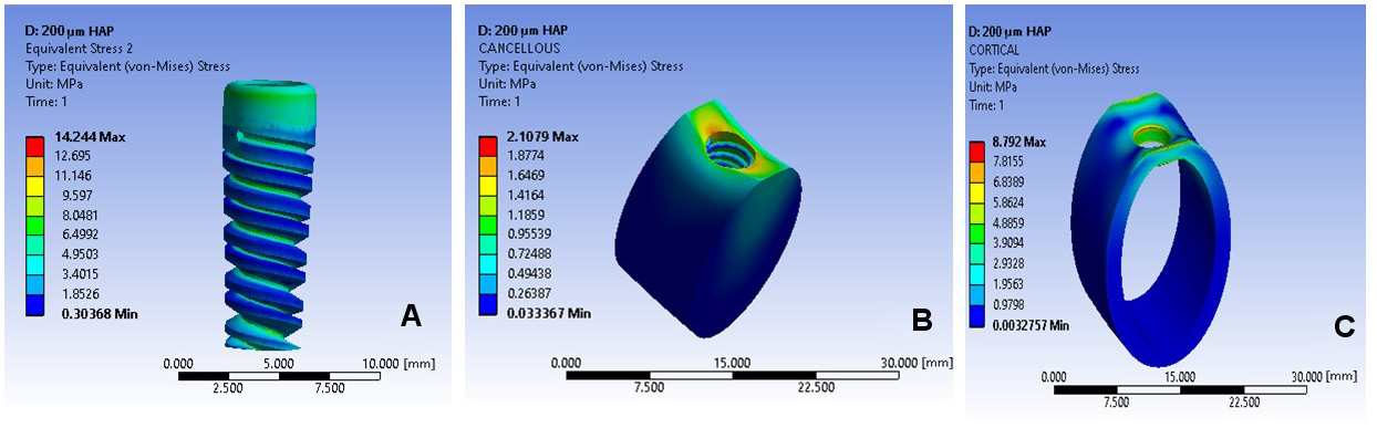
Figure 4 and Figure 5 show the distribution of the induced stress and its effect, respectively, for bone combination 1. It was observed that the von Mises stress in the cortical bone was 83.77 MPa with the HAP coating at a thickness of 50 µm. Meanwhile, in the case of the HAP coating at a thickness of 200 µm, the generated stress was reduced by 87%. On the other hand, the effect of the von Mises stress in the cancellous bone with the HAP coating at a thickness of 200 µm was reduced by 90% (Figure 4 and Figure 5).
Effect of stress on the implant
and the coating layer (combination 1)
The stress value for the 100-micrometer HAP coating layer was 19.43 MPa, while it was reduced by nearly 90% for the 200-micrometer HAP coating layer with regard to 50 μm. The implant stress with the HAP coating was significantly different for combination 1 as compared to MTC and TiN (Figure 5).
Stress induced in the cortical
and cancellous bones (combination 2)
Using combination 2, the maximum stress value in the cortical bone with the HAP coating at a thickness of 50 µm was over 50 MPa. In the cancellous bone, the maximum stress value with the 50-micrometer MTC coating was around 15 MPa. As the thickness of the coating increased, the stress in the cancellous bone decreased, with the 200-micrometer MTC coating reducing it by 78% (Figure 6 and Figure 7).
Effect of stress on the implant
and the coating layer (combination 2)
Figure 7 shows the stress values for combination 2 using coating thicknesses between 50 µm and 200 µm. The stress value for the MTC coating was reduced by 88% at a thickness of 200 µm. As the thickness of the coating layer increased, the implant stress increased as well, reaching as high as 113.81 MPa. The implant material used in this study (Ti-6Al-4V) has a yield strength ranging from 800 MPa to 950 MPa,39 so the stress values were within the safe limits.
Stress induced in the cortical
and cancellous bones (combination 3)
For combination 3, the stress value was high at 50 µm and low at 200 µm (Figure 8 and Figure 9). Using the 200-micrometer HAP coating, the stress was reduced by 87% in the cancellous bone.
Effect of stress on the implant
and the coating layer (combination 3)
Figure 9 shows that as compared to the 50-micrometer coating, the stress was reduced by 81% for the 200-micrometer HAP coating, whereas there was an 83% reduction when using the 200-micrometer MTC coating. In addition, the stress at the implant was similar to that in combination 2, but was within the safe limits.
Stress induced in the cortical
and cancellous bones (combination 4)
For combination 4, increasing the HAP coating thickness from 50 µm to 200 µm reduced the stress by 78% in the cortical bone. In the cancellous bone, the stress value decreased by 91% with the 200-micrometer thickness of the HAP coating as compared to the thickness of 50 µm (Figure 10 and Figure 11).
Effect of stress on the implant
and the coating layer (combination 4)
For the MTC coating material, the stress value was reduced by 84% for the 200-micrometer thickness as compared to 50 µm. Also, the stress at the implant was 87 MPa when using the 50-micrometer coating, and 119 MPa when using the 200-micrometer coating, which was within the safe limits (Figure 11).
Stress induced in the cortical
and cancellous bones (combination 5)
For combination 5, the stress value in the cortical bone was reduced by 77% with the 200-micrometer HAP coating as compared to the 50-micrometer coating (Figure 12 and Figure 13). A similar stress reduction in the cortical bone was found for the MTC coating material, where the stress was decreased by 77% by replacing the 50-micrometer coating with a 200-micrometer layer (Figure 13). In addition, the stress value in the cancellous bone was reduced by 83% when using the 200-micrometer HAP coating as compared to the 50-micrometer coating (Figure 12 and Figure 13).
Effect of stress on the implant
and the coating layer (combination 5)
Figure 13 shows that the HAP and MTC coating materials had a more significant role in decreasing the stress than TiN. Also, the stress was reduced by 72% by increasing the coating thickness from 50 µm to 200 µm. The maximum stress value observed for the HAP-coated implant was 107.83 MPa, although this was rather low as compared to combination 2.
Hypothesis testing
Statistical analysis was carried out on an uncoated implant model, which acted as a control. For combination 1, the mean stress value on coated implants was 87.29 MPa, while it was 89.33 MPa on uncoated implants. For combination 2, the mean stress value was 87.20 MPa on coated implants and 105.00 MPa on uncoated implants. For combination 3, the mean stress value on coated implants was 87.46 MPa and it was 76.00 MPa on uncoated implants. For combination 4, the mean stress value was 87.98 MPa on coated implants and 85.88 MPa on uncoated implants. Meanwhile, for combination 5, the mean stress value on coated implants was 86.84 MPa, and on uncoated implants, it was 84.31 MPa.
Here we accept the null hypothesis H0 and reject the alternative hypothesis Ha. According to hypothesis testing:
H0: µ = µH0 = 1
The cases considered as Ha are as follows:
Ha: µ ≠ µH0 The alternative hypothesis is that the population mean is not 1.
Ha: µ < µH0 The alternative hypothesis is that the population mean is less than 1.
Ha: µ > µH0 The alternative hypothesis is that the population mean is more than 1.
All the values obtained for the coatings ranged between 87 MPa and 88 MPa. Therefore, the difference is 1, which is a minimum value for variations. However, the values for uncoated implants ranged from 76 MPa to 105 MPa. Therefore, the difference in values is not equal to 1. Thus, the null hypothesis value is considered to be 1, and we accept H0 and reject Ha. The decision chart is shown in Table 3.
Discussion
The role of the coating on dental implants is to enhance osteoconduction, to fill gaps, and to create a bond between the bone and the implant. The selection of the coating material depends on various parameters, such as non-toxicity, corrosion resistance, fatigue strength, and durability. Hydroapatite bioceramic coatings have an additional advantage, as they reduce the healing time and have improved fixation strength.39 The HAP coating thickness on dental implants ranges from 0 µm to 150 µm, and increasing the coating thickness reduces the induced stress and stress concentration.33 Monticellite is a bioceramic that can form bone-like structures similar to the cortical bone,40, 41 while TiN coatings have a good load-carrying capacity.42 For the above reasons, these 3 materials were selected as coatings in this modeling study, and were applied at thicknesses of 50 µm, 100 µm, 150 µm, and 200 µm, which is beyond those used previously (150 µm). In the present study, it was found that the optimum change in the induced stress was obtained between a coating thickness of 150 µm and 200 µm. Changing the thickness of any of the coating materials used from 150 µm to 200 µm had a significant impact, as the induced stress values were much lower as compared to coating thicknesses below 150 µm.
Bone quality is one of the parameters that affect stress distribution in the cortical and cancellous bones.43 Furthermore, there is a high stress concentration in the area between the cortical and cancellous bones, as stated previously.44 In the present work, a high stress concentration was observed at the neck area of the uncoated implant, which was reduced by coating the implant with a 200-micrometer layer. Figure 14 shows stress distribution for different bone conditions, using uncoated implants. It was found that the 200-micrometer coating thickness reduced the stress values in the cancellous bone as compared to the uncoated implant by 34.3% (combination 1), 61.8% (combination 2), 149.0% (combination 3), 51.2% (combination 4), and 36.0% (combination 5).
Another important parameter is bone remodeling, which is a process that begins during the healing period.45 It involves an increase in bone mass and high stress during bone formation (BF), while the stress is reduced during bone resorption (BR).46 To ensure a successful BF process, more bone mass is needed. In this study, the microstrain values for coated implants ranged from 2,000 to 2,700 for all bone combinations. In contrast, the microstrain values for uncoated implants exceeded 3,700 for all bone combinations. Therefore, coated implants provide superior bone remodeling as compared to uncoated implants.
Limitations
The current study has several limitations, including the use of homogeneous and isotropic mechanical properties for the cortical and cancellous bones, and the simplified load conditions. Future studies should consider non-linear properties, dynamic loading, fatigue analysis, and contact analysis. Using more realistic material models would enable a better estimation of the mechanical environment at the bone–implant interface.
Conclusions
It can be concluded that the implants coated with HAP at a thickness of 200 µm were relatively superior in all 5 bone combinations. At the 200-micrometer coating thickness, both HAP and MTC gave better results than TiN. The most favorable coating thickness would be in the range of 150–200 µm, since in such conditions, the induced stress values significantly decreased for the cortical bone, the cancellous bone and the coating layer in this patient-specific study. Therefore, FEA could be used to design dental implants with patient-specific coatings.
Ethics approval and consent to participate
Not applicable.
Data availability
The datasets generated and/or analyzed during the current study are available from the corresponding author on reasonable request.
Consent for publication
Not applicable.