Abstract
Background. Oral surgery and surgical interventions in the field of periodontology require the uneventful wound healing of soft tissues to be successful. However, since the oral cavity is exposed to saliva and is a niche for oral microorganisms, wound healing can be delayed, causing pain and discomfort to the patient. Hence, a great deal of interest has shifted to the study of agents that can enhance wound-healing processes.
Objectives. The aim of the present study was to evaluate the oral wound-healing potential of the Actinidia deliciosa (kiwifruit) extract by assessing its effects on the viability, proliferation and migration of human gingival fibroblasts, and its ability to enhance angiogenesis in the chicken chorioallantoic membrane (CAM).
Material and methods. The collection of the A. deliciosa extract and experimentation were carried out in the Maratha Mandal Dental College, Belgavi, India. Human gingival fibroblasts were cultured in a suitable medium and treated with 6.25 μg, 12.5 μg or 25 μg of the ethanolic kiwifruit extract. The MTT cell viability and scratch assays were then carried out in vitro. To determine the angiogenic potential of the extract, solutions of concentrations of 10 μg/mL, 50 μg/mL and 100 μg/mL were transferred to sterile Whatman™ filter paper disks, which were placed on the CAM of 5-day-old Leghorn chicken eggs.
Results. The ethanolic extract of A. deliciosa was not cytotoxic to gingival fibroblasts at a dosage of 6.25 μg/mL. In addition, the scratch assay demonstrated the migration of the fibroblasts and the closure of an artificially created wound within 24 h, which is similar to in vivo wound closure. The CAM assay showed that the kiwifruit extract concentrations of 10 μg/mL and 50 μg/mL showed a moderate pro-angiogenic effect, and a concentration of 100 μg/mL exerted a strong pro-angiogenic effect.
Conclusions. The results demonstrate that the kiwifruit extract is safe to use, can enhance the proliferation and migration of human gingival fibroblasts, and promotes angiogenesis, making it a suitable oral wound-healing agent.
Keywords: wound healing, plant extracts, herbal therapy, angiogenesis stimulators, gingiva
Introduction
Wound healing is a complex process that aims to repair and reconstruct the lost or injured tissues.1 In the oral mucosa, wound healing differs from the healing of the skin, as it proceeds faster and rarely scars.2 Studies have found that the fibroblasts isolated from the oral mucosa and skin have several inherent differences, including their capability to remodel tissues, secretion patterns of certain extracellular matrix (ECM) molecules, cellular migration, cellular adhesion, response to growth factors, and differential expression of ECM receptors.3, 4
Oral surgery and surgical interventions in periodontology include flap surgeries, tooth extractions, implant placement, and the treatment of conditions such as oral abscesses and ulcers. These treatment modalities require the uneventful wound healing of soft tissues to be successful. However, since the oral cavity is exposed to saliva and is a niche for oral microorganisms, wound healing may be delayed, causing pain and discomfort to the patient. Therefore, the main objective of holistic oral wound management is to facilitate healing in the shortest time possible, with minimal pain and discomfort.5 Hence, a great deal of interest has shifted to studying agents that can enhance wound healing.
Therapeutic wound-healing agents can be either natural or synthetic; they are characterized by different levels of efficacy and clinical acceptance. Various agents that promote wound healing and tissue regeneration have come into focus in recent times, such as bone morphogenic proteins (BMPs),6 lactoferrin,7 platelet-rich fibrin,8 vitamin D,9 melatonin, and taurine, among many others. These agents have shown excellent results when implemented after oral and implant surgeries.6, 7, 8, 9 Due to their clinical efficacy, simplicity and affordability, the healing agents derived from plants and herbs have assumed a central role in wound care. Hence, various herbal products have been evaluated as local therapeutic agents for the enhancement of wound healing.10
The kiwifruit belongs to the genus Actinidia (Actinidiaceae). It is rich in vitamin C, carotenoids, tannins, and saponins, among many other active compounds. The nutrient and biologically-active phytochemical content of the fruit has stimulated investigations into its antioxidant and anti-inflammatory potential in preventing cardiovascular disease, cancer, and other degenerative disorders.11, 12, 13 Furthermore, the Actinidia deliciosa extract has been found to be effective against gram-positive and gram-negative bacteria.14, 15
Studies have shown that the kiwifruit extract accelerates scar detachment and helps in the healing of burn wounds. These effects may be attributed to the presence of some compounds in the kiwifruit, such as ascorbic acid, which acts as a scavenger, and the strong protein-degrading enzyme actinidin, which makes it a good anti-escharotic agent, as well as carotenoids, which have strong antioxidant properties.12, 13, 14
The objective of the present study was to evaluate the efficacy of the kiwifruit extract in wound healing by assessing the migration and proliferation of human gingival fibroblasts, and evaluating angiogenesis, with the aim of using this natural agent to improve oral wound healing.
Material and methods
The study design was approved by the Institutional Review Board (IRB) at the Maratha Mandal Dental College, Belgavi, India, before commencement (No. BCD Exam/509/2019-20), and it was performed in accordance with the Declaration of Helsinki ethical standards.
Procurement and preparation
of the extract
Pure A. deliciosa extract was obtained in its powder form from an International Organization of Standardization (ISO) certified natural extract manufacturer (Navchetana Kendra Health Care Private Limited, New Delhi, India). An ethanolic extract of the dried kiwifruit powder was prepared using the maceration technique. A total of 5 g of the extract powder was added to 50 mL of 70% ethanol in a 250-milliliter sterile conical flask, which was plugged with sterile cotton and placed in a shaking incubator (Köttermann, Uetze, Germany) at 200 rpm for 24 h. The filtration process was carried out using a muslin cloth, and it was repeated 3 times to obtain a clear ethanolic solution of the extract.
Human gingival fibroblast cell culture
Human gingival fibroblasts (Adult (HGF) ATCC® PCS-201-018™) were procured (Department of Microbiology, the Maratha Mandal Dental College, Belgavi, India). They were initially centrifuged at 225 g for 5 min and washed in phosphate-buffered saline (PBS). The cells were maintained in Eagle’s Minimal Essential Medium (EMEM) supplemented with 10% fetal calf serum (FCS), antibiotics (5% of a mixture of gentamicin (10 µg), 100 U/mL penicillin and 100 g/mL streptomycin) and L-glutamine (200 mM), and were incubated at 37°C in a humidified atmosphere of 5% carbon dioxide (CO2) and 95% oxygen (O2) for 48 h. The medium was replaced every few hours until confluence was reached.16
MTT cell viability assay
The 3-(4,5-dimethylthiazol-2-yl)-2,5-diphenyl tetrazolium bromide (MTT) assay is a non-radioactive colorimetric system that measures cellular metabolic activity as an indicator of cell viability, proliferation and cytotoxicity. A commercially available cell proliferation kit was purchased (Sigma-Aldrich, Bangalore, India), from which a stock solution was prepared by adding 5 mg of MTT to 1 mL of PBS. The test compound was assessed by the spectrophotometric determination of the conversion of MTT into the formazan blue product by viable cells. Three quantities of the kiwifruit extract – 6.25 µg, 12.5 µg and 25 µg – were diluted in 1 mL of dimethyl sulfoxide (DMSO), and then added to wells containing 100 µL of the previously prepared MTT stock solution. For the control, only DMSO was added to the stock solution, without the addition of the kiwifruit extract. The human gingival fibroblasts were then added to the test solutions and incubated for 4 h. After incubation, the supernatant was carefully aspirated. The precipitated crystals of formazan blue were dissolved by adding 100 µL of DMSO. The optical density (OD) was measured at a wavelength of 570 nm, using an LISA Plus microplate reader (Aspen Medical Group, Inc., Deakin, Australia).17
The mean of 6 readings was used to calculate the results by using the following formula (Equation 1):
where:
OD – optical density.
Scratch assay
The scratch assay was performed to study cell migration and the cell–cell interaction. This was achieved by seeding human gingival fibroblasts onto a 24-well cell culture plate and creating a linear scratch in the confluent cell monolayer with a 200-microliter pipette tip. Cell debris was washed out with plain EMEM. The ethanolic extract of A. deliciosa at concentrations of 6.25 µg/mL, 12.5 µg/mL and 25 µg/mL was used for the experimental groups, and 0.5% DMSO was used for the control group. Following the addition of the extract, images of the cellular gap were captured after 10 h, 12 h, 16 h, and 24 h. The study was completed when the scratch wound was fully closed.18
CAM assay
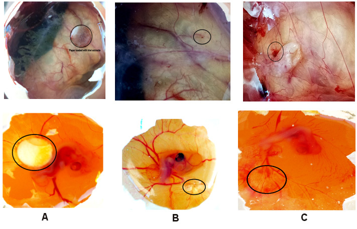
The chicken chorioallantoic membrane (CAM) bioassay was used to evaluate the potential toxicity and pro-angiogenic influence of the ethanolic kiwifruit extract at the vascular level of CAM.15 Fertilized white Leghorn chicken eggs were purchased (Tamil Egg Hatchery, Chennai, India) and incubated in a Multiquip Incubator (E2) (Multiquip Inc., Cypress, USA) at 37°C and 60% humidity for 24 h. The egg tray was automatically tilted at a 450-degree angle every 30 min to mimic natural processes. A small window was made in the shell on the 5th day of the chick embryo development under aseptic conditions, and sterile Whatman™ filter paper disks containing 10 µL of PBS and the ethanolic kiwifruit extract at different concentrations were carefully placed onto the CAM of the eggs. The test compound was prepared at concentrations of 10 μg/mL, 50 μg/mL and 100 μg/mL in 0.2% DMSO. A DMSO control was carried out simultaneously for the comparison with the angiogenic activity of the ethanolic extract. The window was re-sealed with adhesive tape, and the eggs were incubated for 48 h. The windows were re-opened after 48 h of incubation, which was the 8th day of the chick embryo development, and close-up photographs were taken to capture the images of CAM. The images were analyzed for the changes in the angiogenesis process induced by the test compound. All experiments were conducted in duplicate.19
Results
MTT cell viability assay
The kiwifruit extract dosage of 25 µg/mL resulted in the lowest viability (76%) and the highest cell death (24%) of the human gingival fibroblasts. Meanwhile, a dosage of 12.5 µg/mL resulted in 93% cell viability and 7% cell death, and treatment with the 6.25 µg/mL dosage led to 100% cell viability and 0% cell death (Figure 1 and Figure 2, Table 1).
Scratch assay
Adult human gingival fibroblasts were used in the scratch assay. The experimental results showed that 6.25 µg/mL of the extract caused significant cellular mobilization. Indeed, microscopic analysis after 10 h revealed migration distances from the top of the scratch of 235.45 µm and 286.86 µm, and migration distances of 208.86 µm and 290.47 µm from the bottom of the scratch (Figure 3). After 16 h, cell migration increased rapidly, with a nearly full closure of the scratch. A complete closure of the scratch was achieved after 24 h. The time taken to close the gap was plotted against the percentage of cell migration that occurred (Figure 4).
CAM assay
The microscopic examination revealed that blood vessel formation was stimulated in all the eggs loaded with the ethanolic kiwi extract. The stimulation was concentration-dependent, with moderate stimulation achieved at 10 µg/mL and 50 µg/mL, and strong stimulation achieved at 100 µg/mL (Figure 5, Table 2).
Discussion
There has been a recent surge of the evidence supporting the health protective properties of plant-derived compounds, which has resulted in great interest and justifiable scientific assessment.20 Various plant extracts are known to contain carotenoids, polyphenols, vitamin A, and ascorbic acid (vitamin C); they are easy to obtain and economical, and have negligible side effects on humans and the environment.10, 20 As such, natural compounds could enhance wound healing following oral surgery and surgical interventions in periodontology. Therefore, the present study evaluated the effects of the kiwifruit extract on the proliferation of human gingival fibroblasts, as well as its impact on angiogenesis in CAM.
Actinidia deliciosa is known to contain antibacterial agents, scavenging agents and proteolytic enzymes, which have been shown to improve wound healing.17, 21, 22 Furthermore, the A. deliciosa extract acts against a wide spectrum of microorganisms, including Candida albicans, Staphylococcus aureus, Escherichia coli, Pseudomonas aeruginosa, Salmonella enterica, and Salmonella typhi.17, 23 Moreover, the qualitative phytochemical analysis of the kiwifruit extract revealed the presence of carotenoids, alkaloids, flavonoids, saponins, tannins, and terpenoids, which shows its high antioxidant capacity.23
Phytochemicals such as vitamin A, vitamin C and carotenoids are required for the synthesis of collagen, a fibrous protein that helps to reinforce connective tissues, which hold the structures of the body together. They are also highly effective in protecting cells against damage by free radicals. It has been observed that after gingival wounding, the amount of myofibroblasts, a fibroblast subtype that is mainly involved in collagen synthesis and fibrillar remodeling, increases.2, 3 While myofibroblasts are not usually found in healthy, mature connective tissues, their formation is enhanced and modulated by wound-healing factors, such as cytokines, enzymes and vitamins.4 Ascorbic acid, at lower concentrations, increases chondrogenesis and osteogenesis, and enhances the secretion of growth factors, anti-inflammatory cytokines and the factors related to bone metabolism. Therefore, ascorbic acid is indispensable in wound healing. Since the kiwifruit extract is a rich source of vitamin C and carotenoids, it was imperative to analyze its effects on wound healing, and assess the viability and functionality of human gingival fibroblasts by using specialized tests, such as the MTT and scratch assays.
Angiogenesis is an important phase of the wound-healing process, and greatly influences the remodeling stage of tissue regeneration and repair.3, 4, 5 For this reason, the effect of the kiwifruit extract on neovascularization were evaluated using the CAM assay.
This study is the first of its kind to assess the oral healing potential of the kiwifruit extract. Previous studies established its role in the management of cutaneous and burn wounds, but to the best of our knowledge, no study has evaluated the effects of the kiwifruit extract on human gingival fibroblasts with the MTT, scratch and CAM assays.
The results of this study revealed that 6.25 µg/mL of the kiwifruit extract caused no gingival fibroblast cellular destruction, while treatment with 12.5 µg/mL resulted in 93% cell viability and 7% cell death, and 25 µg/mL of the extract led to 76% cell viability and 24% cell death. Therefore, the results demonstrated that the ethanolic kiwifruit extract concentrations ranging from 6.25 µg/mL to 12.5 µg/mL exerted minimal toxic effects on human gingival fibroblasts, whereas 25 µg/mL was toxic to human gingival fibroblasts.
Previous studies showed that the kiwifruit extract had a broad anti-inflammatory effect on human monocytes, and could stimulate cellular proliferation in normal human keratinocytes (NHK) and immortalized human keratinocytes (HaCaT).24, 25 The kiwifruit is well-known for containing compounds such as β-carotene and ascorbic acid, both of which are toxic to fibroblasts at high concentrations (>10 mg/L) and reduce oxidative stress in skin fibroblasts at low concentrations (1 mg/mL).26 This may help to explain the results obtained in the cell viability test, which showed the toxic effect of the kiwifruit extract on human gingival fibroblasts at a higher concentration (25 µg/mL).
The scratch assay demonstrated the migration of fibroblasts and the closure of the artificially created, 606.51-micrometer-wide scratch within 24 h, which is similar to in vivo wound closure. Indeed, cell migration rapidly increased 10 h post-treatment and fibroblasts completely closed the wound at 24 h. The initiation of fibroblast proliferation depends on specific proteins from ECM, including elastin, laminin and collagen, and proteoglycans, such as hyaluronan.27
Studies have demonstrated that ascorbic acid promotes the synthesis of ECM proteins, especially collagen, at lower concentrations, but is toxic to fibroblasts at higher concentrations.26, 28 This correlates with the results of the current study, indicating that the ascorbic acid present in the kiwifruit extract increased the proliferation of human gingival fibroblasts, thereby promoting oral wound healing.
Regarding angiogenesis, the CAM assay showed no negative impact on capillary formation when using minute concentrations of the extract. At concentrations of 10 µg/mL, 50 µg/mL and 100 µg/mL, the extract stimulated abundant blood vessel formation, which progressively decreased with a decreasing concentration, demonstrating a strong pro-angiogenic effect. Low concentrations of plant-derived compounds, specifically β-carotene, β-cryptoxanthin, lutein, and zeaxanthin, are known to stimulate the production of vascular endothelial growth factor (VEGF),10, 25, 29 which improves angiogenesis during wound healing by propagating the growth and migration of endothelial cells through ECM. These compounds are found in abundance in the kiwifruit extract, which may explain the results of the CAM assay.11, 12, 25
It is safe to conclude that a concentration range of 6.25–12.5 µg/mL of the ethanolic kiwifruit extract accelerates fibroblast migration with minimum cytotoxicity and with good pro-angiogenic action, thus indicating its use as an oral wound-healing agent.
In the present study, only the ethanolic kiwifruit extract was used for the MTT cell viability, scratch and CAM assays. The use of different solvents or other kiwifruit extracts may result in different outcomes, as demonstrated in a study on the Piper longum extracts, which showed that different solvents (aqueous, alcohol, acetone) resulted in variable outcomes in the CAM assay.30 Therefore, there is scope for further studies using different solvents to verify and compare the concentration ranges achieved in the present study. The results from this study could be applied in the development of mucosal patches, ointments, gels, and solutions, to enhance wound healing following oral and gingival surgeries.
Conclusions
To conclude, the present study showed that the kiwifruit extract enhanced the proliferation and migration of human gingival fibroblasts, and promoted angiogenesis in vitro. The enhanced wound healing activity may be due to the individual activity or synergistic effects of the bioactive molecules present in the kiwifruit extract. This data opens up the possibility of using the kiwifruit extract to effectively manage oral wounds, especially those caused by oral surgery and periodontal procedures, to prevent patient morbidity and discomfort. However, there were several limitations to this study. The study was conducted in vitro, and further in vivo investigations are required, as is the use of different kiwifruit extract concentrations and solvents. Such studies will confirm the suitability of the kiwifruit extract as an effective oral wound-healing agent, and are required before it can be used for this purpose.
Ethics approval and consent to participate
The study design was approved by the Institutional Review Board (IRB) at the Maratha Mandal Dental College, Belgavi, India, before commencement (No. BCD Exam/509/2019-20), and it was performed in accordance with the Declaration of Helsinki ethical standards.
Data availability
The datasets generated and/or analyzed during the current study are available from the corresponding author on reasonable request.
Consent for publication
Not applicable.