Dental and Medical Problems

Dent Med Probl
JCR Impact Factor (IF) – 2.6
Scopus CiteScore (2022) – 2.9 (CiteScore Tracker 2023 – 4.0)
Index Copernicus (ICV 2022) – 134.48
MNiSW – 70 pts
Average rejection rate (2023) – 82.91%
ISSN 1644-387X (print)
ISSN 2300-9020 (online)
Periodicity – bimonthly


Download original text (EN)

Dental and Medical Problems

2022, vol. 59, nr 4, October-December, p. 573–581

doi: 10.17219/dmp/146195

Publication type: original article

Language: English

License: Creative Commons Attribution 3.0 Unported (CC BY 3.0)

Download citation:

  • BIBTEX (JabRef, Mendeley)
  • RIS (Papers, Reference Manager, RefWorks, Zotero)

Cite as:


K. Wąsacz, M. Chomyszyn-Gajewska, D. Hukowska. Oral health-related quality of life (OHRQoL) in Polish adults with periodontal diseases, oral mucosal diseases and dental caries. Dent Med Probl. 2022;59(4):573–581. doi:10.17219/dmp/146195

Oral health-related quality of life (OHRQoL) in Polish adults with periodontal diseases, oral mucosal diseases and dental caries

Katarzyna Wąsacz1,A,B,C,D,E,F, Maria Chomyszyn-Gajewska1,A,E,F, Dominika Hukowska2,B,E,F

1 Department Of Periodontology, Dental Prophylaxis and Oral Pathology, Jagiellonian University Medical College, Cracow, Poland

2 Private practice, Cracow, Poland

Abstract

Background. With the development of medicine and extending the human lifespan, the next challenge for healthcare providers is to improve the quality of life. Oral Health Impact Profile (OHIP) is a worldwide known questionnaire that is used for assessing oral health-related quality of life (OHRQoL).

Objectives. The aim of the present study was to assess the impact of periodontal diseases, oral mucosal lesions and dental caries on OHRQoL among Polish adults.

Material and methods. A cross-sectional study consisting of an intraoral clinical examination and a questionnaire was conducted among 250 adult patients seeking dental treatment at the University Dental Clinic (UDC) in Cracow, Poland. The obtained clinical data included the number of decayed, filled and missing teeth (DMFT), the presence of fixed or removable dental prostheses, the type and size of oral mucosal diseases, periodontal data based on a visual examination as well as the approximal plaque index (API) and modified sulcus bleeding index (mSBI) scores, and the patient’s dental history. A modified OHIP questionnaire was used, which had been previously validated amongst patients with periodontal and oral mucosal diseases.

Results. In patients reporting problems with oral mucosa, the OHIP-14 scores in relation to oral mucosa and other soft tissues were higher, and the scores in relation to the teeth were lower than in patients who did not suffer from oral mucosal diseases (0.86 (0.25–1.81) vs. 0.29 (0–1.00); p < 0.001, and 0.39 (0.07–1.07) vs. 0.68 (0.29–1.29); p = 0.048, respectively). Among patients looking for treatment due to caries and other dental problems, the OHIP-14 scores relating to dentures were higher and the scores relating to oral mucosa were lower than in patients who did not report such problems (2.07 (0.96–2.15) vs. 0.64 (0–1.38); p = 0.043, and 0.14 (0–0.56) vs. 0.57 (0.14–1.31); p = 0.001, respectively). Among patients noticing prosthetic problems, the OHIP-14 scores relating to dentures were higher than in those who did not suffer from such issues (2.07 (1.23–2.36) vs. 0.64 (0–1.36); p = 0.004).

Conclusions. The symptoms reported by patients with periodontal diseases, oral mucosal lesions and dental caries influenced their OHRQoL. The proper prophylaxis and treatment of these diseases are important to avoid the worsening of OHRQoL.

Keywords: periodontal diseases, oral mucosal diseases, caries, OHIP-14, OHRQoL

Introduction

With the development of medicine and technology, leading to the extension of the human lifespan, the subsequent challenges for healthcare providers in dentistry are to advance diagnostic processes and consider patient well-being. Medical and dental history, a clinical examination, and additional diagnostic tests (e.g., radiography, cone-beam computed tomography (CBCT), ultrasound imaging, and dental photography) allow the dentist to make an adequate diagnosis and plan alternative treatment options.1, 2, 3 Furthermore, simulating the possible treatment outcomes may be advantageous for patient psychological well-being4 and may improve patient–dentist communication. It is particularly important for dentists to consider the development of technologies and devices in each field of dentistry. The use of modern appliances may help to reduce the dose of radiation emitted during the examination and decrease the biological risk related to ionizing radiation.5 The development of non-invasive diagnostic methods, such as ultrasonography (USG), can also help to minimize biological risk to the patient. Owing to scientific progress, it has become possible to overcome the existing limitations of some examinations, and new indications for their use have emerged. Ultrasonography seems to be the best tool for use in the differential diagnosis of bone lesions, such as periapical lesions or tumors,6, 7, 8 and may be useful in planning periodontal and peri-implant surgeries, or for evaluating the stability of tissues after such procedures.5, 9, 10 Furthermore, USG may be an additional tool for the examination of disc displacements in the temporomandibular joint (TMJ).11 The gold standard examination with regard to a disc displacement in TMJ and its differentiation from other diseases, such as coronoid process hypertrophy,12 is magnetic resonance imaging (MRI),13 which is also radiation-free.

In diagnostic and therapeutic processes, the patient’s individual expectations and experience should be taken into consideration in order to respect their well-being. Oral health-related quality of life (OHRQoL) is a complex and multidimensional concept describing the influence of the oral cavity condition on an individual’s well-being and quality of life. To assess OHRQoL properly, it is necessary to use tools developed and validated for a specific population, with regard to its age, native language and diseases. Until now, many such questionnaires in many language versions have been developed for use in children (e.g., Early Childhood Oral Health Impact Scale (ECOHIS),14, 15, 16, 17 Child Perceptions Questionnaire (CPQ)18, 19, 20), adolescents (Oral Impact on Daily Performance (OIDP)21, 22) and adults (Oral Health Impact Profile (OHIP),23 Geriatric Oral Health Assessment Index (GOHAI),24, 25 Liverpool Oral Rehabilitation Questionnaire (LORQ),26 the World Health Organization Quality of Life (WHOQOL) assessment tool27).

The OHIP is a worldwide known questionnaire used for assessing OHRQoL. It was developed and validated in 1994 by Slade and Spencer to investigate the social impact of oral disorders on well-being.23 Initially, the questionnaire consisted of 49 items (OHIP-49), but to facilitate its usage in clinical settings, Slade created a shortened version (OHIP-14) that has been shown to be as reliable as the primary form.28 The OHIP-14 captures 7 dimensions that have affected people’s life and health over the previous 12 months. This includes functional limitations, physical pain, psychological discomfort, physical disability, psychological disability, social disability, and handicap.28 Each dimension is determined by 2 items (yes/no); for example, it may ask participants if they have had any trouble pronouncing any words because of problems with their teeth, mouth or dentures.

To adapt the OHIP questionnaire to other populations, the original English version was translated into other languages, including Polish,29 Spanish,30 Greek,31 Turkish,32 and Korean,33 adapted and validated.

Until now, the link between many specific oral disorders, such as caries,34, 35, 36, 37, 38 oral mucosal diseases,39, 40, 41, 42, 43, 44, 45, 46, 47 periodontal diseases,36, 48, 49, 50, 51, 52, 53, 54, 55 tooth wear,56, 57 tooth loss,58, 59 and temporomandibular disorders (TMD)60, 61, and general diseases, including diabetes,51, 53 rheumatic diseases,62 leukemia,63 and renal diseases,64 and OHRQoL has been thoroughly researched among adults.

The aim of this study was to appraise the impact of periodontal diseases, oral mucosal lesions and dental caries on OHRQoL among Polish adults looking for treatment at a specialized university clinic.

Material and methods

A cross-sectional study was conducted on 250 adult patients seeking dental treatment at the Department of Periodontology, Dental Prophylaxis and Oral Pathology of the University Dental Clinic (UDC) in Cracow, Warsaw. Ethical approval was obtained from the Ethics Committee of the Jagiellonian University Medical College in Cracow (No. 122.6120.354.2016). The patients were given detailed information on the study and informed written consent was obtained from each of the enrolled participants. The exclusion criteria were as follows: under 18 years of age; and the lack of consent for participation in the study.

Data was collected by conducting an intraoral clinical examination and a questionnaire survey. Clinical data was obtained through an examination conducted by one dentist, using artificial light, a dental mirror and a WHO periodontal probe. The obtained data included the number of decayed, filled and missing (due to any reason, excluding third molars) teeth (DMFT), the presence of fixed or removable dental prostheses, the type and size of oral mucosal diseases, periodontal data based on a visual examination as well as the approximal plaque index (API) and modified sulcus bleeding index (mSBI) scores, and the patient’s dental history. More data was collected from the self-completed quality of life (QoL), OHRQoL and OHIP-14 questionnaires, with the results of the latter being analyzed, and patients were asked about their reasons for visiting the UDC.

Statistical analysis

Data analysis was conducted using the R software, v. 4.1.1 (https://www.r-project.org/).65 The significance level for all statistical tests was set at 0.05. The Mann–Whitney U test was used to compare quantitative and ordinal variables between two groups. The relationship between two quantitative and/or ordinal variables was assessed with Spearman’s coefficient of correlation. Quantitative variables were summarized as median (interquartile range) (Me (IQR)).

Results

A group of 250 patients was enrolled in the study (age: 18–82 years; mean age: 52.16 ±15.85 years; females: 65.2%) (Figure 1). They all underwent an intraoral clinical examination and self-completed the questionnaire.

A modified OHIP-14 questionnaire was used, which had been validated amongst patients with periodontal and oral mucosal diseases and caries.66 The adjustments involved: 1) asking about all of the items separately in relation to the teeth (subscale 1), oral mucosa and other soft tissues (e.g., gingiva, the tongue) (subscale 2) and dentures (subscale 3); and 2) adding 2 additional answers, i.e., ‘I don’t know’ and ‘not applicable’. The changes were implemented to explore detailed OHRQoL and, according to the authors’ current knowledge, they had never been used before in studies on the OHIP questionnaire.

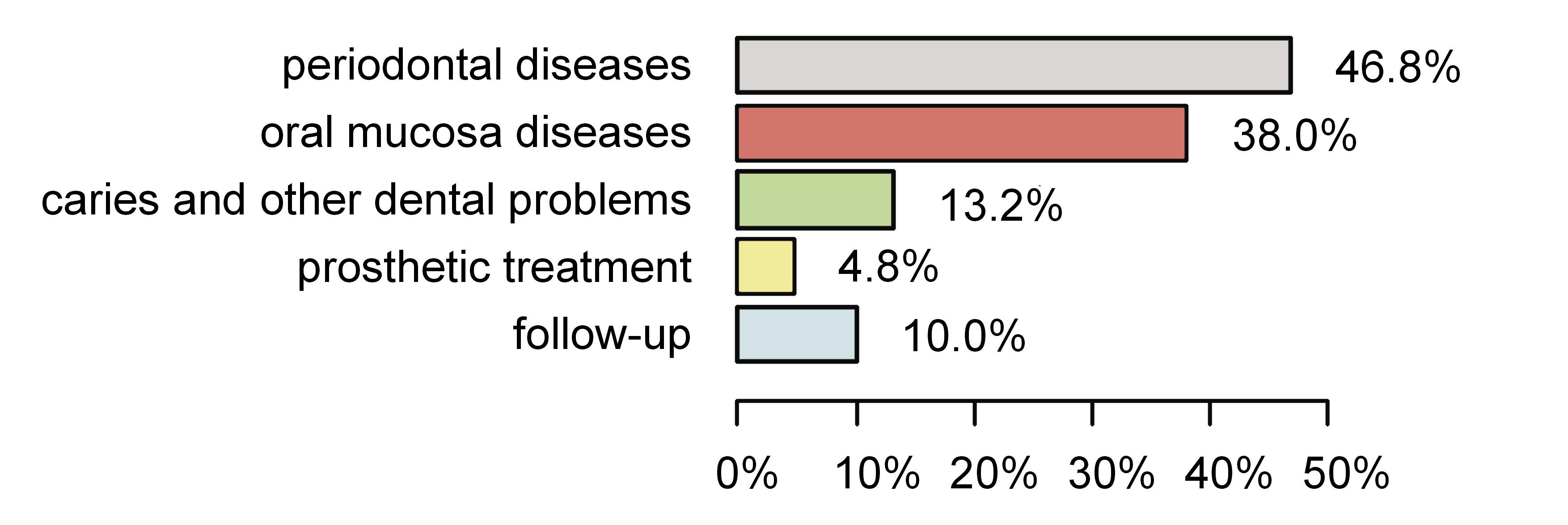
The patients were divided into 5 groups depending on their reason for visiting the UDC: group 1 – periodontal diseases (117 (46.8%)); group 2 – oral mucosal diseases (95 (38.0%)); group 3 – caries and other dental problems (33 (13.2%)); group 4 – prosthetic treatment (12 (4.8%)); and group 5 – follow-up (25 (10.0%)) (Figure 2).

The mean number of missing teeth (due to any reason, excluding third molars) among the respondents was 8.34 ± 8.36, and only 43.7% had all of the missing teeth replaced. The types of prostheses used are shown in Figure 3.

The OHIP-14 items were rated on a Likert-type scale as never (0), almost never (1), sometimes (2), fairly often (3), or almost all of the time (4). The scale referred to the frequency of the occurrence of the symptoms or problems related to the teeth (subscale 1), oral mucosa (subscale 2), or dentures (subscale 3). The higher the OHIP-14 score was, the worse OHRQoL was assumed.

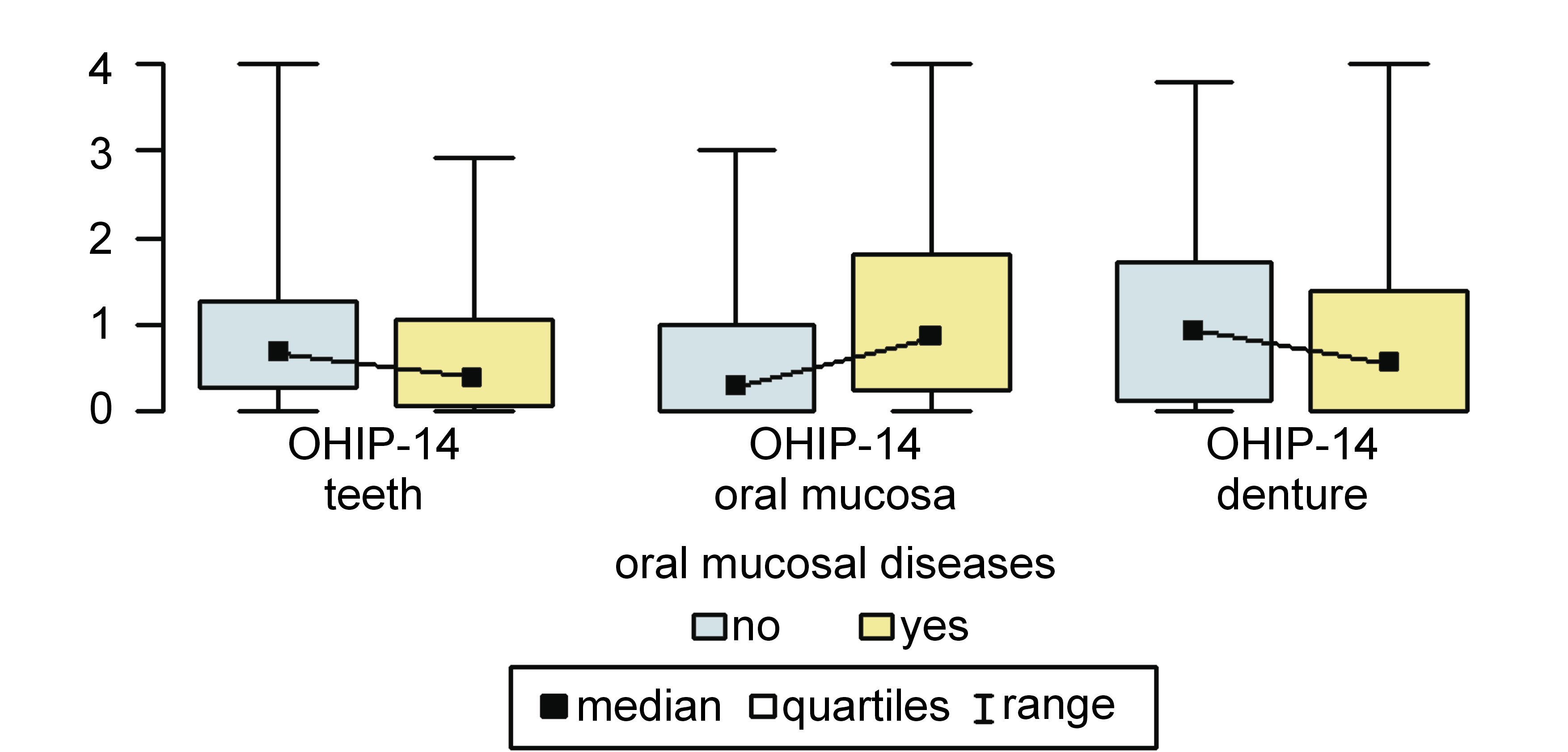
There was a significantly higher subscale 2 score and a significantly lower subscale 1 score in group 2 than in any other group (0.86 (0.25–1.81) vs. 0.29 (0–1.00); p < 0.001, and 0.39 (0.07–1.07) vs. 0.68 (0.29–1.29); p = 0.048, respectively (Figure 4). There was no significant difference in the OHIP-14 scores within subscale 3 between these groups of respondents.

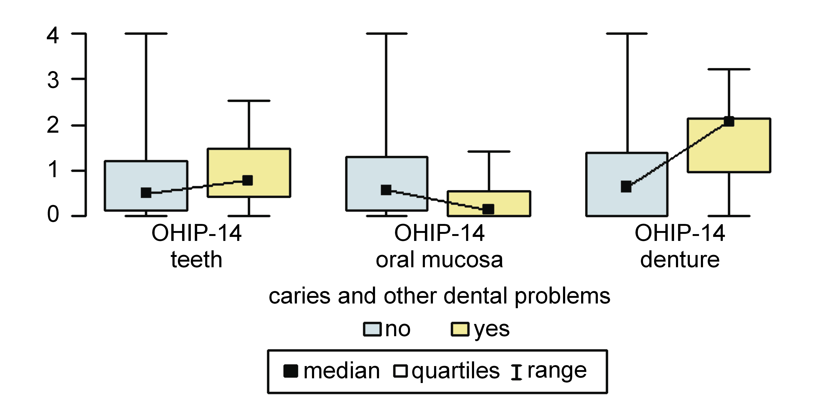
Patients suffering from caries and other dental problems (group 3) had a significantly higher OHIP-14 score within subscale 3 and a significantly lower score within subscale 2 as compared to other respondents (2.07 (0.96–2.15) vs. 0.64 (0–1.38); p = 0.043, and 0.14 (0–0.56) vs. 0.57 (0.14–1.31); p = 0.001) (Figure 5).

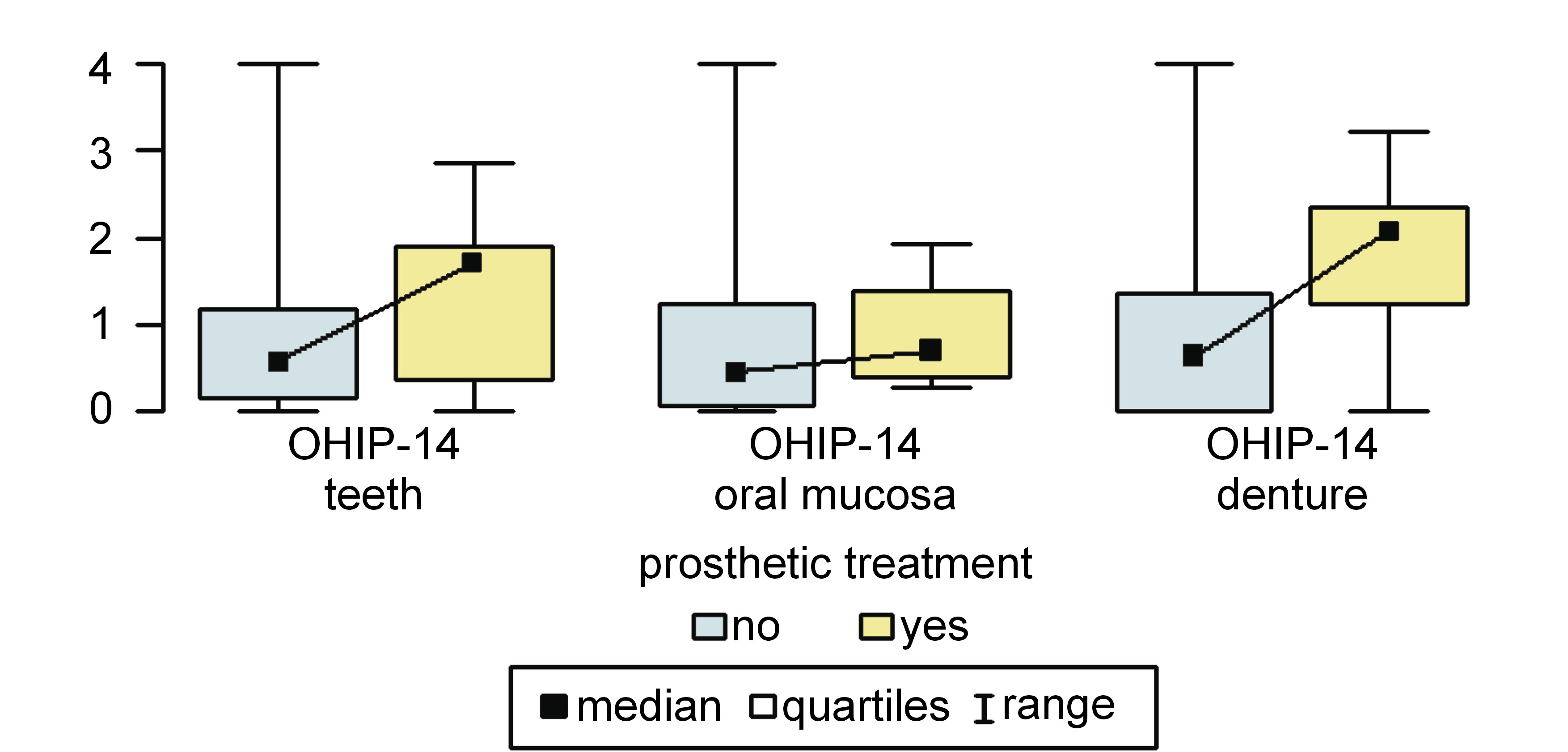
In group 4 (patients looking for prosthetic treatment), the OHIP-14 score within subscale 3 was significantly higher than among other patients (2.07 (1.23–2.36) vs. 0.64 (0–1.36); p = 0.004) (Figure 6).

In group 1 (patients with periodontal diseases) and group 5 (follow-up), there were no statistically significant differences observed within any subscale in comparison with other participants (p > 0.05).

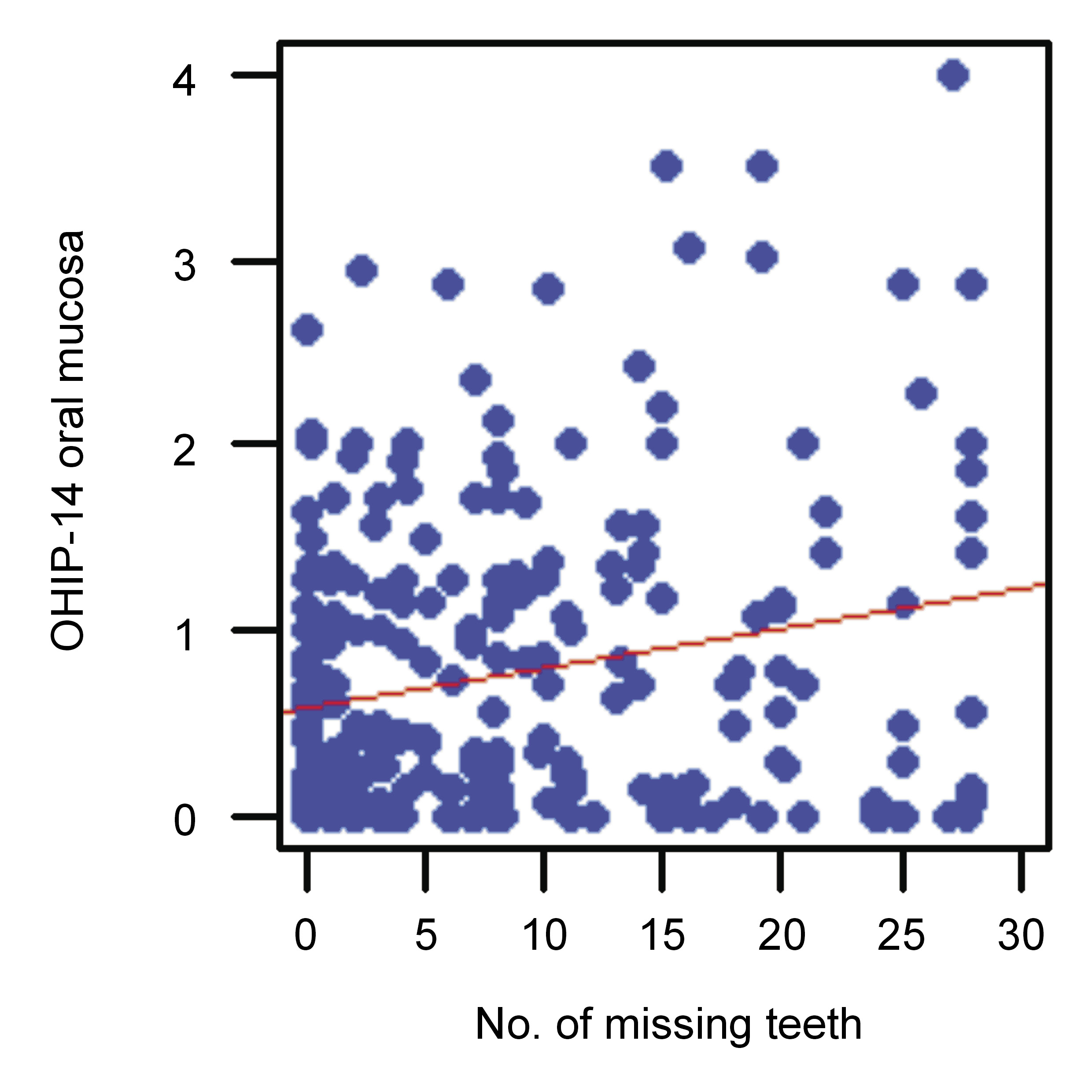
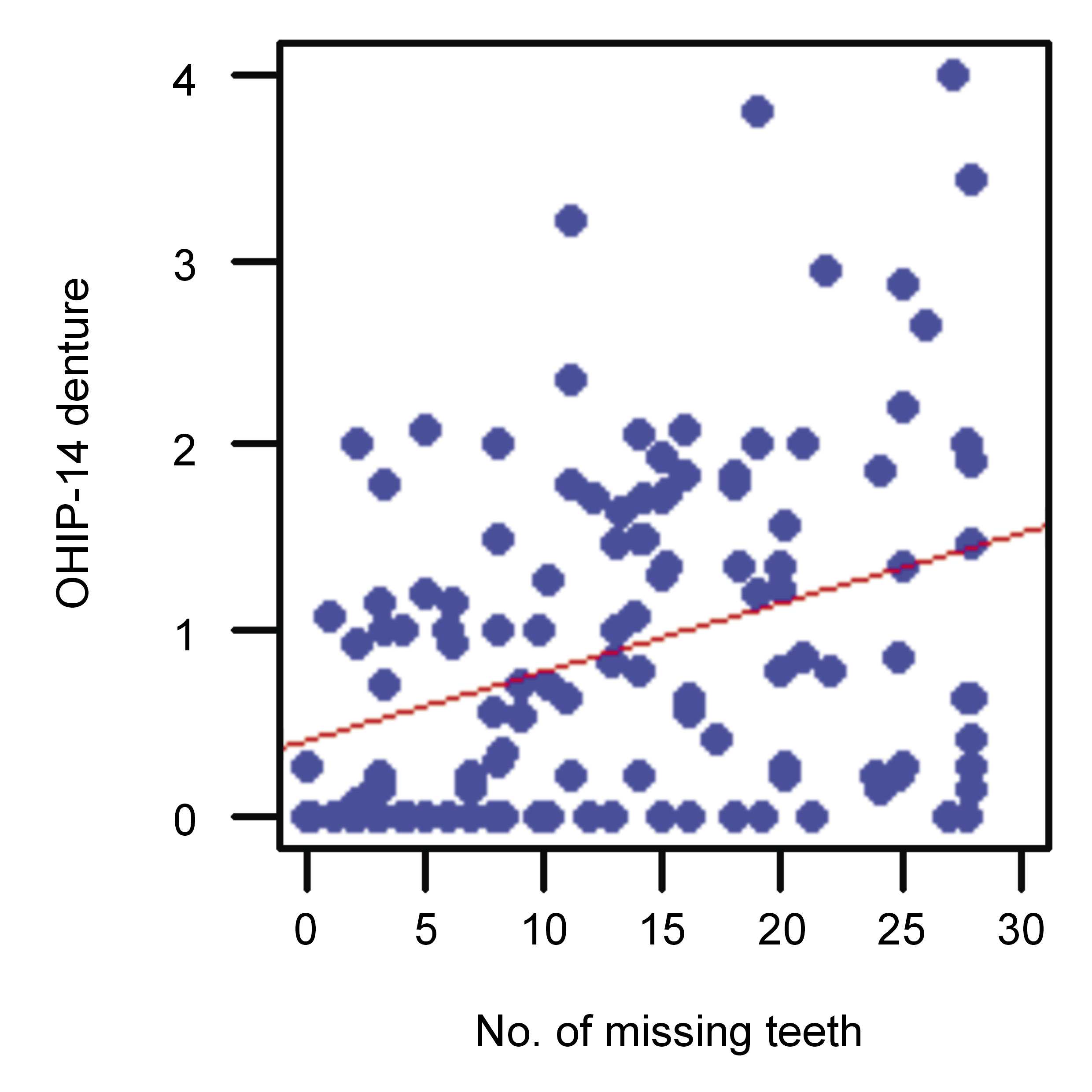
There was a statistically significant (p < 0.05) positive correlation (r > 0) between the number of missing teeth and the OHIP-14 scores within subscales 2 and 3 (Figure 7 and Figure 8).

Discussion

The most prevalent oral diseases are dental caries, periodontal diseases, tooth loss (having 9 or fewer teeth),67 and cancers (of the lips and of the oral cavity).68 They remain a significant global public health concern and have a serious socio-economic impact.68, 69 This is especially true in low-income and middle-income countries, where the prevalence of these diseases remains high and the financing of oral healthcare is low.68, 70 Untreated oral diseases have many negative consequences on personal life, including suffering from pain, impaired quality of life, school absenteeism, and decreased productivity at work.68

Dental caries is a multifactorial disease involving interactions between the hard tissues of the tooth, the biofilms formed on the tooth surface, and sugars that cause tooth demineralization.71 The disease is conditioned by genes and the salivary flow.71 It occurs in 60–90% of children and in nearly 100% of adults.70 Caries may occur in children under 6 years of age, in their deciduous teeth, and is called early childhood caries (ECC).71, 72 In epidemiological studies, the international decayed, missing and filled teeth (DMFT) index is used for assessing caries at the individual and population level.71 Caries is preventable and the application of preventive measures generates significantly lower costs than the treatment of the disease.71, 73 Such measures include proper oral hygiene (tooth brushing and interdental hygiene), the daily and professional use of fluoride, the reduction of the amount and frequency of sugar intake, saliva stimulation, and the use of antibacterial agents.71, 73, 74 Biofilm engineering, through the use of pre- and probiotics, seems to have a long-term anti-cariogenic effect.73

The most common periodontal disease is periodontitis. This is an inflammatory, chronic and multifactorial disease that leads to the destruction of connective tissue and the bone supporting the teeth, and is a result of interactions between host inflammatory immune responses and the bacterial biofilm.75, 76, 77 Untreated periodontal disease leads to bacterial dysbiosis, calculus deposits and periodontal pockets. It may be manifested by bleeding gingiva,78 tooth mobility and the eventual tooth loss, halitosis, and the worsening of oral and dental esthetics. It all causes functional and social difficulties while biting, chewing and speaking, which eventually worsen OHRQoL77, 79, 80, 81, 82, 83 and systemic health.77, 78 By activating inflammatory pathways, periodontitis may influence well-being and health during pregnancy, and can lead to complications such as preeclampsia, which is one of the major causes of maternal and perinatal morbidity and mortality,84 premature delivery, and low birth weight.85, 86 Lipopolysaccharide is one of the most virulent factors of the anaerobic bacterium Porphyromonas gingivalis that is present in periodontitis,87 and it may affect gene expression and diverse cellular processes by increasing the levels of microRNA-155.85, 86 Mahendra et al. suggested that the levels of microRNA-155 could be a genetic diagnostic tool for the rapid identification of preeclampsia.86

Studies have shown that generalized forms of periodontitis have a greater impact on OHRQoL than localized88 and more severe forms.49, 79, 89 Oliveira et al. demonstrated that periodontitis had similar effects on OHRQoL to general diseases, such as end-stage renal disease.89 Other periodontal diseases that negatively affect OHRQoL are gingivitis and gingival recession.83, 90 Most of the patients in this study suffered from chronic forms of periodontitis, which may progress gradually throughout the years. However, unless any exacerbation (e.g., a periodontal abscess) occurs, the patient may not observe any major changes. Of the respondents in this group, 55.3% were continuing treatment, which means that acute complaints had already been mostly treated. The results showed slight differences in the subscale 1 OHIP-14 scores among periodontal patients, but they were not statistically significant (p = 0.054).

Systemic diseases, especially those with a pro-inflammatory component (e.g., type 2 diabetes mellitus, cardiovascular diseases), are a risk factor for the initiation and progression of periodontitis.67, 70 Indeed, periodontitis has been declared the 6th most frequent complication of diabetes mellitus.91 Periodontitis and systemic chronic diseases have the same fundamental risk factors,78 including tobacco use, poor oral hygiene, and dietary and lifestyle behaviors.67

The basis of periodontal therapy is regular home care, and professional subgingival plaque and calculus removal, which is called non-surgical therapy.92 There may be indications for the use of adjunctive measures to improve periodontal health, such as topical antimicrobial agents, antibiotics, antiseptics, or host-modulating drugs.92, 93 With regard to the latter, a recently conducted systematic review suggests that the topical or systemic administration of melatonin might be useful for the management of periodontitis.93 Furthermore, non-surgical periodontal treatment improves OHRQoL by reducing pain, psychological discomfort and physical disability.55, 94

Tooth loss is a serious impairment that causes functional problems while eating, and thus deterioration in OHRQoL.95 Comparing studies conducted in the 1980s and 1990s with the current work, there is a positive trend showing a reduction of edentulism in the Danish population (17.7% vs. 3.4%).96 However, these values remain much higher in developing countries, where dental healthcare is limited to emergency care.70 Tooth loss may be a consequence of caries or a periodontal disease.70 Therefore, access to dental healthcare, regular dental visits, and patient education about disease prophylaxis and their compliance are some of the factors that can reduce the risk of tooth loss.

Nowadays, and particularly following the coronavirus disease 2019 (COVID-19) pandemic, telemedicine is rapidly developing, enabling patients to keep in contact with healthcare workers. This allows the detection of the early symptoms of the disease or the worsening of the health condition.97 Social media are widely used, especially by younger people, and they may prove advantageous to develop appropriate and valuable health content on such platforms, and thus to broaden patient knowledge about prophylaxis and the proper treatment of oral and general diseases.98, 99

Oral mucosal diseases comprise many disorders, such as oral leukoplakia (OL), oral erythroplakia (OE), burning mouth syndrome (BMS), oral lichen planus (OLP), oral submucous fibrosis (OSF), xerostomia, candidiasis, Sjögren’s syndrome (SS), recurrent aphthous stomatitis (RAS), and autoimmune bullous diseases. Some of them (OL, OE, OLP, OSF, and oral epithelial dysplasia) are considered as potentially premalignant oral epithelial lesions (PPOELs)100 or oral potentially malignant disorders (OPMDs),101 of which leukoplakia is the most frequently occurring.70 The rate of their malignant transformation to oral squamous cell carcinoma (OSCC) varies depending on the type and site of the lesion, and it is the highest for OE, with an average of 26.3% (ranging from 14.3% to 50.0%).102 It is important to emphasize that prevention, early detection and treatment may halt malignant transformation to OSCC.100, 103 The main risk factors for developing OPMDs and their malignant transformation are tobacco and alcohol use. Depending on the type of disease, its severity and course (active or inactive), the reported symptoms may be more painful for the patient,39, 104 may have a social impact42 or may reduce the secretion of saliva, as observed in SS,105 all of which eventually worsen OHRQoL.39, 41 The proper treatment of these complaints may decrease pain and anxiety, and improve OHRQoL.106, 107 In this cross-sectional study, the OHRQoL of the group of patients with oral mucosal diseases was most influenced by their oral mucosal disease. In other patients, OHRQoL was affected to a greater degree by the condition of their teeth. This means that oral mucosal diseases are perceived by patients as much more serious and that they may affect their life in multiple ways.

In patients looking for restorative or prosthetic treatment, the use of dentures significantly worsened OHRQoL. In this study, almost half of the patients were using removable prostheses and 34.59% had only fixed dentures. The UDC is a specialized clinic in which most patients are treated within the National Healthcare Fund, which only refunds acrylic dentures. Nonetheless, prosthetic treatment improves OHRQoL regardless of the treatment used.108 Fixed tooth-supported dentures are set on the teeth, which need appropriate preparation, so it is important to properly assess the condition of the teeth before prosthetic treatment to avoid complications and failures. A significant advantage of such an approach is the reduction of prosthesis extension and the preservation of bone volume. Removable dental prostheses are supported on crowns (e.g., cast partial dentures, overdentures109). They can be carefully cleaned outside the mouth and can have teeth added. This may be important for older people with decreased fine motor skills, or for periodontal patients, in whom the risk of tooth loss is greater.110, 111 With the use of removable dental prostheses, financial constraints may be overcome; the dentures may also be used as temporary ones.111 Another treatment option are implant-supported fixed or removable prostheses, which expand the prosthetic treatment opportunities. A proper surgical technique and the presence of an implant in the bone may influence bone volume in the short and long term.112 Before prosthetic treatment, all indications, contraindications and patient expectations need to be considered.

Yoshimoto et al. reported a strong association between OHRQoL and masticatory satisfaction (defined as the ability to eat comfortably) among individuals using removable partial dentures,113 whereas Inoue et al. claimed that the quality of removable dentures (stability and esthetics) had a minimal effect on the OHIP score.114 On the contrary, according to Kurosaki et al., only implant-supported fixed dentures improved OHRQoL 6 years after prosthetic treatment, and there was no significant improvement in patients wearing fixed or removable partial dentures.115 Meanwhile, according to Ali et al., both tooth- and implant-supported fixed prostheses positively affected OHRQoL in the short and long term.116

In this cross-sectional study, there was a significant positive correlation between the number of missing teeth and impact on OHRQoL in relation to oral mucosa and dentures. This means that the greater the number of lost teeth, the greater the problems for the patients referred regarding their oral mucosal disease and denture use, and the greater the necessity for a dental visit.

Conclusions

To prevent the worsening of OHRQoL, the proper prophylaxis of caries and its complications (pain, tooth destruction and extraction) is very important. It delays any need for prosthetic treatment. Proper patient education, prophylaxis and treatment of oral mucosal diseases and periodontal diseases are crucial for the preservation of OHRQoL.

Ethics approval and consent to participate

The present research was approved by the Ethics Committee of the Jagiellonian University Medical College in Cracow, Poland (No. 122.6120.354.2016). Informed written consent was obtained from all the participants.

Data availability

The datasets generated and/or analyzed during the current study are available from the corresponding author on reasonable request.

Consent for publication

Not applicable.

Figures


Fig. 1. Age structure of the patients
Fig. 2. Reason for visiting the University Dental Clinic (UDC)
Percentages do not sum up to 100%, as each patient could have several reasons for the visit (a multiple-choice question).
Fig. 3. Prostheses used in the respondents with missing teeth
Fig. 4. Modified Oral Health Impact Profile (OHIP-14) scores in the group of patients reporting oral mucosal diseases vs. patients without such complaints
Fig. 5. Modified Oral Health Impact Profile (OHIP-14) scores in the group of patients suffering from caries and other dental problems vs. patients without such complaints
Fig. 6. Modified Oral Health Impact Profile (OHIP-14) in the group of patients looking for prosthetic treatment vs. patients without such needs
Fig. 7. Correlation between the number of missing teeth and the OHIP-14 score within subscale 2 (oral mucosa)
Fig. 8. Correlation between the number of missing teeth and the OHIP-14 score within subscale 3 (denture)

References (116)

  1. McCance AM, Moss JP, Wright WR, Linney AD, James DR. A three-dimensional soft tissue analysis of 16 skeletal class III patients following bimaxillary surgery. Br J Oral Maxillofac Surg. 1992;30(4):221–232. doi:10.1016/0266-4356(92)90264-j
  2. Moss JP, McCance AM, Fright WR, Linney AD, James DR. A three-dimensional soft tissue analysis of fifteen patients with Class II, Division 1 malocclusions after bimaxillary surgery. Am J Orthod Dentofacial Orthop. 1994;105(5):430–437. doi:10.1016/S0889-5406(94)70002-8
  3. Minervini G, Lucchese A, Perillo L, Serpico R, Minervini G. Unilateral superior condylar neck fracture with dislocation in a child treated with an acrylic splint in the upper arch for functional repositioning of the mandible. Cranio. 2017;35(5):337–341. doi:10.1080/08869634.2016.1203560
  4. Alhammadi MS, Al-Mashraqi AA, Alnami RH, et al. Accuracy and reproducibility of facial measurements of digital photographs and wrapped cone beam computed tomography (CBCT) photographs. Diagnostics (Basel). 2021;11(5):757. doi:10.3390/diagnostics11050757
  5. Reda R, Zanza A, Cicconetti A, et al. Ultrasound imaging in dentistry: A literature overview. J Imaging. 2021;7(11):238. doi:10.3390/jimaging7110238
  6. Patil S, Alkahtani A, Bhandi S, et al. Ultrasound imaging versus radiographs in differentiating periapical lesions: A systematic review. Diagnostics (Basel). 2021;11(7):1208. doi:10.3390/diagnostics11071208
  7. Natanasabapathy V, Arul B, Mishra A, et al. Ultrasound imaging for the differential diagnosis of periapical lesions of endodontic origin in comparison with histopathology – a systematic review and meta-analysis. Int Endod J. 2021;54(5):693–711. doi:10.1111/iej.13465
  8. Musu D, Rossi-Fedele G, Campisi G, Cotti E. Ultrasonography in the diagnosis of bone lesions of the jaws: A systematic review. Oral Surg Oral Med Oral Pathol Oral Radiol. 2016;122(1):e19–e29. doi:10.1016/j.oooo.2016.03.022
  9. Chan HL, Sinjab K, Li J, Chen Z, Wang HL, Kripfgans OD. Ultrasonography for noninvasive and real-time evaluation of peri-implant tissue dimensions. J Clin Periodontol. 2018;45(8):986–995. doi:10.1111/jcpe.12918
  10. Bohner L, Habor D, Tortamano P, Radermacher K, Wolfart S, Marotti J. Assessment of buccal bone surrounding dental implants using a high-frequency ultrasound scanner. Ultrasound Med Biol. 2019;45(6):1427–1434. doi:10.1016/j.ultrasmedbio.2019.02.002
  11. Su N, van Wijk AJ, Visscher CM, Lobbezoo F, van der Heijden GJ. Diagnostic value of ultrasonography for the detection of disc displacements in the temporomandibular joint: A systematic review and meta-analysis. Clin Oral Investig. 2018;22(7):2599–2614. doi:10.1007/s00784-018-2359-4
  12. d’Apuzzo F, Minervini G, Grassia V, Rotolo RP, Perillo L, Nucci L. Mandibular coronoid process hypertrophy: Diagnosis and 20-year follow-up with CBCT, MRI and EMG evaluations. Appl Sci. 2021;11(10):4504. doi:10.3390/app11104504
  13. Minervini G, Nucci L, Lanza A, Femiano F, Contaldo M, Grassia V. Temporomandibular disc displacement with reduction treated with anterior repositioning splint: A 2-year clinical and magnetic resonance imaging (MRI) follow-up. J Biol Regul Homeost Agents. 2020;34(1 Suppl 1):151–160. PMID:32064850.
  14. Pahel BT, Rozier RG, Slade GD. Parental perceptions of children’s oral health: The Early Childhood Oral Health Impact Scale (ECOHIS). Health Qual Life Outcomes. 2007;5:6. doi:10.1186/1477-7525-5-6
  15. Contaldo M, Della Vella F, Raimondo E, et al. Early Childhood Oral Health Impact Scale (ECOHIS): Literature review and Italian validation. Int J Dent Hyg. 2020;18(4):396–402. doi:10.1111/idh.12451
  16. Li S, Veronneau J, Allison PJ. Validation of a French language version of the Early Childhood Oral Health Impact Scale (ECOHIS). Health Qual Life Outcomes. 2008;6:9. doi:10.1186/1477-7525-6-9
  17. Sheen MH, Hsiao SY, Huang ST. Translation and validation of Taiwanese version of the Early Childhood Oral Health Impact Scale (ECOHIS). J Dent Sci. 2020;15(4):513–518. doi:10.1016/j.jds.2020.05.029
  18. Foster Page LA, Thomson WM, Jokovic A, Locker D. Validation of the Child Perceptions Questionnaire (CPQ 11–14). J Dent Res. 2005;84(7):649–652. doi:10.1177/154405910508400713
  19. Jokovic A, Locker D, Guyatt G. Short forms of the Child Perceptions Questionnaire for 11–14-year-old children (CPQ11–14): Development and initial evaluation. Heal Qual Life Outcome. 2006;4:4. doi:10.1186/1477-7525-4-4
  20. Bekes K, John MT, Schaller HG, Hirsch C. The German version of the child perceptions questionnaire on oral health-related quality of life (CPQ-G11–14): Population-based norm values. J Orofac Orthop. 2011;72(3):223–233. doi:10.1007/s00056-011-0027-2
  21. Astrøm AN, Okullo I. Validity and reliability of the Oral Impacts on Daily Performance (OIDP) frequency scale: A cross-sectional study of adolescents in Uganda. BMC Oral Health. 2003;3(1):5. doi:10.1186/1472-6831-3-5
  22. Cortés-Martinicorena FJ, Rosel-Gallardo E, Artazcoz-Osés J, Bravo M, Tsakos G. Adaptation and validation for Spain of the Child-Oral Impact on Daily Performance (C-OIDP) for use with adolescents. Med Oral Patol Oral Cir Bucal. 2010;15(1):e106–e111. doi:10.4317/medoral.15.e106
  23. Slade GD, Spencer AJ. Development and evaluation of the Oral Health Impact Profile. Community Dent Health. 1994;11(1):3–11. PMID:8193981.
  24. Atchison KA, Dolan TA. Development of the Geriatric Oral Health Assessment Index. J Dent Educ. 1990;54(11):680–687. PMID:2229624.
  25. Niesten D, Witter D, Bronkhorst E, Creugers N. Validation of a Dutch version of the Geriatric Oral Health Assessment Index (GOHAI-NL) in care-dependent and care-independent older people. BMC Geriatr. 2016;16:53. doi:10.1186/s12877-016-0227-0
  26. Pace-Balzan A, Cawood JI, Howell R, Butterworth CJ, Lowe D, Rogers SN. The further development and validation of the Liverpool Oral Rehabilitation Questionnaire: A cross-sectional survey of patients attending for oral rehabilitation and general dental practice. Int J Oral Maxillofac Surg. 2006;35(1):72–78. doi:10.1016/j.ijom.2005.07.004
  27. The World Health Organization Quality of Life assessment (WHOQOL): Position paper from the World Health Organization. Soc Sci Med. 1995;41(10):1403–1409. doi:10.1016/0277-9536(95)00112-k
  28. Slade G. Derivation and validation of a short-form oral health impact profile. Community Dent Oral Epidemiol. 1997;25(4):284–290. doi:10.1111/j.1600-0528.1997.tb00941.x
  29. Skośkiewicz-Malinowska K, Kaczmarek U, Ziętek M, Malicka B. Validation of the Polish version of the oral health impact profile-14. Adv Clin Exp Med. 2015;24(1):129–137. doi:10.17219/acem/35476
  30. Montero-Martín J, Bravo-Pérez M, Albaladejo-Martínez A, Hernández-Martín LA, Rosel-Gallardo EM. Validation the Oral Health Impact Profile (OHIP-14sp) for adults in Spain. Med Oral Patol Oral Cir Bucal. 2009;14(1):E44–E50. PMID:19114956.
  31. Papagiannopoulou V, Oulis CJ, Papaioannou W, Antonogeorgos G, Yfantopoulos J. Validation of a Greek version of the oral health impact profile (OHIP-14) for use among adults. Health Qual Life Outcomes. 2012;10:7. doi:10.1186/1477-7525-10-7
  32. Balci N, Alkan N, Gurgan CA. Psychometric properties of a Turkish version of the oral health impact profile-14. Niger J Clin Pract. 2017;20(1):19–24. doi:10.4103/1119-3077.164353
  33. Bae KH, Kim HD, Jung SH, et al. Validation of the Korean version of the oral health impact profile among the Korean elderly. Community Dent Oral Epidemiol. 2007;35(1):73–79. doi:10.1111/j.1600-0528.2007.00331.x
  34. Aimée NR, Damé-Teixeira N, Alves LS, et al. Responsiveness of oral health-related quality of life questionnaires to dental caries interventions: Systematic review and meta-analysis. Caries Res. 2019;53(6):585–598. doi:10.1159/000500855
  35. Bahho LA, Thomson WM, Foster Page LA, Drummond BK. Dental trauma experience and oral-health-related quality of life among university students. Aust Dent J. 2020;65(3):220–224. doi:10.1111/adj.12774
  36. Carvalho JC, Mestrinho HD, Stevens S, van Wijk AJ. Do oral health conditions adversely impact young adults? Caries Res. 2015;49(3):266–274. doi:10.1159/000375377
  37. Brignardello-Petersen R. Active caries, consequences of untreated caries, and tooth pain relate to only a small decrease in older adults’ quality of life. J Am Dent Assoc. 2017;148(5):e62. doi:10.1016/j.adaj.2017.02.046
  38. Shao R, Hu T, Zhong YS, et al. Socio-demographic factors, dental status and health-related behaviors associated with geriatric oral health-related quality of life in Southwestern China. Health Qual Life Outcomes. 2018;16(1):98. doi:10.1186/s12955-018-0925-8
  39. Tadakamadla J, Kumar S, Lalloo R, Gandhi Babu DB, Johnson NW. Impact of oral potentially malignant disorders on quality of life. J Oral Pathol Med. 2018;47(1):60–65. doi:10.1111/jop.12620
  40. Flink H, Tegelberg Å, Arnetz JE, Birkhed D. Self-reported oral and general health related to xerostomia, hyposalivation, and quality of life among caries active younger adults. Acta Odontol Scand. 2020;78(3):229–235. doi:10.1080/00016357.2019.1690677
  41. Skośkiewicz-Malinowska K, Malicka B, Ziętek M, Kaczmarek U. Does oral dryness influence quality of life? Current perspectives in elderly dental care. Adv Clin Exp Med. 2019;28(9):1209–1216. doi:10.17219/acem/104601
  42. Parlatescu I, Tovaru M, Nicolae CL, Sfeatcu R, Didilescu AC. Oral health-related quality of life in different clinical forms of oral lichen planus. Clin Oral Investig. 2020;24(1):301–308. doi:10.1007/s00784-019-02951-8
  43. Radwan-Oczko M, Zwyrtek E, Owczarek JE, Szcześniak D. Psychopathological profile and quality of life of patients with oral lichen planus. J Appl Oral Sci. 2018;26:e20170146. doi:10.1590/1678-7757-2017-0146
  44. Suliman NM, Johannessen AC, Ali RW, Salman H, Astrøm AN. Influence of oral mucosal lesions and oral symptoms on oral health related quality of life in dermatological patients: A cross sectional study in Sudan. BMC Oral Health. 2012;12:19. doi:10.1186/1472-6831-12-19
  45. Lu HX, Chen XL, Wong MCM, Zhu C, Ye W. Oral health impact of halitosis in Chinese adults. Int J Dent Hyg. 2017;15(4):e85–e92. doi:10.1111/idh.12242
  46. Carbone M, Goss E, Carrozzo M, et al. Systemic and topical corticosteroid treatment of oral lichen planus: A comparative study with long-term follow-up. J Oral Pathol Med. 2003;32(6):323–329. doi:10.1034/j.1600-0714.2003.00173.x
  47. Di Stasio D, Lauritano D, Gritti P, et al. Psychiatric disorders in oral lichen planus: A preliminary case control study. J Biol Regul Homeost Agents. 2018;32(2 Suppl 1):97–100. PMID:29460524.
  48. Paśnik-Chwalik B, Konopka T. Impact of periodontitis on the Oral Health Impact Profile : A systematic review and meta-analysis. Dent Med Probl. 2020;57(4):423–431. doi:10.17219/dmp/125028
  49. Ferreira MC, Dias-Pereira AC, Branco-de-Almeida LS, Martins CC, Paiva SM. Impact of periodontal disease on quality of life: A systematic review. J Periodontal Res. 2017;52(4):651–665. doi:10.1111/jre.12436
  50. Geevarghese A, Baskaradoss JK, Sarma PS. Oral health-related quality of life and periodontal status of pregnant women. Matern Child Health J. 2017;21(8):1634–1642. doi:10.1007/s10995-016-2255-y
  51. Hsu YJ, Lin KD, Chen JH, et al. Periodontal treatment experience associated with oral health-related quality of life in patients with poor glycemic control in type 2 diabetes: A case–control study. Int J Environ Res Public Health. 2019;16(20):4011. doi:10.3390/ijerph16204011
  52. Masood M, Younis LT, Masood Y, Bakri NN, Christian B. Relationship of periodontal disease and domains of oral health-related quality of life. J Clin Periodontol. 2019;46(2):170–180. doi:10.1111/jcpe.13072
  53. Cortelli SC, Costa FO, Gargioni-Filho A, et al. Impact of gingivitis treatment for diabetic patients on quality of life related to periodontal objective parameters: A randomized controlled clinical trial. Arch Oral Biol. 2018;86:80–86. doi:10.1016/j.archoralbio.2017.11.010
  54. Buset SL, Walter C, Friedmann A, Weiger R, Borgnakke WS, Zitzmann NU. Are periodontal diseases really silent? A systematic review of their effect on quality of life. J Clin Periodontol. 2016;43(4):333–344. doi:10.1111/jcpe.12517
  55. Khan S, Khalid T, Bettiol S, Crocombe LA. Non-surgical periodontal therapy effectively improves patient-reported outcomes: A systematic review. Int J Dent Hyg. 2021;19(1):18–28. doi:10.1111/idh.12450
  56. Mehta SB, Loomans BA, Banerji S, Bronkhorst EM, Bartlett D. An investigation into the impact of tooth wear on the oral health related quality of life amongst adult dental patients in the United Kingdom, Malta and Australia. J Dent. 2020;99:103409. doi:10.1016/j.jdent.2020.103409
  57. Patel J, Baker SR. Is toothwear associated with oral health related quality of life in adults in the UK? Community Dent Health. 2020;37(3):174–179. doi:10.1922/CDH_00026Patel06
  58. Schierz O, Baba K, Fueki K. Functional oral health‐related quality of life impact: A systematic review in populations with tooth loss. J Oral Rehabil. 2021;48(3):256–270. doi:10.1111/joor.12984
  59. Batista MJ, Lawrence HP, de Sousa M da LR. Impact of tooth loss related to number and position on oral health quality of life among adults. Health Qual Life Outcomes. 2014;12:165. doi:10.1186/s12955-014-0165-5
  60. Dahlström L, Carlsson GE. Temporomandibular disorders and oral health-related quality of life. A systematic review. Acta Odontol Scand. 2010;68(2):80–85. doi:10.3109/00016350903431118
  61. Moccia S, Nucci L, Spagnuolo C, d’Apuzzo F, Grazia M, Minervini G. Polyphenols as potential agents in the management of temporomandibular disorders. Appl Sci. 2020;10(15):5305. doi:10.3390/app10155305
  62. Schmalz G, Patschan S, Patschan D, Ziebolz D. Oral-health-related quality of life in Adult patients with rheumatic diseases – a systematic review. J Clin Med. 2020;9(4):1172. doi:10.3390/jcm9041172
  63. Schmalz G, Busjan R, Dietl M, Hasenkamp J, Trümper L, Ziebolz D. Oral health-related quality of life in adult patients with newly diagnosed acute leukaemia. Oral Health Prev Dent. 2020;18(1):461–466. doi:10.3290/j.ohpd.a44685
  64. Oduncuoğlu BF, Alaaddinoğlu EE, Çolak T, Akdur A, Haberal M. Effects of renal transplantation and hemodialysis on patient’s general health perception and oral health-related quality of life: A single-center cross-sectional study. Transplant Proc. 2020;52(3):785–792. doi:10.1016/j.transproceed.2020.01.016
  65. R Core Team. R: A language and environment for statistical computing. R Foundation for Statistical Computing, Vienna, Austria. 2021. https://www.r-project.org/. Accessed October 29, 2021.
  66. Wąsacz K, Pac A, Darczuk D, Chomyszyn-Gajewska M. Validation of a modified Oral Health Impact Profile scale (OHIP-14) in patients with oral mucosa lesions or periodontal disease. Dent Med Probl. 2019;56(3):231–237. doi:10.17219/dmp/109388
  67. Al-Nasser L, Lamster IB. Prevention and management of periodontal diseases and dental caries in the older adults. Periodontol 2000. 2020;84(1):69–83. doi:10.1111/prd.12338
  68. Peres MA, Macpherson LM, Weyant RJ, et al. Oral diseases: A global public health challenge. Lancet. 2019;394(10194):249–260. doi:10.1016/S0140-6736(19)31146-8
  69. Jin LJ, Lamster IB, Greenspan JS, Pitts NB, Scully C, Warnakulasuriya S. Global burden of oral diseases: Emerging concepts, management and interplay with systemic health. Oral Dis. 2016;22(7):609–619. doi:10.1111/odi.12428
  70. Petersen PE, Bourgeois D, Ogawa H, Estupinan-Day S, Ndiaye C. The global burden of oral diseases and risks to oral health. Bull World Health Organ. 2005;83(9):661–669. PMID:16211157. PMCID:PMC2626328.
  71. Pitts NB, Zero DT, Marsh PD, et al. Dental caries. Nat Rev Dis Primers. 2017;3:17030. doi:10.1038/nrdp.2017.30
  72. Mathur VP, Dhillon JK. Dental caries: A disease which needs attention. Indian J Pediatr. 2018;85(3):202–206. doi:10.1007/s12098-017-2381-6
  73. Twetman S. Prevention of dental caries as a non-communicable disease. Eur J Oral Sci. 2018;126(Suppl 1):19–25. doi:10.1111/eos.12528
  74. van Loveren C. Sugar restriction for caries prevention: Amount and frequency. Which is more important? Caries Res. 2019;53(2):168–175. doi:10.1159/000489571
  75. Manresa C, Sanz-Miralles EC, Twigg J, Bravo M. Supportive periodontal therapy (SPT) for maintaining the dentition in adults treated for periodontitis. Cochrane Database Syst Rev. 2018;1(1):CD009376. doi:10.1002/14651858.CD009376.pub2
  76. Meyle J, Chapple I. Molecular aspects of the pathogenesis of periodontitis. Periodontol 2000. 2015;69(1):7–17. doi:10.1111/prd.12104
  77. Hegde R, Awan KH. Effects of periodontal disease on systemic health. Dis Mon. 2019;65(6):185–192. doi:10.1016/j.disamonth.2018.09.011
  78. Petersen PE, Ogawa H. The global burden of periodontal disease: Towards integration with chronic disease prevention and control. Periodontol 2000. 2012;60(1):15–39. doi:10.1111/j.1600-0757.2011.00425.x
  79. Needleman I, McGrath C, Floyd P, Biddle A. Impact of oral health on the life quality of periodontal patients. J Clin Periodontol. 2004;31(6):454–457. doi:10.1111/j.1600-051X.2004.00498.x
  80. da Silva Araújo AC, Gusmão ES, Mazza Batista JE, Cimões R. Impact of periodontal disease on quality of life. Quintessence Int. 2010;41(6):e111–e118. PMID:20490384.
  81. Bernabé E, Marcenes W. Periodontal disease and quality of life in British adults. J Clin Periodontol. 2010;37(11):968–972. doi:10.1111/j.1600-051X.2010.01627.x
  82. Fuller J, Donos N, Suvan J, Tsakos G, Nibali L. Association of oral health-related quality of life measures with aggressive and chronic periodontitis. J Periodontal Res. 2020;55(4):574–580. doi:10.1111/jre.12745
  83. Eltas A, Uslu MO, Eltas SD. Association of oral health-related quality of life with periodontal status and treatment needs. Oral Health Prev Dent. 2016;14(4):339–347. doi:10.3290/j.ohpd.a35613
  84. Kuklina EV, Ayala C, Callaghan WM. Hypertensive disorders and severe obstetric morbidity in the United States. Obstet Gynecol. 2009;113(6):1299–1306. doi:10.1097/AOG.0b013e3181a45b25
  85. Dai Y, Diao Z, Sun H, Li R, Qiu Z, Hu Y. MicroRNA-155 is involved in the remodelling of human-trophoblast-derived HTR-8/SVneo cells induced by lipopolysaccharides. Hum Reprod. 2011;26(7):1882–1891. doi:10.1093/humrep/der118
  86. Mahendra J, Mahendra L, Mugri MH, et al. Role of periodontal bacteria, viruses, and placental mir155 in chronic periodontitis and preeclampsia – a genetic microbiological study. Curr Issues Mol Biol. 2021;43(2):831–844. doi:10.3390/cimb43020060
  87. How KY, Song KP, Chan KG. Porphyromonas gingivalis: An overview of periodontopathic pathogen below the gum line. Front Microbiol. 2016;7:53. doi:10.3389/fmicb.2016.00053
  88. Llanos AH, Benítez Silva CG, Ichimura KT, et al. Impact of aggressive periodontitis and chronic periodontitis on oral health-related quality of life. Braz Oral Res. 2018;32:e006. doi:10.1590/1807-3107bor-2018.vol32.0006
  89. Oliveira LM, Sari D, Schöffer C, Santi SS, Antoniazzi RP, Zanatta FB. Periodontitis is associated with oral health-related quality of life in individuals with end-stage renal disease. J Clin Periodontol. 2020;47(3):319–329. doi:10.1111/jcpe.13233
  90. Wagner TP, Costa RS, Rios FS, et al. Gingival recession and oral health-related quality of life: Aa population-based cross-sectional study in Brazil. Community Dent Oral Epidemiol. 2016;44(4):390–399. doi:10.1111/cdoe.12226
  91. Löe H. Periodontal disease. The sixth complication of diabetes mellitus. Diabetes Care. 1993;16(1):329–334. PMID:8422804.
  92. Drisko CH. Nonsurgical periodontal therapy. Periodontol 2000. 2001;25:77–88. doi:10.1034/j.1600-0757.2001.22250106.x
  93. Balaji TM, Varadarajan S, Jagannathan R, et al. Melatonin as a topical/systemic formulation for the management of periodontitis: A systematic review. Materials (Basel). 2021;14(9):2417. doi:10.3390/ma14092417
  94. Botelho J, Machado V, Proença L, et al. The impact of nonsurgical periodontal treatment on oral health-related quality of life: A systematic review and meta-analysis. Clin Oral Investig. 2020;24(2):585–596. doi:10.1007/s00784-019-03188-1
  95. Gerritsen AE, Allen PF, Witter DJ, Bronkhorst EM, Creugers NH. Tooth loss and oral health-related quality of life: A systematic review and meta-analysis. Heal Qual Life Outcomes. 2010;8:126. doi:10.1186/1477-7525-8-126
  96. Petersen PE, Davidsen M, Jensen HR, Ekholm O, Christensen AI. Trends in dentate status and preventive dental visits of the adult population in Denmark over 30 years (1987–2017). Eur J Oral Sci. 2021;129(5):e12809. doi:10.1111/eos.12809
  97. Ricciardi D, Casagrande S, Iodice F, et al. Myasthenia gravis and telemedicine: A lesson from COVID-19 pandemic. Neurol Sci. 2021;42(12):4889–4892. doi:10.1007/s10072-021-05566-8
  98. Di Stasio D, Romano AN, Paparella RS, et al. How social media meet patients’ questions: YouTube™ review for children oral thrush. J Biol Regul Homeost Agents. 2018;32(2 Suppl 1):101-106.
  99. Di Stasio D, Romano A, Paparella RS, et al. How social media meet patients’ questions: YouTube™ review for mouth sores in children. J Biol Regul Homeost Agents. 2018;32(2 Suppl 1):117–121. PMID:29460528.
  100. Awadallah M, Idle M, Patel K, Kademani D. Management update of potentially premalignant oral epithelial lesions. Oral Surg Oral Med Oral Pathol Oral Radiol. 2018;125(6):628–636. doi:10.1016/j.oooo.2018.03.010
  101. Wetzel SL, Wollenberg J. Oral potentially malignant disorders. Dent Clin North Am. 2020;64(1):25–37. doi:10.1016/j.cden.2019.08.004
  102. Reddi SP, Shafer AT. Oral premalignant lesions: Management considerations. Oral Maxillofac Surg Clin North Am. 2006;18(4):425–433. doi:10.1016/j.coms.2006.08.002
  103. Di Stasio D, Romano A, Gentile C, et al. Systemic and topical photodynamic therapy (PDT) on oral mucosa lesions: An overview. J Biol Regul Homeost Agents. 2018;32(2 Suppl 1):123–126. PMID:29460529.
  104. Bilgic A, Aydin F, Sumer P, et al. Oral health related quality of life and disease severity in autoimmune bullous diseases. Niger J Clin Pract. 2020;23(2):159–164. doi:10.4103/njcp.njcp_216_19
  105. Azuma N, Katada Y, Yoshikawa T, et al. Evaluation of changes in oral health-related quality of life over time in patients with Sjögren’s syndrome. Mod Rheumatol. 2021;31(3):669–677. doi:10.1080/14397595.2020.1795391
  106. Adamo D, Pecoraro G, Fortuna G, et al. Assessment of oral health-related quality of life, measured by OHIP-14 and GOHAI, and psychological profiling in burning mouth syndrome: A case–control clinical study. J Oral Rehabil. 2020;47(1):42–52. doi:10.1111/joor.12864
  107. Pereira da Mata AD, de Almeida Rato Amaral JP, Thomson WM, et al. Patient-related outcomes in Sjögren syndrome treated with stimulants of salivary secretion: Randomized clinical trial. Oral Dis. 2020;26(2):313–324. doi:10.1111/odi.13251
  108. John MT, Slade GD, Szentpétery A, Setz JM. Oral health-related quality of life in patients treated with fixed, removable, and complete dentures 1 month and 6 to 12 months after treatment. Int J Prosthodont. 2004;17(5):503–511. PMID:15543905.
  109. Minervini G, Romano A, Petruzzi M, et al. Telescopic overdenture on natural teeth: Prosthetic rehabilitation on (OFD) syndromic patient and a review on available literature. J Biol Regul Homeost Agents. 2018;32(2 Suppl 1):131–134. PMID:29460531.
  110. Friel T, Waia S. Removable partial dentures for older adults. Prim Dent J. 2020;9(3):34–39. doi:10.1177/2050168420943435
  111. Campbell SD, Cooper L, Craddock H, et al. Removable partial dentures: The clinical need for innovation. J Prosthet Dent. 2017;118(3):273–280. doi:10.1016/j.prosdent.2017.01.008
  112. Antonelli A, Bennardo F, Brancaccio Y, et al. Can bone compaction improve primary implant stability? An in vitro comparative study with osseodensification technique. Appl Sci. 2020;10(23):8623. doi:10.3390/app10238623
  113. Yoshimoto T, Hasegawa Y, Salazar S, Kikuchi S, Hori K, Ono T. Factors affecting masticatory satisfaction in patients with removable partial dentures. Int J Environ Res Public Health. 2021;18(12):6620. doi:10.3390/ijerph18126620
  114. Inoue M, John MT, Tsukasaki H, Furuyama C, Baba K. Denture quality has a minimal effect on health-related quality of life in patients with removable dentures. J Oral Rehabil. 2011;38(11):818–826. doi:10.1111/j.1365-2842.2011.02222.x
  115. Kurosaki Y, Kimura-Ono A, Mino T, et al. Six-year follow-up assessment of prosthesis survival and oral health-related quality of life in individuals with partial edentulism treated with three types of prosthodontic rehabilitation. J Prosthodont Res. 2021;65(3):332–339. doi:10.2186/jpr.JPR_D_20_00095
  116. Ali Z, Baker SR, Shahrbaf S, Martin N, Vettore MV. Oral health-related quality of life after prosthodontic treatment for patients with partial edentulism: A systematic review and meta-analysis. J Prosthet Dent. 2019;121(1):59–68.e3. doi:10.1016/j.prosdent.2018.03.003