Abstract
Background. The masseter muscle can be evaluated in various ways to examine its condition in healthy individuals or to identify pathological changes in the muscle.
Objectives. This study aimed to examine the tone and viscoelastic parameters of the masseter muscle, which is the focal muscle of various pathologies, to reveal its relationship with age and gender, and to determine the reference values of this muscle in healthy individuals.
Material and methods. Individuals aged 18–50 years were evaluated. They were divided into 3 groups in terms of age. A total of 389 individuals participated in the study (18–28 years: 131 males, 104 females; 29–39 years: 29 males, 56 females; and 40–50 years: 30 males, 39 females). The tone and viscoelastic properties of the masseter muscle were evaluated bilaterally in the supine position.
Results. The mean age of all individuals was 28.64 ±9.68 years. The masseter muscle tone was found to be higher in men than in women. The elasticity of the muscle was higher in women (p < 0.05). It was determined that the masseter muscle tone and stiffness increased, whilst its elasticity decreased with aging (p < 0.05). A weak positive correlation was found between the right and left masseter muscle tone and age (r = 0.307 and r = 0.325, respectively; p = 0.001). There was a moderate positive correlation between the right and left masseter muscle stiffness and age (r = 0.507 and r = 0.511, respectively; p = 0.001). A strong positive correlation was observed between the right and left masseter muscle elasticity and age (r = 0.614 and r = 0.645, respectively; p = 0.001).
Conclusions. The data obtained in this study may assist clinicians in evaluating the treatment of the pathological conditions related to the masseter muscle as well as in the planning of treatment and pre- and post-operation evaluations.
Keywords: viscosity, aging, gender identity, muscle tone, masseter muscle
Introduction
The masseter is one of the muscles of mastication, primarily responsible for the elevation and some protraction of the mandible. The masseter muscle thickness is extensively studied, as it is related to the craniofacial mechanisms. The activity of this muscle is also associated with chewing, swallowing, temporomandibular joint disorders (TMD), craniomandibular dysfunctions, bruxism, and orofacial pain.1, 2, 3 In a study by Ariji and Ariji, the formation of intramuscular echogenic bands (structures that reflect sound waves in ultrasound) was observed and the bands in the masseter muscle were thickened in patients with TMD.4 The masseter muscle can be evaluated in various ways to examine the condition of the muscle in healthy individuals or to identify pathological changes in the muscle. In recent studies, the masseter muscle was evaluated by means of electromyography (EMG),5, 6 ultrasonography (USG)7 and palpation.8 The muscle activity can be determined by placing EMG electrodes over the skin or EMG needles into the muscle. This method is invasive and can be painful.7 Modalities such as elastography, shear-wave elastography (SWE) or free oscillation techniques are valid and reliable in the evaluation of the mechanical properties of muscles and tendons. However, these modalities may have limited availability in clinics because of high purchasing and maintenance costs, and the requirement of technical expertise.9 Thus, there is a need for easy-to-use, cost-effective, objective, reliable, and valid methods to evaluate the mechanical properties of the musculoskeletal system.10 Palpation is a prominent and easy method for the evaluation of cases when digital measurements are not available.11 Masseter muscle palpation is a subjective and qualitative evaluation. On the other hand, the objective measurement of the viscoelastic properties of the muscle with Myoton® PRO (Myoton, Tallinn, Estonia) has high test-retest reliability.12, 13 It has been observed that the myotonometric measurements obtained with EMG and USG show a high level of correlation with the measurements obtained with the Myoton PRO device.14
It has been stated in studies that the masseter muscle thickness, the masticatory function and the craniofacial mechanisms are related. Aging affects facial morphology, the muscle thickness, occlusal morphology, and bite force. It has also been reported that muscle anatomy is related to the physiognomy and anthropometric variables of individuals. There are some suggestions that the muscle thickness is related to facial morphology. Some authors emphasized that the thickness of the head and neck muscles, muscle pain, and facial morphology could be associated with bite force and occlusal factors. In addition, there seems to be a relationship between the masseter muscle thickness and various features of dental arches, such as the alveolar process thickness and the maxillary dental arch width.3, 4, 7 The activity of the muscle is associated with the chewing behavior and swallowing, and impaired activity may be associated with TMD. The masseter is the largest jaw-raising muscle and also provides the greatest contribution to jaw closure, and its size is closely related to bite force. It is known that variations in the size of this muscle may be a critical factor regarding individual differences in oral functions.3, 7 We believe that the data obtained in this study may assist clinicians in evaluating the treatment of the pathological conditions related to the masseter muscle as well as in the planning of treatment and pre- and post-operation evaluations. There are several studies on chewing muscles that have evaluated the muscles with the use of the Myoton PRO device, but studies that focus on the masseter muscle are rare. This study aimed to examine the tone and viscoelastic parameters of the masseter muscle, which is the focal muscle of various pathologies, to reveal its relationship with age and gender, and to determine the reference values of this muscle in healthy individuals.
Material and methods
Participants and the study design
In our study, 420 individuals aged 18–50 years were randomly selected from the students and staff of the Hasan Kalyoncu University in Gaziantep, Turkey, and evaluated.
All subjects were initially screened using the Fonseca anamnestic index (FAI), which has been validated in the Turkish population and assesses factors such as chewing, parafunctional habits, movement limitations, joint noise, and dizziness.15 A total of 389 individuals who were considered to have no dysfunction in the temporomandibular joint (i.e., had scores between 0 and 15), no primary and secondary headache, no toothache because of dental disease, and no denture problems, including pain or ill-fitting dentures, were included in the study. Individuals who had history of trauma to the face or the temporomandibular joint, history of whiplash, rheumatic disease, joint hypermobility, previous orthodontic or prosthodontic treatment, sleep-related conditions (e.g., obstructive sleep apnea syndrome), cognitive incapacity, medical disorders, or severe malocclusion or other malformations that may affect occlusion were excluded. They were divided into 3 groups in terms of age. Individuals aged 18–28 years (131 males, 104 females), 29–39 years (29 males, 56 females) and 40–50 years (30 males, 39 females) were evaluated. The study was conducted between June 2018 and June 2019.
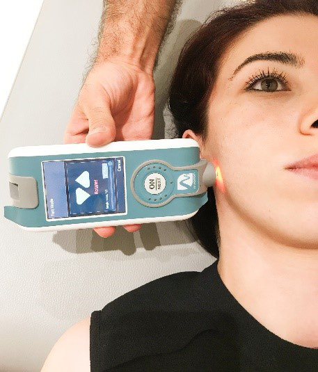
The physical characteristics and demographic information concerning the individuals were recorded before the test. The tone and viscoelastic properties of the masseter muscle were evaluated bilaterally in the supine position with the Myoton PRO device (Figure 1). The mean value was calculated after taking 3 consecutive measurements at the measurement site for each parameter. The measurement site was assumed as the highest point of the muscle belly and was marked with a water-based pen to provide an anatomical reference point. The mandible was placed centrally with no contact between the upper and lower teeth.16
Ethical approval
Ethical approval was obtained from the ethics committee of the Hasan Kalyoncu University (decision No. 2019/61). The subjects were informed about the purpose and content of the study. Written consent was obtained from all individuals.
Measurement tools
Myoton® PRO device
The Myoton PRO device is a non-invasive, portable, hand-held myotonometer used to evaluate the viscoelastic properties of soft tissues.17 This device is known as cost-effective, reliable, valid, and easy-to-use in evaluating the mechanical properties of the musculoskeletal system.18, 19 The muscle tone and viscoelastic parameters (stiffness and elasticity) were assessed by taking myotonometric measurements bilaterally from the skin overlying the masseter by means of Myoton PRO. The examiner marked the masseter with a small ink dot.
This device has shown good-to-excellent reliability in healthy individuals,20 the elderly,12 cancer21 and stroke patients,22 athletes,23 and patients with neurodegenerative disease.24 The device measures the mechanical oscillations of the assessed soft tissues, induced by a mechanical impulse that is of a short duration (15 ms) and involves constant mechanical force (up to 0.6 N). Measuring the mechanical oscillations occurring due to the mechanical impulse yields the following data: oscillation frequency [Hz]; logarithmic decrement (elasticity); and stiffness [N/m].25 Resting muscle tone is the elastic and/or viscoelastic stiffness in the absence of contractile activity. Elasticity is the property whereby a body, when deformed by the applied load, recovers its previous configuration when the load is removed. The applied force is proportional to the strain within the elastic limit. Stiffness is a biomechanical feature of a muscle that characterizes its resistance to contraction or an external force that disrupts its initial state.26
Fonseca anamnestic index
Fonseca et al. developed the Fonseca anamnestic index (FAI), which is a 10-item instrument that allows the assessment of jaw function limitation, pain frequency, psychological distress, and the parafunctional behaviors related to TMD.27 The cross-cultural adaptation of the English version of FAI was completed in 2020 by Kaynak et al.15 The specificity was 83.7% and the sensitivity – 93.57%. They found that FAI had a high level of accuracy (AUC (area under curve) = 0.928; 95% CI (confidence interval) = 0.890–20.964), a high sensitivity value (131 individuals with TMD diagnosed by means of FAI, compared to 140 individuals with TMD diagnosed by the dentist), and a high specificity value (54 individuals with no TMD diagnosed by means of FAI, compared to 65 individuals with no TMD diagnosedby the dentist and the physical therapist). The results demonstrated that the Turkish version of FAI (FAI-T) had good-to-excellent test-retest reliability and a high level of internal consistency, and provided considerable evidence that FAI-T could be used as a screening tool for the identification of TMD.15 Stasiak et al. stated that FAI was very sensitive (97.21%) in identifying patients who actually had TMD, but not very specific (26%) in identifying non-TMD patients, thus being indicated for use in the initial screening of patients only.28
Data analyses
The power analysis was performed with the use of the G*Power software, v. 3.1.9.2 (www.psycho.uni-duesseldorf.de/abteilungen/aap/gpower3), based on the expectation of a large effect size (f = 0.40) for comparisons among 3 age groups in terms of numerical variables for each gender (α = 0.05; 1-β = 0.80). The minimum required total sample size was estimated as 66 for 3 groups and 22 for each age group. The IBM SPSS Statistics for Windows software, v. 24.0 (IBM Corp., Armonk, USA), was used to analyze the data. The normal distribution of the data was determined with the Shapiro–Wilk test. The Mann–Whitney U test was used to make comparisons between 2 groups. Since the number of groups was more than 2, the Kruskal–Wallis test was used to compare non-normally distributed variables. If, according to the Kruskal–Wallis test, the p-value was found to be significant, multiple comparison tests were used to determine the source of the difference. Also, Spearman’s rank correlation test was used to determine the relationships between numerical variables. A p-value of less than 0.05 was considered statistically significant.
Results
The mean age of all individuals was 28.64 ±9.68 years. The demographic characteristics of the participants are given in Table 1.
It was found that there were differences between genders in terms of viscoelastic measurements and tone of the right and left masseter muscles. The tone of the right and left masseter muscles was higher in men than in women (p = 0.185 and p = 0.035, respectively). The measurements on both sides were similar in both genders in terms of stiffness (p > 0.05). The elasticity of the muscle, in terms of logarithmic decrement, on the right and left sides was higher in females (p < 0.05) (Table 2).
Mechanical properties of the masseter muscle in all individuals
According to the results of Dunn’s multiple comparison test, the right and left masseter muscle tone and stiffness were significantly lower in the 18–28 years age group (group 1) as compared to the other 2 groups (p < 0.05) except for the right masseter muscle tone, where the difference was insignificant (p = 0.051), and there was no significant difference between the age groups of 29–39 years (group 2) and 40–50 years (group 3) (p > 0.05). The right and left masseter muscle elasticity was significantly higher in group 1 than in the other 2 groups (p < 0.05), for the right masseter muscle, it was similar between groups 2 and 3 (p > 0.05), whereas for the left masseter muscle, it was significantly higher in group 2 than in group 3 (p < 0.05). In light of these results, it can be said that as age increases, the masseter muscle tone and stiffness increase, and the elasticity of the muscle (logarithmic decrement – if the value increases, elasticity decreases) decreases (Table 3 and Table 4).
Mechanical properties of the masseter muscle in male individuals
Based on Dunn’s multiple comparison test results for males, it was observed that the group 1 values for the right and left masseter muscle tone were significantly lower than in group 2 and group 3 (p < 0.05). For the right masseter muscle, the tone values were significantly higher in group 2 than in group 3 (p < 0.05), whereas there was no significant difference between groups 2 and 3 for the left masseter muscle in terms of tone (p > 0.05). The right and left masseter muscle stiffness values in group 1 were significantly lower than in the other 2 groups (p < 0.05), and there was no significant difference between group 2 and group 3 (p > 0.05). The right and left masseter muscle elasticity in group 1 was significantly higher than in the other 2 groups (p < 0.05). Also, the right and left masseter muscle elasticity was significantly lower in group 3 than in group 2 (p < 0.05) (Table 3 and Table 4).
Mechanical properties of the masseter muscle in female individuals
Based on Dunn’s multiple comparison test results for females, the right and left masseter muscle tone was significantly lower in group 1 than in the other 2 groups (p < 0.05), and both values were significantly lower in group 2 than in group 3 (p < 0.05). The right and left masseter stiffness values in group 1 were significantly lower than in the other 2 groups (p < 0.05), and there was no significant difference between group 2 and group 3 (p > 0.05). The right and left masseter muscle elasticity in group 1 was significantly higher than in the other 2 groups (p < 0.05), and the values in group 2 did not differ significantly from those in group 3 (p > 0.05) (Table 3 and Table 4).
A weak positive correlation was found between the right and left masseter muscle tone and age (r = 0.307 and r = 0.325, respectively; p = 0.001). There was a moderate positive correlation between the right and left masseter muscle stiffness and age (r = 0.507 and r = 0.511, respectively; p = 0.001). A strong positive correlation was observed between the right and left masseter muscle elasticity and age (r = 0.614 and r = 0.645, respectively; p = 0.001).
Discussion
The major findings of this study, in which we examined the tone and viscoelastic parameters of the masseter muscle in healthy individuals as well as their relationship with age and gender, and aimed to specify the reference values for the muscle, are that the tone of the masseter muscle is higher in males, its elasticity (logarithmic decrement) is higher in females, the stiffness of the muscle is similar in both genders, and the elasticity of the muscle decreases with aging.
It is unavoidable that some changes occur in the muscle structure due to aging. One of the changes is the reduction of the contractile areas of the muscle due to the transformation of contractible fibers into non-contractile areas. However, muscle stiffness tends to increase with aging. Changes in the distribution of muscle fibers are also observed with aging. These changes are characterized by the loss of type II fibers and an increase in type I fibers. As a result, an increase in muscle stiffness occurs, as type I fibers cause more stiffness than type II fibers. These age-related changes have been investigated in several studies.29, 30 Following the previous findings in the literature, we also found that the tone and stiffness of the muscle increase, and its elasticity decreases with aging.
It is known that the total muscle mass in women is 15–20% lower than in men. At the same time, women have weaker muscle tone and strength.31, 32 In terms of jaw muscles, it has been reported that the jaw muscles in men are stronger than in women, and there is a positive correlation between testosterone levels and mouth closing force.33 In an experimental mouse model study, it was shown that the masseter muscle mass increases by nearly 38% with the supplementation of testosterone.34 It has been noted in studies that testosterone has a significant effect on the masseter muscle mass.34 In our study, the tone of the masseter muscle was found to be higher in males than in females, and we think that this result may be due to the effects of sex hormones.
Muscle tendons are weaker and looser in women; this situation causes more joint mobility in women. In other words, elasticity and joint mobility are higher in women.32 We think that the higher elasticity (logarithmic decrement) of the masseter muscle in the female group in our study can be explained by the differences in the musculoskeletal system of males and females.
Myotonometric measurement results may vary, depending on the position (horizontal or vertical) of the muscles.24 However, in a study evaluating the stiffness of the masseter muscle in the sitting position, no difference was found between genders.35 In our study, the measurements were made in the supine position, where there is no activity against gravity. In accordance with the literature, we found that the stiffness of the masseter muscle was similar in both genders.
There are studies in the literature that observed a difference or no difference between the tone and viscoelastic values of the right and left masseter muscle.36, 37 In our study, differences were observed between the values of some parameters of the right and left masseter muscles. We think that these differences could be explained by dominance.
Limitations
The fact that the temporal muscle was not included in the measurements of our study could be viewed as a limitation. Also, all participants came from the same university, and this selection method from the same participant pool was a limitation of our study with regard to sample selection. Although FAI is a simple, quick and easy way to determine myogenic TMD, it is not the gold standard for the clinical evaluation. Bruxism and TMD cases were excluded from the study; they were determined through the FAI questions based upon people’s statements. Individuals diagnosed with sleep problems, such as obstructive sleep apnea, were not included in the study. However, since such problems are difficult to diagnose even by a physician due to their complex etiology, their presence was determined based upon people’s statements. These are the limitations of our study. In the future, multidisciplinary studies can be designed based on the samples diagnosed by dentists.
Conclusions
The tone and viscoelastic parameters of the masseter muscle can be examined using different evaluation methods to compare pre- and post-treatment changes in the muscle in fields such as plastic surgery, dentistry, physical therapy and rehabilitation, and speech and language therapy. We hope that the results of our study can provide the reference values for studies in these fields.