Abstract
The aim of the review was to systematically synthesize and critically appraise the available evidence from systematic reviews and meta-analyses evaluating the clinical effectiveness, safety and applicability of laser-based therapies in the management of oral leukoplakia (OL), and to develop evidence-based clinical recommendations of the World Federation for Laser Dentistry (WFLD). The search of PubMed/MEDLINE, Embase, Scopus, and the Cochrane Library was conducted to identify systematic reviews and meta-analyses published between 2016 and 2026. Eligible reviews assessed laser-based interventions for OL and reported clinically relevant outcomes. Methodological quality was evaluated using a laser-adapted nine-domain risk-of-bias assessment tool. Ten reviews were included, comprising systematic reviews, meta-analyses and network meta-analyses. Evaluated modalities included CO2, erbium-doped yttrium aluminium garnet (Er:YAG), erbium, chromium-doped yttrium, scandium, gallium, and garnet (Er,Cr:YSGG), diode, neodymium-doped yttrium aluminium garnet (Nd:YAG), and potassium titanyl phosphate (KTP) lasers, as well as laser-assisted photodynamic therapy (PDT). Overall, laser-based interventions demonstrated recurrence rates comparable to or lower than those associated with conventional surgery, with favorable postoperative outcomes including reduced pain and improved hemostasis. All included reviews were classified as having a low (n = 6) or moderate (n = 4) risk of bias according to the study-specific assessment framework, although substantial heterogeneity in laser parameters and outcome reporting was observed. Laser-based therapies represent clinically justified options for the management of OL when appropriately selected and integrated with histopathological assessment, adequate margin control, complete lesion removal, and long-term surveillance. However, current evidence does not demonstrate a consistent reduction in the risk of malignant transformation or definitive superiority of any specific laser modality. Heterogeneity in surgical procedures, outcome definitions and study designs underscores the need for standardized reporting and high-quality prospective studies.
Keywords: recurrence, laser therapy, malignant transformation, oral potentially malignant disorders, leukoplakia
Introduction
Oral leukoplakia (OL) is one of the most common oral potentially malignant disorders (OPMDs), defined as a white plaque of questionable risk after the exclusion of other known diseases not associated with an increased risk of cancer.1, 2, 3 Its management should be considered within the broader OPMD framework, encompassing oncologic risk reduction, histopathological risk stratification, adequate surgical management, recurrence prevention, and long-term surveillance. With a global prevalence of 0.1–4.5%, OL represents a significant public health concern due to its potential for malignant transformation into oral squamous cell carcinoma.2, 3, 4 Meta-analytic evidence indicates that OL is associated with significantly increased odds of developing oral cancer (summary odds ratio (SOR): 3.35; 95% confidence interval (95% CI): 3.21–3.50).5 Malignant transformation rates vary according to clinical characteristics, anatomical site and lesion subtype, with particularly high rates reported for proliferative verrucous leukoplakia (PVL), which may approach 50%.3, 6, 7 Although available management strategies include active surveillance, conventional surgical excision, pharmacological interventions, and laser-based therapies,8, 9 no universally accepted, evidence-based management protocol has yet been established.10, 11 Laser therapy offers potential advantages over conventional surgery, including improved hemostasis, reduced postoperative morbidity, minimal scarring, and the elimination of the need for sutures, which may prevent distortion of the surgical site and enhance surgical precision, particularly for extensive or multifocal lesions.12, 13 Systems in clinical use include CO2, erbium-doped yttrium aluminium garnet (Er:YAG), erbium, chromium-doped yttrium, scandium, gallium, and garnet (Er,Cr:YSGG), neodymium-doped yttrium aluminium garnet (Nd:YAG), and diode lasers, each with distinct wavelength-specific tissue interaction properties.12, 13, 14 Several clinical and experimental studies have contributed to our understanding of laser–tissue interactions in OPMDs.15, 16, 17 Despite the increasing availability of laser-based treatments, the evidence remains heterogeneous and methodologically limited. Available systematic reviews suggest that laser-based treatment is associated with lower recurrence rates than conventional surgery, but has no consistent effect on malignant transformation.12 Core clinical determinants, including lesion characteristics, histopathology and the adequacy of surgical margins, are consistent across treatment modalities, supporting a principle-based rather than parameter-driven approach.13, 18 Several clinically relevant questions remain unresolved: whether laser therapy confers advantages beyond its technical surgical benefits, whether recurrence is driven primarily by treatment modality or lesion biology, and how surgical margins, ablation depth and surgical procedures should be optimized according to histopathological risk.
Objectives
This narrative review, developed by the World Federation for Laser Dentistry (WFLD) study group through literature synthesis and expert consensus, aims to evaluate the clinical effectiveness, safety and applicability of laser-based treatments in the management of OL and to provide evidence-informed clinical recommendations. The specific objectives are to: (1) evaluate the clinical effectiveness of laser treatments, including lesion resolution, recurrence, symptom control, and malignant transformation risk, in comparison with conventional surgical and non-surgical approaches; (2) identify and compare laser modalities and technical procedures; (3) assess safety profiles, postoperative morbidity, and functional and aesthetic outcomes; (4) appraise methodological quality, heterogeneity and knowledge gaps; and (5) establish an initial framework for consensus-based clinical recommendations to guide standardized and safe use of lasers in OL management.
Methodology
This study was designed as a narrative review supported by an analysis of systematic reviews and meta-analyses. Selected methodological principles from the Joanna Briggs Institute (JBI) umbrella review framework were applied to ensure transparency, and reporting followed the PRISMA (Preferred Reporting Items for Systematic reviews and Meta-Analyses) 2020 guidelines.19, 20, 21, 22 The protocol was registered in the PROSPERO (International Prospective Register of Systematic Reviews) database (registration No. CRD420261287719).
PICO question
In patients with oral leukoplakia, does laser-based surgical treatment (excision, ablation or vaporization), compared with conventional surgery, pharmacological treatment or no intervention, improve lesion resolution, recurrence rates, postoperative morbidity, the risk of malignant transformation, and functional or aesthetic outcomes?
Search strategy and study selection
A structured literature search was conducted in PubMed/MEDLINE, Embase, Scopus, and the Cochrane Library using predefined strategies informed by the PICO framework and combining controlled vocabulary, where applicable, with free-text terms related to OL and laser-based interventions. The search covered studies published from January 1, 2016 to January 31, 2026. Broad review filters were applied, where available, in the respective databases. Eligibility was restricted during screening to systematic reviews and meta-analyses reporting extractable data relevant to OL. Records were screened independently by multiple reviewers in 2 stages: first, title and abstract screening, followed by full-text assessment. Disagreements were resolved through consensus. The complete database-specific search strategies and the number of records retrieved are presented in Table 1.
Data extraction
Three reviewers extracted data independently using a standardized, pilot-tested form. Extracted review-level information included study characteristics, patient populations, lesion characteristics, as well as anatomical sites, interventions, comparators, and follow-up periods. Laser-related data, where reported, included the laser system (CO2, Er:YAG, Er,Cr:YSGG, diode, Nd:YAG, or potassium titanyl phosphate (KTP)), wavelength, power output, energy or radiant exposure, application mode, treatment duration, number of sessions, surgical margins, and ablation or excision depth. For laser-assisted photodynamic therapy (PDT), information on the photosensitizer, light source, wavelength, irradiation parameters, and the number of treatment sessions was also extracted, where available. Outcomes included lesion response or resolution, recurrence, malignant transformation, postoperative pain and complications, bleeding and hemostasis, functional and aesthetic outcomes, patient-reported symptoms, and adverse events. Discrepancies were resolved through consensus, with arbitration by an additional senior reviewer when necessary. Missing information was sought in supplementary materials or by contacting the corresponding authors and was recorded as “not reported” when it could not be retrieved. The extracted data was collated to assess the consistency and clinical applicability of the available evidence and to inform the development of the WFLD clinical recommendations.
Risk of bias and quality assessment
The methodological quality of each included review was assessed independently by 3 reviewers using a structured framework developed for this study, informed by AMSTAR-2 (A MeaSurement Tool to Assess systematic Reviews, version 2) and adapted to incorporate laser-specific domains relevant to reproducibility, technical transparency and clinical interpretation.
The assessment criteria included: (1) description of the laser system and its parameters (wavelength, power, fluence, and application mode); (2) definition of treatment protocols, the number of sessions, and a priori case selection criteria, including lesion subtype, clinical and histopathological features, and patient-related factors; (3) specification of outcomes, including lesion resolution, recurrence, postoperative morbidity, and malignant transformation; (4) inclusion of appropriate comparator groups; (5) transparent inclusion criteria and characterization of the patient population; (6) measures to control bias, including randomization, allocation concealment and blinding; (7) appropriateness and reproducibility of statistical analyses; (8) completeness of outcome and safety reporting, including follow-up duration; and (9) disclosure of funding and conflicts of interest.
Each domain was scored dichotomously (1 = fulfilled, 0 = not fulfilled), yielding a total score of 0–9. Reviews were categorized as having a low (7–9), moderate (4–6) or high (0–3) risk of bias. Disagreements were resolved through discussion or arbitration by a fourth reviewer. The framework was informed by the Cochrane Handbook for Systematic Reviews of Interventions and adapted to address heterogeneity in laser-specific reporting.21
Consensus development process
The evidence synthesis and consensus process was conducted by the WFLD study group, comprising clinicians and researchers with expertise in oral surgery, oral medicine, oral pathology, laser dentistry, and OPMDs. A structured review of the available evidence was combined with iterative expert discussions focused on clinically relevant principles and their practical implementation. Disagreements were resolved through further discussion until consensus was reached among the contributing experts. When evidence was limited or heterogeneous, clinical statements were intentionally formulated as conditional, principle-based recommendations rather than prescriptive treatment protocols. This evidence-informed and consensus-based approach was consistent with the methodology previously applied in the development of the official WFLD clinical recommendations for the laser-assisted management of other complex oral conditions. The objective was to provide a clinically oriented framework for the safe and appropriate use of lasers in the management of OL.
Results
Study selection
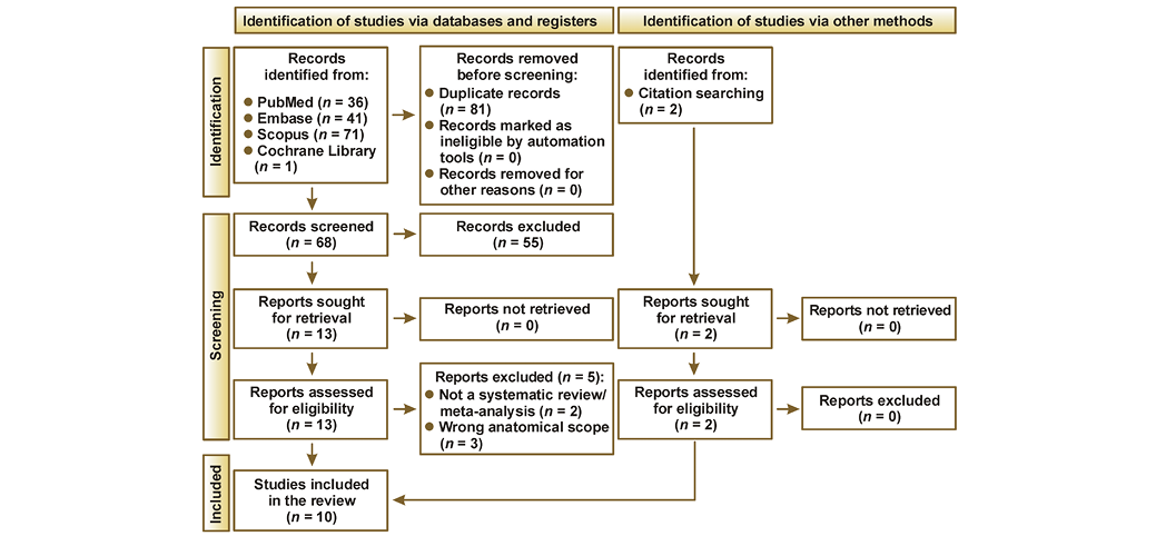
The database search identified 149 records (PubMed, n = 36; Embase, n = 41; Scopus, n = 71; and the Cochrane Library, n = 1). After the removal of 81 duplicates, 68 records underwent title and abstract screening, of which 55 were excluded. The remaining 13 reports were retrieved and assessed for eligibility. Two additional reports were identified through citation searching and assessed for eligibility. Of the 15 full-text reports assessed, 5 were excluded: 2 because they were not systematic reviews or meta-analyses; and 3 because they addressed laryngeal or vocal fold leukoplakia rather than OL. Ultimately, 10 systematic reviews and meta-analyses met the eligibility criteria and were included in the review (Figure 1).
Risk of bias and quality assessment
Six reviews were classified as having a low risk of bias and 4 as having a moderate risk of bias; none was classified as having a high risk of bias. Reviews categorized as having a low risk of bias generally demonstrated transparent methodology, adequate reporting of intervention parameters, and appropriate statistical analyses.12, 13, 23, 24, 25, 26 Studies categorized as having a moderate risk of bias showed incomplete reporting of irradiation parameters, insufficient lesion characterization, absent or inadequately defined comparator groups, and limited assessment of potential confounding factors.27, 28, 29, 30, 31 Common limitations across the included reviews encompassed substantial heterogeneity in treatment protocols, inconsistent outcome definitions, limited availability of randomized controlled trials (RCTs), and a potential overlap among primary studies. The extent of overlap among primary studies was not formally quantified. Detailed results are presented in Table 2.
Characteristics of the included reviews
The 10 included reviews evaluated CO2, Er:YAG, Er,Cr:YSGG, Nd:YAG, diode, and KTP lasers, as well as laser-assisted PDT. Individual reviews included between 11 and 80 primary studies, although not all primary studies specifically addressed OL in reviews with broader eligibility criteria.12, 13, 23, 24, 25, 26, 27, 28, 29, 31 The CO2 laser was the most extensively investigated modality and was evaluated in systematic reviews addressing recurrence, malignant transformation, postoperative outcomes, and comparisons with conventional surgery or other treatment modalities.12, 13, 23, 28, 31 Although some analyses suggested favorable recurrence outcomes following CO2 laser treatment, the findings were heterogeneous, and no consistent reduction in the risk of malignant transformation compared with conventional treatment was demonstrated.12, 28 Combined laser excision and vaporization were associated with particularly favorable recurrence outcomes in 1 large systematic review and meta-analysis.23
Evidence concerning Er:YAG and Er,Cr:YSGG lasers was derived primarily from a network meta-analysis and a limited number of comparative primary studies. These modalities achieved favorable rankings for selected outcomes, including recurrence, intraoperative bleeding and postoperative pain; however, the certainty and generalizability of these findings remained limited.25 Evidence for diode, Nd:YAG and KTP lasers was comparatively sparse and was generally derived from small subsets of primary studies included in broader reviews.12, 23, 25, 31
Three reviews evaluated PDT using photosensitizers such as 5-aminolevulinic acid (ALA), methylene blue (MB), Photofrin, Photosan, temoporfin (mTHPC; Foscan), hematoporphyrin derivatives, and chlorin e6.24, 27, 29 Reported response rates varied substantially based on lesion type, photosensitizer, irradiation protocol, and outcome definition, limiting direct comparisons and the extrapolation of findings specifically to OL. The review addressing PVL reported a high pooled recurrence rate and found insufficient evidence that any available treatment reliably prevents recurrence in this particularly aggressive subtype.26 Overall, substantial heterogeneity in lesion classification, intervention protocols, comparators, follow-up periods, and outcome definitions limited comparability across the included reviews. The characteristics of the included reviews are presented in Table 3.
Discussion
Clinical effectiveness of laser therapies
This narrative review contextualizes laser-assisted interventions within the broader principles governing OPMD management rather than serving as a purely technical review. Across the available evidence, recurrence control, adequate surgical margins, histopathological risk stratification, and long-term surveillance represent more clinically relevant determinants of outcome than the specific laser modality.
Laser-based therapies were most consistently associated with effective intraoperative hemostasis, reduced postoperative morbidity, and recurrence rates comparable to or lower than those observed following conventional surgery.8, 32, 33, 34 Potential advantages for the management of extensive or multifocal lesions have been suggested, although these observations are derived predominantly from heterogeneous observational data and therefore remain speculative. Lesion characteristics, dysplasia grade, multifocality, anatomical location, and margin adequacy were identified across multiple reviews as important determinants of recurrence and oncologic outcomes, irrespective of the laser modality used.12, 23, 26, 31
Mechanisms of action and tissue interaction
CO2 lasers (10,600 nm) exhibit high absorption in tissue water, enabling precise superficial ablation with limited peripheral thermal spread, and remain the most extensively studied modality for OL.13, 31, 32 Er:YAG (2,940 nm) and Er,Cr:YSGG (2,780 nm) lasers exhibit even greater absorption in water, resulting in narrower thermal damage zones than those associated with CO2 lasers (median (Me): 30.9 µm vs. 72.6 µm), with potential advantages for tissue preservation and healing. However, clinical evidence remains limited.35, 36, 37 Diode (808–980 nm) and Nd:YAG (1,064 nm) lasers exhibit lower absorption in water and deeper tissue penetration, promoting hemostasis while potentially increasing the risk of collateral thermal damage if treatment parameters are not carefully controlled.38 In anatomically sensitive areas, such as the gingiva and palate, a tangential rather than perpendicular approach may help minimize thermal injury to the periosteum, bone or dental structures.14, 39
Recurrence rates
A 2024 systematic review and meta-analysis of 80 studies involving 7,614 samples reported a pooled recurrence proportion of approx. 22% for laser-based approaches, with a network meta-analysis indicating that combined protocols (excision followed by vaporization) ranked among the most favorable strategies for reducing recurrence.23 A 2025 meta-analysis on CO2 laser treatment reported a trend toward lower recurrence rates compared with other modalities (odds ratio (OR): 0.36, 95% CI: 0.12–1.04), although this difference did not reach statistical significance (p = 0.06).13 Overall, the available data suggests a possible benefit of laser-assisted treatment in reducing recurrence rates, but the findings remain heterogeneous and inconclusive.
Postoperative outcomes
Laser-based therapies are generally associated with lower pain, improved hemostasis and reduced intraoperative bleeding compared with conventional surgery. An RCT reported significantly greater pain and swelling following cold blade surgery during the first 3 postoperative days (p < 0.05),40 while another RCT comparing CO2 laser, Er:YAG and conventional scalpel surgery demonstrated significantly shorter operative times with laser-based approaches (p < 0.001) and less intraoperative bleeding with the CO2 laser (p < 0.001).37 However, a 2025 meta-analysis found no significant differences in postoperative pain between CO2 laser treatment and other modalities (standardized mean difference (SMD): −0.16, 95% CI: −1.37–1.05),13 reflecting substantial variability among the studies. These findings should be interpreted with caution given the heterogeneity in study designs and the limited availability of high-quality randomized evidence.14, 37, 39, 40, 41, 42
Lesion resolution
Complete response rates following laser therapy vary considerably, ranging from approx. 50% to over 90%, depending on lesion characteristics, the degree of dysplasia, multifocality, and treatment modality. A prospective study using a high-power diode laser (808 nm) reported a complete response in 93.5% of cases, with a low recurrence rate during follow-up,14 while Er:YAG laser treatment demonstrated complete and partial responses in a substantial proportion of lesions, although recurrence remained a concern.14, 35 A retrospective study using a CO2 laser noted a permanent treatment success rate of 97.8% at 6-year follow-up after complete excision to a depth of ≥1 mm with a 3-mm margin of healthy-appearing tissue. However, the cohort comprised predominantly homogeneous leukoplakias without dysplasia, suggesting that the favorable outcomes may reflect lesion characteristics rather than the technical superiority of the laser modality.15
Photodynamic therapy as an adjunctive approach
Photodynamic therapy combines a photosensitizing agent, commonly ALA or MB, a light source operating in the red spectrum (approx. 630–660 nm), and molecular oxygen to generate reactive oxygen species (ROS) that exert cytotoxic effects on dysplastic cells. Systematic reviews have reported substantial heterogeneity in photosensitizers, irradiation parameters, treatment schedules, lesion characteristics, and outcome definitions.24, 27, 29 A systematic review addressing OL reported pooled complete and partial response rates of 32.9% and 43.2%, respectively, corresponding to an overall response rate of 76.1%.29 Ablative fractional laser-assisted PDT achieved an overall positive response rate of 87.5%, including a complete response rate of 62.5%, in a retrospective study with a 3-year follow-up period.43 Photodynamic therapy with ALA has also demonstrated promising clinical outcomes, suggesting that laser pre-treatment may improve photosensitizer penetration in selected lesions.34 Nevertheless, the available evidence remains heterogeneous, and these findings do not establish the superiority of any specific photosensitizer, light source or laser-assisted PDT protocol.24, 25, 27, 29
Malignant transformation
The available evidence has not demonstrated a consistent or statistically significant difference in malignant transformation rates following laser therapy compared with conventional treatment approaches. Reported rates range from approx. 1% to 15%, with longer follow-up generally associated with higher cumulative rates.12, 32, 44, 45, 46, 47, 48 Recurrent leukoplakia is associated with an approx. 7-fold higher risk of malignant transformation compared with non-recurrent lesions,23 highlighting the clinical importance of long-term disease monitoring. Established risk factors include dysplasia severity, lesion heterogeneity, anatomical location, and recurrence status.28, 49, 50 The risk of malignant transformation appears to be influenced primarily by lesion biology and patient-related factors rather than the surgical modality employed.
Comparative effectiveness of laser devices
The CO2 laser has the largest evidence base among the laser modalities used for OL, with studies reporting favorable recurrence control, effective hemostasis and acceptable postoperative outcomes. Nevertheless, the available evidence remains heterogeneous, and neither consistent superiority over other surgical approaches nor a reduction in the risk of malignant transformation has been demonstrated.12, 13, 23, 28, 31
The Er:YAG laser produces a narrower zone of thermal damage than the CO2 laser, which may be advantageous for tissue preservation and histopathological assessment.37 In a retrospective comparative study, Er:YAG treatment was associated with a significantly better clinical outcome than conventional scalpel surgery (p = 0.015). However, no significant overall differences were observed among the 5 treatment modalities evaluated.45 The consistent superiority of Er:YAG laser treatment over CO2 laser treatment or conventional surgery has not been established in systematic reviews.12, 25, 31
Diode lasers provide effective hemostasis and may facilitate outpatient excision or ablation of OL. However, the available evidence is derived predominantly from small observational or non-comparative studies, and robust conclusions regarding recurrence control and oncologic outcomes cannot be drawn.14, 38, 51, 52, 53
Evidence concerning the KTP laser remains limited. A retrospective comparative study reported a lower recurrence rate following KTP laser treatment than after CO2 laser treatment.52 However, the available systematic reviews and limited comparative evidence are insufficient to establish the superiority of the KTP laser over the CO2 laser or other laser modalities.12, 31, 52 The selection of a laser modality should therefore be guided by lesion characteristics, anatomical location, the intended surgical procedure, histopathological requirements, and operator experience.
Technical considerations and optimal parameters
Adequate surgical margin width is a critical determinant of recurrence rates in homogeneous OL, with favorable and sustained outcomes reported even during long-term follow-up.15 Surgical margins of ≤3 mm have been associated with higher recurrence rates, whereas margins of 3–5 mm may reduce the risk of recurrence.54, 55, 56 However, this relationship varies considerably according to lesion subtype. Proliferative verrucous leukoplakia exhibits high recurrence rates even with surgical margins that exceed 3 mm. In general, a margin extension of 5 mm is considered optimal.57 For vaporization, a safety margin of approx. 3 mm beyond the clinically visible lesion boundary has been associated with lower recurrence rates (p < 0.05) (Table 4).15, 58, 59, 60, 61
Collectively, these findings support lesion-specific surgical planning rather than uniform excision protocols.
Diagnostic utility
Incisional biopsy with histopathological evaluation remains mandatory before any treatment of OL, regardless of the surgical modality used. Laser-induced thermal effects may alter tissue architecture, potentially affecting histopathological interpretation and the assessment of surgical margins. The intraoperative and postoperative clinical advantages of laser surgery, including improved hemostasis and reduced morbidity, are well documented. However, current evidence does not support the diagnostic superiority of lasers over conventional biopsy techniques. These considerations highlight the importance of appropriate surgical technique and wavelength selection when laser-assisted biopsy is performed.61, 62, 63, 64, 65
Safety and complications
The findings of this review indicate that laser-based therapies for OL are generally well tolerated and associated with low complication rates. Serious adverse events were infrequently reported, with most studies noting minimal postoperative morbidity.25, 32, 34 Common transient side effects include mild to moderate pain, swelling and temporary limitation of mouth opening, typically peaking within the first few days after treatment.48 Wound healing is generally satisfactory, with minimal scarring, although variability in healing time has been reported, with some cases requiring several weeks for complete re-epithelialization.60 Rare complications, including pyogenic granuloma formation, fibrosis and persistent discomfort have been described.39, 45, 60 The reporting of adverse events remains inconsistent across studies, which may limit the accuracy of safety assessments.12, 32, 33, 46 As expected, no association between surgical modality (laser-based or conventional) and the risk of malignant transformation has been demonstrated. The risk of malignant transformation appears to be influenced by the biological and histological characteristics of the lesions and a history of head and neck cancer.66 Nevertheless, these findings must be interpreted with caution due to the limited availability of long-term oncologic data (Table 5).12, 32, 33, 45, 67
Limitations of the evidence
This narrative review confirms that the overall quality of the available evidence remains limited. Substantial clinical and methodological heterogeneity in laser devices, treatment parameters and protocols precludes reliable quantitative synthesis. Outcome definitions, including those for recurrence rates, lesion resolution and malignant transformation, varied considerably, and long-term follow-up was frequently insufficient. The majority of the studies were small, non-randomized and inadequately controlled. These limitations underscore the need for well-designed RCTs with standardized protocols, uniform outcome evaluation and long-term follow-up.
Limitations of the review process
The search was restricted to English-language publications, and gray literature was excluded, introducing a potential risk of language and publication bias. Relevant clinical data was frequently unavailable, preventing subgroup analyses according to dysplasia grade, lesion site or history of exposure to relevant risk factors. Future studies may benefit from integrating an expert consensus with structured evidence synthesis to minimize subjective interpretation and facilitate the development of clear recommendations based strictly on clinical and histopathological considerations.
Implications for practice, policy and future research
Laser-based therapies are a valid treatment option for selected patients with OL, particularly when lesion extent, multifocality or anatomical constraints limit conventional surgery. Laser excision enables precise lesion removal with effective hemostasis, whereas laser ablation may be considered for superficial or anatomically challenging lesions. The use of lasers does not eliminate the need for histopathological assessment, risk-stratified patient selection or long-term surveillance, all of which remain essential components of OL management. Consensus-based frameworks addressing indications for excision versus vaporization, safety margins, the documentation of laser parameters, and follow-up standards are urgently needed, alongside structured training and credentialing. Future RCTs should compare laser modalities with conventional surgery, incorporate extended follow-up periods, and stratify patients according to dysplasia grade and lesion subtype, with endpoints including recurrence-free survival, malignant transformation and quality of life.
Conclusions
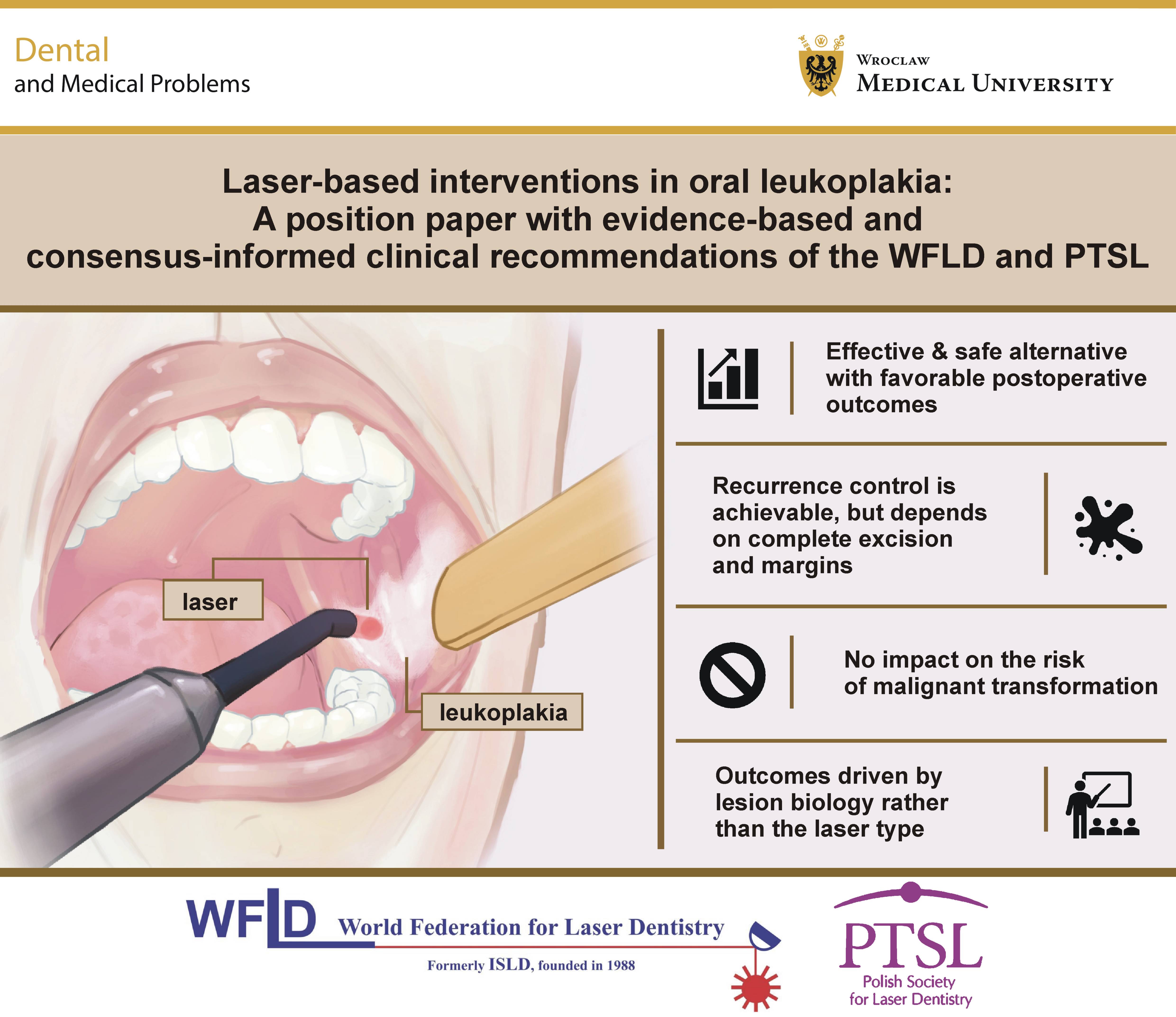
Laser-based management of OL offers surgical precision, tissue preservation and reduced postoperative morbidity, with recurrence rates comparable to or lower than those associated with conventional surgery when adequate surgical margins and complete removal of the lesion to an adequate depth are achieved.
Current evidence does not demonstrate a consistent effect of laser treatment on the risk of malignant transformation in OL, which appears to be influenced by lesion biology and patient-related factors. Clinical management should integrate histopathological findings, lesion characteristics and the patient’s individual risk profile, with an emphasis on surgical margin control, case selection and long-term follow-up. Within the limitations of the available evidence, laser-based therapies constitute a clinically justified component of contemporary OL management when applied within a structured framework, encompassing diagnosis, adequate procedures and surveillance.
General principles for the clinical management of oral leukoplakia according to the WFLD study group
Laser-based interventions in OL may serve as: (A)adjuncts to clinical examination; (B) diagnostic support through laser-assisted biopsy; and (C) surgical management with adequate margin control and complete lesion depth ablation control. The consensus-informed clinical principles developed by the WFLD study group are summarized in Table 6.
General therapeutic principles
1. Histopathological assessment is mandatory before laser management. Following histopathological assessment, laser ablation may be considered for appropriately selected lesions.
2. Laser treatment represents a surgical modality rather than a biological modifier. It does not eliminate field cancerization or the intrinsic malignant potential of the lesion.
3. Risk stratification is essential, incorporating lesion subtype, size, location, dysplasia grade, multifocality, history of recurrence, prior squamous cell carcinoma of the upper aerodigestive tract, and patient-related risk factors, including tobacco use, alcohol consumption and relevant comorbidities.
4. Recurrence prevention requires complete lesion removal, with adequate surgical margins tailored to the histopathological risk:
• ≥3 mm of clinically healthy-appearing marginal mucosa, with complete lesion ablation to an appropriate depth, for lesions without dysplasia;
• ≥5 mm for lesions with mild to moderate dysplasia;
• ≥10 mm for lesions with severe dysplasia or carcinoma in situ. When microinvasive carcinoma is suspected, preoperative magnetic resonance imaging (MRI) is recommended. Surgical margins of ≥1 cm are required, and osteotomy should be considered for gingival or palatal localizations.
Safety and operator requirements
Procedures should be performed by clinicians with appropriate training in laser dentistry and OPMD management, in accordance with established laser safety standards. Patients must be informed that laser therapy does not eliminate the need for long-term surveillance.
Trial registration
The study was registered prospectively in the PROSPERO database (registration No. CRD420261287719).
Ethics approval and consent to participate
Not applicable.
Data availability
The datasets generated and/or analyzed during the current study are available from the corresponding author on reasonable request.
Consent for publication
Not applicable.
Use of AI and AI-assisted technologies
AI-assisted tool (Grammarly AI; Superhuman Platform Inc., San Francisco, USA) was used exclusively for language editing and improvement of readability. No AI tools were used for data analysis, interpretation or generation of scientific conclusions. All content was reviewed and approved by the authors.