Abstract
Background. Tooth decay is a common problem, particularly among young patients. Compomer materials combine favorable aesthetic properties and durability with antibacterial activity resulting from fluoride release. The incorporation of silver (Ag0) and copper oxide (CuO) particles may further enhance their antimicrobial properties, reducing the risk of complications such as secondary caries and pulpitis.
Objectives. The aim of the study was to evaluate the cytotoxic and antibacterial properties of compomer materials doped with 0.125 wt%, 0.25 wt% and 0.5 wt% CuO and Ag0 particles using both static and dynamic methods in the Centers for Disease Control and Prevention (CDC) biofilm reactor.
Material and methods. Reference microbial strains were stored at −80°C in tryptic soy broth (TSB) supplemented with 15 wt% glycerol and cultured under strain-specific conditions before the experiments. Streptococcus mutans, Lactobacillus rhamnosus, Candida albicans, and Staphylococcus epidermidis were grown on appropriate media under aerobic, anaerobic or elevated CO2 conditions. Microbiological analysis involved preparing standardized microbial suspensions (0.5 McFarland), incubating them with the tested biomaterials under appropriate conditions. After incubation, biofilms were detached using saponin, serially diluted and plated on selective media, after which colony-forming units (CFU/mL and CFU/cm2) were calculated. The experiments were performed in 6 replicates using both static and dynamic (flow reactor) biofilm models. Cytotoxicity was evaluated according to the ISO 10993 standard using L929 fibroblasts and BALB/3T3 cells exposed for 24 h to extracts or disks containing Ag0 and CuO at concentrations of 0.125 wt%, 0.25 wt% and 0.5 wt%.
Results. Both Ag0 and CuO exhibited statistically significant dose-dependent antibacterial activity against S. mutans and S. epidermidis. Silver nanoparticles at concentrations of 0.25 wt% and 0.5 wt% demonstrated the highest cytotoxicity in both BALB/3T3 and L929 cell lines.
Conclusions. The incorporation of both CuO and Ag0 particles into compomer materials enhanced their antimicrobial activity against S. mutans and S. epidermidis.
Keywords: copper oxide, biofilms, biocompatible materials, silver nanoparticles, dental materials
Introduction
Compomers, also known as polyacid-modified composite resins, were introduced into dentistry in the early 1990s. They were designed to combine the favorable properties of composite and glass ionomer materials. The former provide good aesthetics and durability, while the latter release fluoride, which is widely utilized and valued in dentistry. Owing to these characteristics, compomers are widely used, particularly in pediatric dentistry, where treatment may be complicated by limited patient cooperation. However, compomers also exhibit certain drawbacks, including greater leakage and lower bonding strength.1, 2 The amount of fluoride released varies among different groups of materials. In a study by Sagmak et al., glass ionomer materials, resin-modified glass ionomer materials, and compomers were compared with respect to fluoride release.3 After 7 days, the mean fluoride release was 24.36 mg/L for glass ionomer materials, 3.17–23.53 mg/L for compomers, and 34.53–67.98 mg/L for resin-modified glass ionomer materials. Compomers exhibited the weakest antimicrobial activity against S. mutans, which was considered insufficient to effectively inhibit advanced carious lesions.3, 4 Compomer materials are frequently used in pediatric dentistry due to poor patient compliance and difficulty maintaining appropriate surgical conditions. Increasing antibacterial and antifungal properties of the material may reduce the risk of complications, such as secondary caries or pulpitis. The incorporation of various particles, including silver (Ag), copper oxide (CuO), zinc oxide (ZnO), and titanium dioxide (TiO2), can enhance antimicrobial properties. Nanoparticles range in size from 10 nm to 100 nm, while submicron particles are smaller than 1 μm.5, 6
Copper oxide has been widely used in medicine due to its antibacterial and anticancer properties. In dentistry, CuO-containing coatings have been developed. However, scientists point to the potential cytotoxicity of CuO.7, 8, 9 The exact antimicrobial mechanism of CuO nanoparticles is not fully understood. A commonly reported mechanism is the increased production of reactive oxygen species (ROS) in bacterial cells, especially in CuO–water suspensions. The antibacterial activity of CuO nanoparticles depends not only on bacterial characteristics, such as cell wall structure, but also on the ratio of surface area to particle size. In general, a larger surface area-to-volume ratio enhances antimicrobial activity. Copper oxide nanoparticles exhibit high toxicity in both in vitro and in vivo models. Their toxicity is mainly determined by copper bioavailability and is influenced by particle size, surface charge and dissolution behavior. Numerous studies involving algae, protozoa, invertebrates, vertebrates, and mammalian cells have shown that CuO nanoparticles induce oxidative stress, mitochondrial dysfunction, DNA damage, and reduced cell proliferation.7, 10 As with cytotoxicity, the higher the concentration of CuO, the greater its antimicrobial activity. However, CuO does not affect all microorganisms equally. A concentration associated with acceptable cytotoxicity does not always reduce the number of microbes.11, 12
Due to their antiviral, antifungal and antibacterial properties, Ag nanoparticles are widely used in medicine, veterinary medicine and the food industry. Similar to other nanoparticles, their properties are influenced by particle size and shape. The generation of ROS, disruption of cell membrane structure, alterations in cell signaling pathways, and damage to DNA and proteins are among the proposed antimicrobial mechanisms. The cytotoxicity and antimicrobial activity of Ag nanoparticles are related to particle size and concentration. Therefore, it is important to identify a concentration that ensures antimicrobial activity while maintaining biocompatibility and minimizing cytotoxic effects on surrounding tissues. Maintaining antimicrobial activity at lower concentrations of Ag nanoparticles can be achieved by combining them with other particles that possess antimicrobial properties.7, 13, 14 It should be noted that Ag nanoparticles are not equally effective against all microorganisms. Their effectiveness depends on their concentration, which influences their cytotoxicity. Another important factor is the method used to produce the particles. Chemically synthesized particles exhibit greater cytotoxicity than particles produced using green synthesis.15
The Centers for Disease Control and Prevention (CDC) biofilm reactor is a device that simulates conditions found in the oral cavity. The tested material, in the form of discs, is immersed in a medium containing selected microbes that flow through the container. A biofilm is a complex structure composed of various microorganisms. It is characterized by high resistance to antimicrobial agents and the immune system. The formation of biofilm significantly increases the risk of developing caries and periodontitis.16, 17, 18 Unlike the static method, the dynamic method continuously supplies nutrients while simultaneously removing toxins and metabolites, and the presence of shear stress makes these conditions more similar to those found in the oral cavity. It also allows greater control over the experimental process, including the addition of various agents. Due to these properties, the dynamic method provides a more accurate representation of oral conditions for both monospecies and multispecies biofilms.19, 20, 21
The aim of the study was to evaluate the cytotoxic, antibacterial and antifungal properties of compomer materials modified with different concentrations of Ag0 nanoparticles and CuO particles using both a static in vitro model and a CDC biofilm reactor. The hypothesis of the study was that the incorporation of Ag0 nanoparticles and CuO particles into the compomer material would enhance its antimicrobial properties while maintaining cytotoxicity similar to or lower than that of the unmodified compomer.
Material and methods
Preparation of nanoparticles
Silver nanoparticles were synthesized using a hydrothermal reduction method, with silver nitrate (AgNO3; Chempur, Piekary Śląskie, Poland) as the silver precursor. Silver nitrate was first dissolved in distilled water, after which aqueous ammonia (Chempur) was added dropwise until a clear diamminesilver(I) complex was formed, [Ag(NH3)2]+. Alpha-D-Glucose (Sigma-Aldrich, St. Louis, USA) and TWEEN 100 (POCH S.A., Gliwice, Poland) were added to the solution. The prepared reaction mixture was transferred to a hydrothermal reactor (Magnum II; ERTEC-Poland, Wrocław, Poland). The reactor was heated to 115–120°C and maintained at approx. 30 bar for 60 min. After the hydrothermal reaction, the autoclave was allowed to cool to room temperature. The synthesized Ag0 particles were purified by repeated washing with a 1:1 mixture of 96% ethanol and distilled water. The suspension was centrifuged at 15,000 rpm for 10 min, and the washing–centrifugation cycle was performed 3 times to ensure complete removal of residual reagents and impurities.
Copper oxide was synthesized using copper(II) chloride (CuCl2) and L-ascorbic acid as a reducing agent via a hydrothermal method followed by thermal treatment. In a typical procedure, an aqueous solution of CuCl2 was prepared by dissolving CuCl2·2H2O in deionized water under constant stirring. Subsequently, a stoichiometric amount of L-ascorbic acid (C6H8O6) was added to the solution. The prepared reaction mixture was transferred to a hydrothermal reactor. The reactor was heated to 115–120°C and maintained at approx. 30 bar for 60 min. After completion of the hydrothermal reaction, the reactor was allowed to cool naturally to room temperature. The synthesized CuO particles were purified by repeated washing in a 1:1 mixture of 96 wt% ethanol and distilled water. The suspension was centrifuged at 15,000 rpm for 10 min, and the washing–centrifugation cycle was performed 3 times to ensure complete removal of residual reagents and impurities. The obtained product was calcined in air at 500°C for 3 h and characterized using scanning electron microscopy (SEM).
Preparation of specimens
The synthesized Ag0 and CuO particles were incorporated into a compomer matrix, and disc-shaped specimens were prepared using standardized molds. The fabricated discs were subsequently finished, and excess material was removed using a polishing rubber. Finally, the specimens were subjected to steam sterilization at 121°C under a pressure of 2 bar for 45 min. The study was conducted using reference strains obtained from the American Type Culture Collection (ATCC): Candida albicans (ATCC 90028); Streptococcus mutans (ATCC 25175); Lactobacillus rhamnosus (ATCC 9595); and Staphylococcus epidermidis (ATCC 35984). The last bacterial strain served as a positive control due to its strong adhesive properties.
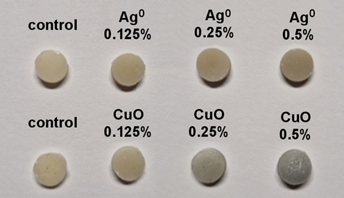
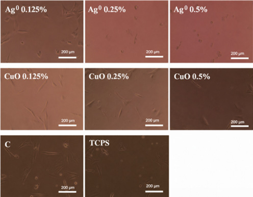
The materials analyzed included an unmodified compomer (Dyract® flow A2; Dentsply Sirona, Charlotte, USA) and its modifications containing CuO and Ag0 in the form of discs with a diameter of 12.7 mm and a thickness of 3.8 mm (Figure 1).
Description of experimental groups
In both the microbiological and cytotoxicity studies, the compomer material (Dyract® flow A2) and its modifications containing Ag0 and CuO particles were tested. Each modification contained 0.125 wt%, 0.25 wt% or 0.5 wt% of the respective particles.
In the microbiological study, both the dynamic method using the CDC biofilm reactor and the static method were applied to each of the 7 experimental groups. Their effect on the formation of a multispecies biofilm consisting of S. mutans, L. rhamnosus and C. albicans was examined, whereas the effect on the formation of a monospecies biofilm of S. epidermidis was assessed separately. In total, 7 groups of materials were tested against 2 different biofilm models using both the static and dynamic methods. Each group was tested in 6 replicates, resulting in a total of 294 samples, corresponding to 42 samples for each tested material.
For the cytotoxicity study using the L929 cell line, extracts from each of the 7 material groups were tested. Each group was analyzed in 4 replicates. For the BALB/3T3 cell line, 2 samples from each of the 7 groups were tested.
Preparation of microbiological experiments
The study was conducted using reference strains obtained from the American Type Culture Collection (ATCC): C. albicans (ATCC 90028); S. mutans (ATCC 25175); L. rhamnosus (ATCC 9595); and S. epidermidis (ATCC 35984).
The reference strains were stored at −80°C in tryptic soy broth (TSB) supplemented with 15 wt% glycerol. Each strain was stored in several replicates. Before each experimental cycle, the strains were cultured under conditions appropriate for each microorganism:
• S. mutans was cultured on Brain Heart Infusion (BHI) broth (BioMaxima S.A., Lublin, Poland) at 37°C for 48 h, under conditions of elevated CO2 levels;
• L. rhamnosus was cultured on De Man–Rogosa–Sharpe (MRS) agar (BioMaxima S.A.) at 37°C for 48 h under anaerobic conditions;
• S. epidermidis was cultured on Tryptic Soy Agar (TSA) (BioMaxima S.A.) at 37°C for 48 h under aerobic conditions;
• C. albicans was cultured on Sabouraud Dextrose Agar (BioMaxima S.A.) at 37°C for 48 h under aerobic conditions.
Methodology of microbiological studies
Static method
A suspension with a density of 0.5 McFarland was prepared from fresh cultures of the analyzed strains in liquid BHI broth supplemented with 5% sucrose (BioMaxima S.A.) for C. albicans, S. mutans and L. rhamnosus. Subsequently, a mixed microbial suspension, consisting of 200 µL of S. mutans, 100 µL of L. rhamnosus, 200 µL of C. albicans, and 1,500 µL of BHI broth supplemented with sucrose, was prepared, resulting in a final volume of 2 mL. After the specimens had been placed in the microbial suspension, the entire mixture was incubated at 37°C under microaerophilic conditions for 24 h.
For S. epidermidis, 200 µL of a 0.5 McFarland suspension prepared in BHI broth supplemented with 5% sucrose was mixed with 1,800 µL of BHI broth containing sucrose and incubated with the specimens under aerobic conditions at 37°C for 24 h.22, 23 After the incubation period, the specimens were rinsed 3 times with 0.9% NaCl and shaken for 1 min in a 0.5% saponin solution (Sigma-Aldrich). The resulting suspension (100 µL) was diluted in geometric progression and seeded on solid BHI agar (3-species biofilm). For S. epidermidis, the diluted suspension was plated on TSA.
Dynamic method
The experiments were performed according to a standard protocol.24 The coupons (test samples) were placed in the reactor. A 1-mL inoculum containing the 3 microorganisms (S. mutans, C. albicans and L. rhamnosus) or S. epidermidis alone was introduced into a reactor containing 340 mL of 40% BHI broth (14.8 g/L) supplemented with 5% sucrose. The reactor was maintained at 37°C during a 4-h stationary phase under continuous stirring at 120 rpm. Subsequently, 20% BHI broth supplemented with sucrose was supplied to the reactor for 24 h using a peristaltic pump (HV-77913-70 Masterflex®; Cole-Parmer Company, Montreal, Canada) at a constant flow rate of 11.67 mL/min. After the incubation period, the specimens were rinsed 3 times with 0.9% NaCl and shaken for 1 min in a 0.5% saponin solution (Sigma-Aldrich). The subsequent procedure was identical to that used for the static biofilm model. The experiment was performed in 6 independent replicates.
Following incubation at 37°C using both methods (24 h; elevated CO2 conditions for the multispecies biofilm and aerobic conditions for the monospecies biofilm), the colonies were counted, and the colony forming units were calculated (CFU/mL) and expressed per coupon surface area. The experiments were performed in 6 independent replicates.22, 23, 24
Calculation of colony-forming units
Colony-forming units were calculated using the following formula:
CFU/mL = average number of colonies × reciprocal of dilution × dilution factor
To determine the number of bacteria per unit surface area, the following equation was used:
where:
X – average number of CFU;
B – plated volume (0.02 mL);
V – suspension volume after shaking (1 mL);
A – sample surface area (4.1 cm2);
D – dilution factor (50; i.e., 1,000/20).
Imaging of samples using scanning electron microscopy
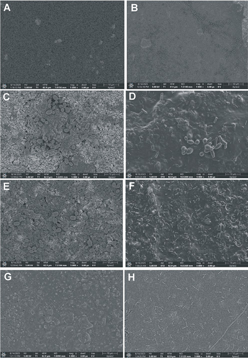
Scanning electron microscopy (Nova NanoSEM 200; FEI Company, Hillsboro, USA) was used to evaluate the structure of the biofilm formed under flow conditions on the coupons. After the incubation period, the samples were rinsed with NaCl, immersed in a 2.5 wt% glutaraldehyde solution (Chempur), and stored for 2 h at 4°C. The specimens were then dehydrated using ethyl alcohol solutions (25 wt%, 50 wt%, 70 wt%, 90 wt%, 95 wt%). Dehydration was performed for 10 min at each concentration at room temperature. Subsequently, the samples were sputter-coated and examined using SEM. Observations were performed using Everhart–Thornley detector (ETD) and in-lens (T1) detectors under high-vacuum conditions at an accelerating voltage of 5 kV. Representative SEM images are shown in Figure 2.
Methodology of cytotoxicity studies
The BALB/3T3 cells were cultured in Dulbecco’s Modified Eagle Medium (DMEM; Gibco, Waltham, USA) supplemented with 10 wt% fetal bovine serum (FBS; Sigma-Aldrich) and 1% antibiotic–antimycotic solution. The cells were maintained at 37°C in a humidified atmosphere containing 5% CO2 and subcultured at 70–80% confluence using trypsin–ethylenediaminetetraacetic acid (EDTA). The cells were harvested, counted and seeded into 6-well plates at a density of 1.5 × 10⁵ cells per well. They were allowed to adhere and stabilize for 24 h under standard culture conditions. The test samples consisted of disks containing Ag0 and CuO at concentrations of 0.125 wt%, 0.25 wt% and 0.5 wt%. After 24 h, sterile disks were gently placed into each well to ensure direct contact with the cell layer. The cells were then incubated with the materials for an additional 24 h under previously described conditions.
L929 fibroblasts (NCTC clone 929; ATCC, Manassas, USA) were also used. The cells were seeded into 96-well plates at a density of 104 cells/well in 100 µL of DMEM (Gibco) supplemented with 10% FBS (Sigma-Aldrich) and 1% penicillin/streptomycin (P/S; Sigma-Aldrich). After 24 h, the medium was removed and replaced with 100 µL of the test extract.
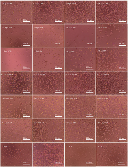
The extracts were prepared according to the ISO 10993 standard. The samples were immersed in cell culture medium at a ratio of 0.1 g/10 mL and incubated at 37°C for 24 h. A high-density polyethylene extract (HDPE) served as the negative control. A sodium lauryl sulfate (SLS) solution in DMEM (0.2 mg/mL), prepared at dilutions of 1:1, 1:2 and 1:4, was used as the positive control. The cells were incubated with the extracts for 24 h, after which photographs were taken.
To assess fibroblast viability, a colorimetric MTT assay was performed in accordance with EN ISO 10993-5:2009, the standard for the biological evaluation of medical devices. The MTT solution was prepared by dissolving 50 mg of MTT salt (3[4,5-dimethyl-2-yl]-2,5-diphenyltetrazolium) in 50 mL of phosphate-buffered saline (PBS) to obtain a concentration of 1 mg/mL. The solution was then sterilized using a syringe filter. After removal of the culture medium, 100 µL of the MTT solution was added to each well. The cells were incubated for 2 h at 37°C in a 5% CO2 environment. The MTT solution was then removed and replaced with 100 µL of isopropanol per well. The plate was shaken for 30 min to ensure complete dissolution of purple formazan crystals. Aliquots of 100 µL were transferred to a 96-well plate, and the absorbance was measured at 570 nm using a spectrophotometer (BioTek Epoch 2; Agilent Technologies, Santa Clara, USA). The absorbance values served as a direct indicator of metabolic activity, allowing for a comparative analysis of cell survival rates across the tested samples.
Statistical analysis
Statistical analyses were performed using the R software (v. 4.2; R Foundation for Statistical Computing, Vienna, Austria). For each experimental group, the normality of the analysis of variance (ANOVA) residuals was assessed using the Shapiro–Wilk test, and homogeneity of variance was evaluated using Levene’s test. When both assumptions were satisfied, a one-way ANOVA followed by Tukey’s honestly significant difference (HSD) post hoc test was performed. Where the normality assumption was violated, the Kruskal–Wallis test followed by Dunn’s test with Bonferroni correction was used instead. The same procedure was applied to the cytotoxicity data (L929, MTT assay), for which the Kruskal–Wallis test followed by Dunn–Bonferroni post hoc analysis was selected because the normality assumption was not met. All pairwise comparisons were performed against the unmodified compomer control. Statistical significance was set at α = 0.05.
Results
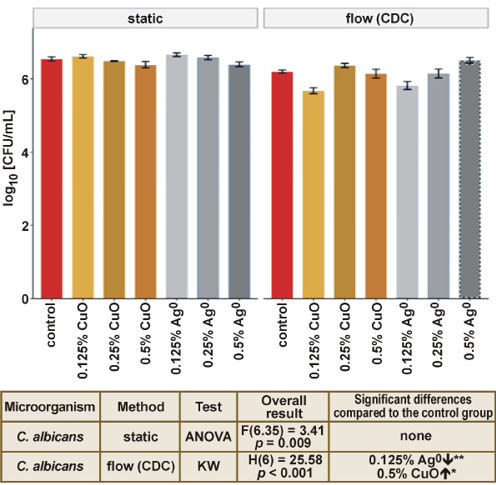
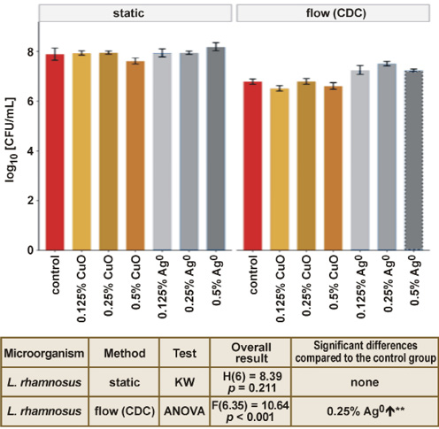
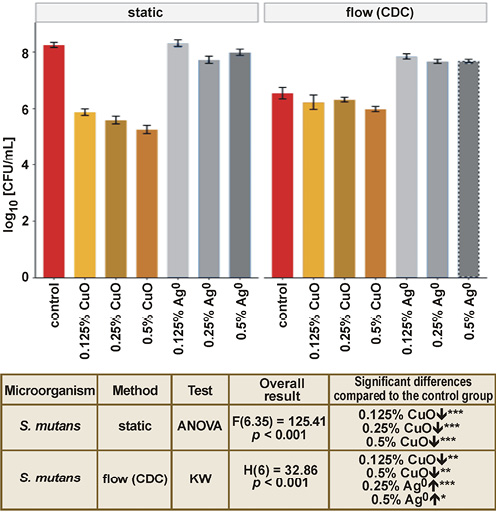
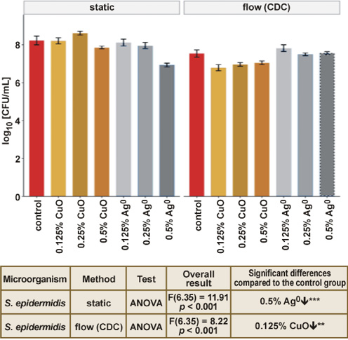
Figure 3, Figure 4, Figure 5, Figure 6 present the results of the static and dynamic microbiological tests.
As shown in Fig. 3, Tukey’s HSD post hoc test did not reveal any significant differences between the individual experimental groups and the control group for C. albicans under static conditions. Neither CuO nor Ag0 at any of the tested concentrations significantly affected the number of C. albicans compared with the unmodified compomer. Under flow conditions, the data was not normally distributed, and Dunn’s post hoc test identified 2 significant comparisons relative to the control group. The 0.125 wt% Ag0 group showed significantly lower values than the control, and the 0.5 wt% CuO group demonstrated slightly higher values than the control group.
The results for L. rhamnosus under static conditions were not statistically significant for any concentration of CuO and Ag0. Under flow conditions, Tukey’s HSD post hoc test identified a significant difference in the 0.25 wt% Ag0 group, with values that were higher than those observed in the control group (Figure 4).
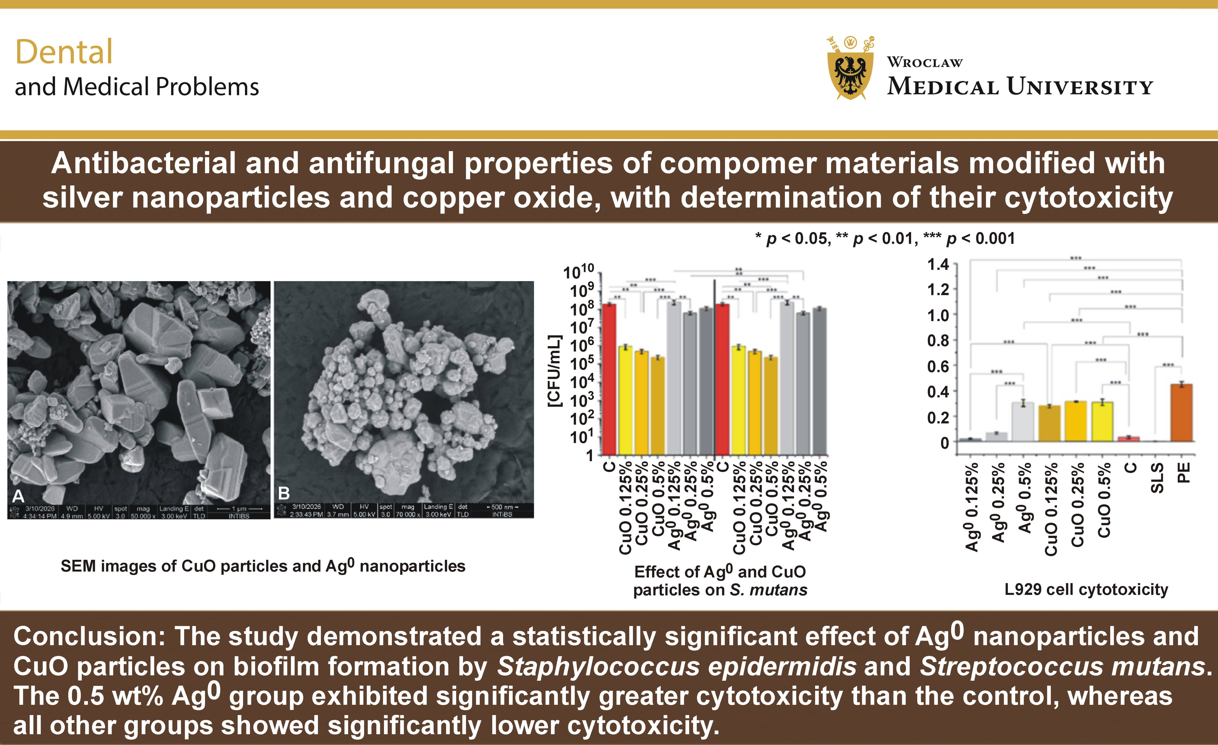
The findings for S. mutans under static conditions revealed that all 3 CuO-modified groups had significantly lower values than the control group (Tukey’s HSD post hoc test). None of the Ag0-modified groups differed significantly from the control group. Under flow conditions, Dunn’s post hoc test with Bonferroni correction showed that the 0.125 wt% CuO and 0.5 wt% CuO groups were significantly lower than the control group, whereas the 0.25 wt% Ag0 and 0.5 wt% Ag0 groups were significantly higher than the control group.
Figure 6 presents the results for S. epidermidis. Tukey’s HSD post hoc analysis showed that only the 0.5 wt% Ag0 group had significantly lower values than the control group. None of the CuO-modified groups differed significantly from the control group. Under flow conditions, Tukey’s HSD post hoc test showed that only the 0.125 wt% CuO group differed significantly from the control group.
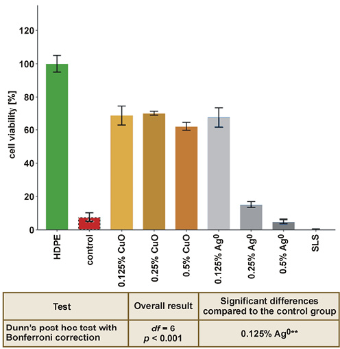
Figure 7 and Figure 8 present the results of the L929 cytotoxicity assay. Dunn’s post hoc test with Bonferroni correction identified a statistically significant differences relative to the unmodified compomer control. The 0.125 wt% Ag0 group showed significantly higher cell viability, which reflected the very low baseline viability of the unmodified compomer rather than the cytotoxic effect of the additive itself. All CuO-modified groups exhibited significantly higher cell viability than that of the control. In contrast, the 0.25 wt% Ag0 and 0.5 wt% Ag0 groups showed significantly reduced cell viability, comparable to that observed for the SLS positive control. In the Ag0 groups, a marked dose-dependent decrease in cell viability was observed, whereas CuO exhibited moderate, concentration-independent cytotoxicity across the entire tested concentration range. Figure 9 presents the results of the cytotoxicity assay performed using the BALB/3T3 cells. Similar to the L929 assay, the highest cytotoxicity was observed for samples containing 0.25 wt% Ag0 and 0.5 wt% Ag0.
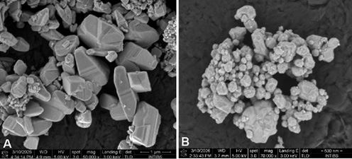
Figure 10 presents representative SEM images of the synthesized particles. Figure 10A shows the copper(II) oxide sample, which consists of distinctly angular, faceted, polyhedral, and plate-like structures with flat surfaces and sharp edges. The sample is composed of large, block-like crystals measuring from 1 µm to over 2 µm along their longest axis. These larger structures are accompanied by numerous smaller, irregularly shaped particles distributed around and directly on the surfaces of the larger crystals. Figure 10B presents the Ag0 nanoparticles that exhibit predominantly spherical and quasi-spherical morphologies. The primary particles are grouped into dense, three-dimensional clusters and larger aggregates. The individual particles visible within these structures vary considerably in size, ranging from less than 100 nm to approx. 300 nm in diameter.
Discussion
Many different types of restorative materials are used in dentistry, and newer, more advanced materials, including those modified with various particles, are constantly being developed. These materials are enhanced with additives that provide antibacterial and antifungal properties, thereby reducing the risk of biofilm formation and secondary caries.
Nanoparticle additives are commonly used in dental materials. However, their effectiveness depends on their concentration in the material. In general, higher concentrations produce stronger antibacterial and antifungal effects but are also associated with increased cytotoxicity. Owing to their small size, nanoparticles can easily penetrate human cells. Yang et al. pointed out the potential impact of nanoparticles on the central nervous system and emphasized the need for further research.25
There are relatively few studies on modifications of compomer materials. However, numerous studies have investigated the incorporation of nanoparticles into other dental materials, such as composites and glass ionomers. Guo et al. demonstrated, among other findings, that modifications with both Ag nanowires and Ag nanoparticles reduced bacterial biofilm formation, including that of L. rhamnosus and S. mutans.26 Cytotoxicity testing in human gingival fibroblasts revealed no statistically significant differences between glass ionomer cements modified with 0.05%, 0.1%, 0.3%, or 0.5% Ag nanowires and those modified with 0.5% Ag nanoparticles.26 In contrast, studies investigating the effects of Ag nanoparticles on human periodontal fibroblasts have shown that particles smaller than 20 nm exert significant negative effects on the cells. It should be noted that these studies evaluated relatively low concentrations ranging from 0 μM to 1,000 μM, whereas most studies have investigated concentrations between 0.1 wt% and 1 wt%.27 In a comprehensive study evaluating the toxicity and antibacterial properties of Ag nanoparticles, Niska et al. demonstrated, using human gingival fibroblasts (HGF-1), that cytotoxicity was not statistically significant at concentrations up to 20 µg/mL (0.002 wt%). Within this concentration range, Ag nanoparticles were also effective against S. epidermidis and S. mutans. It should be noted that the tested Ag nanoparticles consisted of 10-nm particles suspended in solution, and only monospecies, 1-day-old biofilms were evaluated. The authors also reported that the cytotoxicity of Ag nanoparticles could be reduced using capping agents, such as polyethylene glycol, lipoic acid and tannic acid, while maintaining antimicrobial properties. However, uncapped Ag nanoparticles exhibited a broader antimicrobial spectrum.28 Hernández-Sierra et al. demonstrated statistically significant cytotoxicity in human periodontal fibroblast monolayers at a concentration of 25 mg/mL (0.0025 wt%).27 Similar conclusions regarding cytotoxicity in L929 cells were reported by Takamiya et al.29 In a study by Eslami et al., the cytotoxic effects of various colloidal solutions, including Ag nanoparticles (0.4 wt%) and CuO nanoparticles (0.1 wt%), were compared.30 A higher percentage of apoptosis was observed for CuO nanoparticles. It is worth noting that the level of apoptosis was only slightly lower than that observed for chlorhexidine, which served as the positive control.30 Shaban et al. incorporated Ag0 and CuO nanoparticles into an acrylic resin denture base and investigated their effects on C. albicans.31 Complete eradication was achieved at concentrations as low as 0.3 wt% (Ag0) and 0.5 wt% (CuO).31 Another study investigated the effects of Ag and CuO incorporated at concentrations of 0.5 wt% and 1 wt% into orthodontic composites against S. mutans.32 Biofilm formation was assessed after 3, 15 and 30 days. Statistically significant differences were observed after 3 and 15 days, but not after 30 days. It should be noted that the nanoparticle concentrations used in that study were higher than those evaluated in most previous reports.32 Prior publications have also confirmed the effectiveness of CuO nanoparticles against S. mutans and C. albicans in soft denture liners and suspensions.33, 34 Copper oxide nanoparticles derived from Nilgirianthus ciliatus leaf extract demonstrated low cytotoxicity toward L929 cells.35 The cytotoxicity of Ag and CuO nanoparticles depends on both particle concentration and size. In general, smaller particles exhibit greater antibacterial activity, which is also associated with increased cytotoxicity. In the present study, Ag0 nanoparticles ranged in size from 100 nm to 300 nm, whereas CuO particles measured approx. 1,000–2,000 nm. The relatively large size of the CuO particles may explain their lower cytotoxicity.36, 37, 38, 39 In the L929 assay, the CuO-modified materials exhibited significantly lower cytotoxicity than the Ag0-modified materials, which may be related to the particle size.36, 37, 38, 39 In a study using the MDPC-23 cell line, the addition of 0.1 wt% and 0.2 wt% Ag0 nanoparticles had no statistically significant effect on the cytotoxicity of glass ionomer cements. It should be noted, however, that Vitrebond exhibited cytotoxicity even in the absence of nanoparticle additives. Similarly, in a study investigating solutions containing Ag (4,000 ppm) and CuO nanoparticles (1,000 ppm), Ag nanoparticles showed lower cytotoxicity toward human gingival fibroblasts.30, 40 Karkehabadi et al. also demonstrated, using stem cells from the apical papilla (SCAPs), that CuO nanoparticle concentrations exceeding 10 μg/mL exhibit cytotoxic properties.41
The literature indicates that approx. 500 bacterial species inhabit the oral cavity. In the present study, 1 multispecies biofilm and 1 monospecies S. epidermidis biofilm, representing a species with strong adhesive properties, were investigated. Compared with studies evaluating monospecies biofilms or planktonic microorganisms, the antimicrobial effect observed in the present study was considerably weaker, particularly for C. albicans.28, 34 Numerous factors influence biofilm formation on dental materials, including ion release, the shape of the filling, and its interaction with dental tissues. Tooth anatomy is highly complex and varies among individuals, as does the quality of restoration placement. Maintaining good oral hygiene remains the foundation for preventing biofilm formation, and, consequently, secondary caries. For the samples used in the study, the authors will present the results of fluoride release in a separate publication. The CDC biofilm reactor enables simulation of oral conditions, and studies using dentin–composite models have confirmed the detrimental effects of biofilm on restorations. Therefore, inhibition of biofilm growth through the incorporation of metal compounds and their ions may increase restoration longevity.42
The results of the present study demonstrated differences between the static and dynamic methods. Copper oxide consistently inhibited S. mutans under static conditions (all concentrations) and under flow conditions (0.125 wt% and 0.5 wt%). It also inhibited S. epidermidis under flow conditions at 0.125 wt%. No inhibitory effect was observed against C. albicans and L. rhamnosus. Silver exhibited a significant inhibitory effect only against S. epidermidis under static conditions. In contrast, stimulatory rather than inhibitory effects were observed for S. mutans and L. rhamnosus. Therefore, neither CuO nor Ag0 demonstrated a consistent or clinically meaningful antifungal effect against L. rhamnosus. Studies using both L929 and BALB/3T3 cells showed lower cytotoxicity than the control group for all modified materials except the sample containing 0.5 wt% Ag0. However, the sample containing 0.25 wt% Ag0 also exhibited high cytotoxicity, indicating a dose-dependent relationship between Ag0 concentration and cytotoxicity. In contrast, the considerably larger CuO particles showed lower cytotoxicity.
Oral biofilm is a complex microbial community comprising numerous interacting species, making its inhibition substantially more challenging. In the present study, the biofilm consisted of 3 species. Although CuO and Ag0 possess antimicrobial properties, most previous studies have evaluated either monospecies biofilms or nanoparticles suspended in a solution rather than particles incorporated into restorative materials, which may substantially influence their antimicrobial efficacy.43, 44 The present study did not demonstrate that CuO and Ag0 particles are equally effective against all tested microorganisms. Once again, the importance of particle size should be emphasized. A study evaluating 10-nm Ag particles against planktonic S. mutans reported a minimum inhibitory concentration (MIC) of 4 ppm, whereas statistically significant cytotoxicity toward human dermal fibroblasts occurred at concentrations above 10 ppm.45 The study also used a CDC biofilm reactor, in which bacterial reduction was observed only at a concentration of 100 ppm. These findings illustrate the significant impact of biofilm on the effectiveness of Ag nanoparticles.45
In the present study, the specimens were prepared for testing using steam sterilization. However, sterilization itself may affect the structure of the material. Farrugia et al. reported the presence of bubbles on the surface following steam sterilization of compomers.46 In addition, the microhardness of the compomer increased after sterilization. Although the specimens in the present study were polished before sterilization, possible structural changes resulting in increased porosity may have influenced biofilm formation.46
Another limitation is illustrated in Figure 2, which depicts the samples tested in the experiment. A visible color change occurred with increasing particle content, particularly in the CuO-modified materials. An advantage of compomer materials is their favorable aesthetics, which were compromised by the addition of 0.25 wt% and 0.5 wt% CuO. Similar esthetic limitations have been reported for other restorative materials modified with antimicrobial additives. Therefore, the selection of an optimal particle concentration should consider not only cytotoxicity and antimicrobial activity but also esthetic properties, which are of considerable importance to patients.47, 48
Further studies evaluating fluoride release and physicochemical properties are needed to determine the optimal concentration and particle size of Ag0 and CuO without compromising the mechanical properties of the compomer. Modified compomer materials should meet the same performance standards as the original material to ensure long-term clinical durability with minimal risk of complications.
Conclusions
Compomer materials are frequently used in young patients who are at an increased risk of complications associated with microleakage and microbial growth. Enhancing their antimicrobial properties through the incorporation of selected particles may reduce biofilm formation and thereby decrease the risk of secondary complications. The present study demonstrated that the incorporation of CuO inhibited the growth of S. mutans at all tested concentrations under static conditions and at 0.125 wt% and 0.5 wt% under flow conditions. Staphylococcus epidermidis was inhibited only by 0.5 wt% Ag0 under static conditions; however, this concentration exhibited unacceptable cytotoxicity. Additionally, the 0.125 wt% CuO group showed significant inhibition under flow conditions.
The clinical applicability of the modified materials also depends on their aesthetic properties, which were adversely affected by the addition of CuO at concentrations of 0.25 wt% and 0.5 wt%. The study demonstrated a dose-dependent increase in cytotoxicity, particularly for Ag0-containing materials. Although both Ag0 and CuO exhibited antimicrobial activity, their overall effectiveness was limited. Therefore, identifying an optimal particle concentration that provides antimicrobial efficacy while maintaining acceptable cytotoxicity remains essential.
Ethics approval and consent to participate
Not applicable.
Data availability
The datasets generated and/or analyzed during the current study are available from the corresponding author on reasonable request.
Consent for publication
Not applicable.
Use of AI and AI-assisted technologies
Not applicable.