



Abstract
Background. Temporomandibular disorders (TMD) cause pain and dysfunction in the temporomandibular joints (TMJs) and masticatory muscles and significantly impact quality of life. Temporomandibular disorder-related pain is associated with sleep disorders and affects heart rate (HR) parameters; however, data from polysomnographic studies remains inconclusive.
Objectives. The aim of the study was to evaluate the impact of TMD on sleep parameters recorded using video-polysomnography (vPSG) and heart rate variability (HRV) monitored with the use of a 24-hour electrocardiogram (ECG).
Material and methods. A cross-sectional study included 93 patients with TMD diagnosed according to the Diagnostic Criteria for Temporomandibular Disorders (DC/TMD). Video-polysomnography and 24-hour ECG monitoring were conducted. Pain was assessed using the numeric rating scale (NRS) (0–10) and categorized as mild (≤5) or severe (>5).
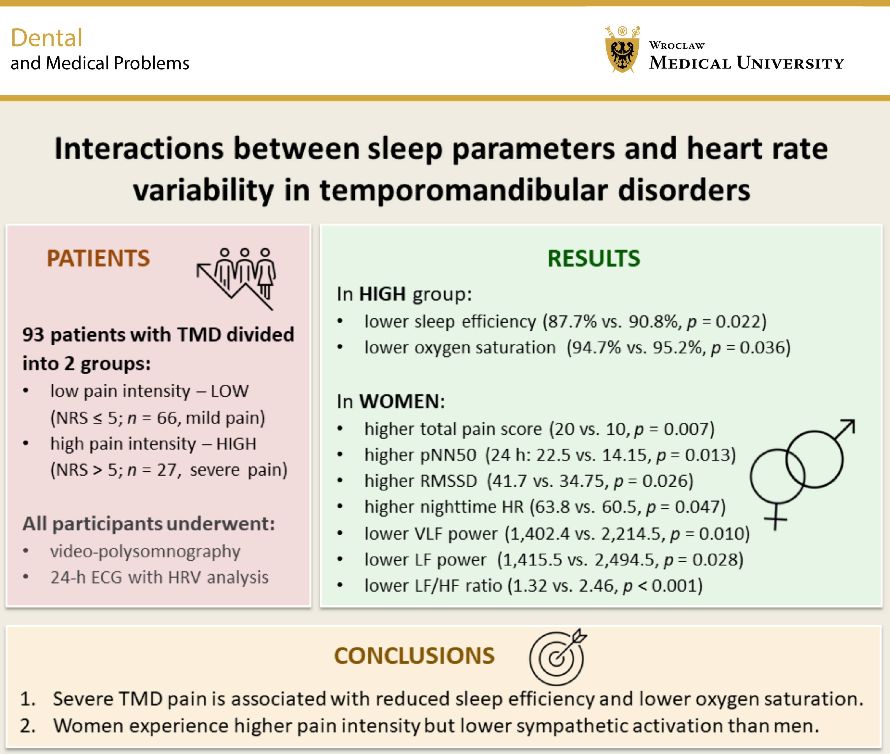
Results. The study included 93 patients with TMD, divided into groups of low pain intensity (n = 66) and high pain intensity (n = 27). The group with high pain intensity demonstrated lower sleep efficiency (87.7% vs. 90.8%, p = 0.022) and lower oxygen saturation (94.7% vs. 95.2%, p = 0.036). Heart rate variability parameters were similar in both groups. Women reported a higher total pain score (20 vs. 10, p = 0.007) and had a higher percentage of normal-to-normal (NN) intervals differing by more than 50 ms (pNN50) (24 h: 22.5% vs. 14.2%, p = 0.013; daytime: 18.0% vs. 9.8%, p = 0.002), higher daytime root mean square of successive differences (RMSSD) (41.7 vs. 34.8, p = 0.026), and a higher nighttime HR (63.8 vs. 60.5, p = 0.047). Additionally, women demonstrated lower values for very low frequency (VLF) power (1,402.4 vs. 2,214.5, p = 0.010), low frequency (LF) power (1,415.5 vs. 2,494.5, p = 0.028), and the low frequency/high frequency (LF/HF) ratio (1.3 vs. 2.5, p < 0.001).
Conclusions. High TMD-related pain intensity is associated with reduced sleep efficiency and lower mean oxygen saturation during sleep. Women experience higher pain intensity but lower sympathetic activation than men.
Keywords: sleep, heart rate variability, temporomandibular disorders
Introduction
Temporomandibular disorders (TMD) encompass a broad category of painful conditions and dysfunctions that affect the masticatory muscles and temporomandibular joints (TMJs). Temporomandibular disorders represent the second most common pain-related condition in the orofacial region, with a complex etiology. Contributing factors include biomechanical, neuromuscular, biological, and biopsychosocial elements. According to the Diagnostic Criteria for Temporomandibular Disorders (DC/TMD), it is estimated that TMD affect approx. 34% of the global population,1 although rates depend strongly on case definitions and population characteristics. The DC/TMD assessment is divided into Axis I (physical) and Axis II (psychosocial). Temporomandibular disorders commonly present as pain-related disorders (muscle or joint pain, headaches) or TMJ-related conditions (disc displacements, degenerative changes).2 Key mechanisms sustaining pain involve sensitization of supraspinal neurons and second-order neurons in the dorsal horn of the spinal cord or the trigeminal nucleus, as well as an impaired balance of analgesic mechanisms.3
Numerous studies indicate a relationship between TMD and obstructive sleep apnea (OSA).4 However, even in TMD patients without OSA, fragmented, non-restorative sleep and frequent nocturnal awakenings are observed. Conversely, chronic sleep disturbances exacerbate pain symptoms, partly by increasing the sympathetic nervous system (SNS) activity and activating inflammatory processes.5, 6 A statistically significant correlation has been found between TMD-related pain and poorer subjective sleep quality, as measured by the Pittsburgh sleep quality index (PSQI).7, 8 Furthermore, TMD-associated headache, along with factors such as older age, female sex and excessive daytime sleepiness, is a predictor of worsening sleep quality.9 Findings from polysomnographic studies vary and often show small differences that correlate more strongly with mood than with pain intensity.10
Heart rate variability (HRV) is an indicator of fluctuations in the time intervals between consecutive heartbeats, primarily resulting from the dynamic regulation of the sinoatrial node by the autonomic nervous system (ANS).11 High HRV reflects effective autonomic regulation and good adaptive capacity of the body. A relationship between HRV, cognitive performance and the functioning of the prefrontal cortex, which is relevant to both physical and mental health, has been demonstrated.12 For HRV analysis, time-domain, frequency-domain and geometric parameters are utilized. Certain parameters are considered markers of the activity of different components of the ANS. Some studies have suggested the association between specific frequency bands and the ANS activity, linking low-frequency (LF) bands to the SNS activity and high-frequency (HF) bands and the root mean square of successive differences (RMSSD) to the parasympathetic nervous system (PNS) activity. The LF/HF ratio has been treated as an indicator of balance between these systems.13 However, the recent data has shown that these parameters are not direct or quantitative measures of sympathovagal balance, as they can be influenced by other factors.14 Higher LF power and a higher LF/HF ratio have been shown to correlate with an increased risk of cardiovasular disease.15 Lower HRV, a higher heart rate (HR) and impaired baroreceptor sensitivity are associated with the presence of painful forms of TMD16 and may indicate sympathetic dominance and impaired parasympathetic regulation.17, 18
Given the high prevalence of TMD and its association with subjectively perceived poorer sleep quality, it seems justified to investigate the impact of these conditions on sleep quality and HRV parameters using objective recording methods. Therefore, our study aimed to analyze sleep architecture and quality, as well as HRV, in patients with TMD using 24-hour electrocardiographic (ECG) monitoring with the assessment of HRV and video-polysomnography (vPSG).
Material and methods
Study design
This single-center, cross-sectional observational study was conducted at the Department of Experimental Dentistry, Wroclaw Medical University, Poland. The assessment of the TMJs and masticatory muscles was performed in accordance with the Axis I of the DC/TMD.2 Individuals presenting to the Outpatient Clinic for Temporomandibular Disorders with complaints of masticatory muscle pain and/or dysfunction, TMJ pain and/or dysfunction, or TMD-related headache between 2017 and 2024 were included in the study. Patients were recruited consecutively throughout the study period, and all individuals meeting the eligibility criteria were invited to participate. The examination was conducted by a dentist with at least 5 years of experience in diagnosing and treating TMD. Patients with a confirmed TMD diagnosis were referred to the Sleep Laboratory of the Clinical Department of Diabetology, Hypertension and Internal Diseases (Wroclaw Medical University), where vPSG and 24-hour ECG monitoring with HRV assessment were performed. The study was conducted in accordance with the principles of the Declaration of Helsinki, and approval was obtained from the Ethics Committee of Wroclaw Medical University (approval No. KB-794/2019). All participants provided written informed consent to participate in the study. Information on clinical trial registration is available in the ClinicalTrials.gov database (identifier No. NCT04214561).
Inclusion and exclusion criteria
The study included 93 patients aged 18 years and older with a confirmed diagnosis of TMD in accordance with the DC/TMD. Exclusion criteria encompassed genetic disorders, inability to undergo polysomnography, neurological disorders or neuropathic pain (including primary headaches), coexisting respiratory failure or active inflammatory processes, active malignant neoplasms, severe psychiatric disorders, significant intellectual disability, pregnancy, the postpartum period, the use of or addiction to analgesics and/or medications affecting the nervous system, muscles or respiration, as well as lack of consent to participate in the study.
Assessment of masticatory muscle pain intensity
The intensity of pain in the temporomandibular region was assessed separately for the right and left temporalis muscles and the right and left masseter muscles. During palpation with a force of 1 kg for 2 s at 9 points on each muscle, participants rated their pain using the numeric rating scale (NRS), which ranged from 0 to 10, where 0 indicated no pain and 10 indicated unbearable pain or an equivalent description. Pain intensity was categorized into 2 groups: mild (NRS ≤ 5); and severe (NRS > 5).19 The average NRS value for each muscle was calculated based on all examined points. The total pain score represented the sum of the mean pain ratings across 4 muscles.
Polysomnographic assessment
Video-polysomnography was conducted using the Nox A1 device (Nox Medical, Reykjavík, Iceland) from 10 pm to 6 am the following day. The examination included electroencephalography, ECG, electrooculography, and electromyography (EMG) of the submental muscles and bilateral masseter muscles, as well as recordings of thoracic and abdominal respiratory movements, assessment of position, and audio and video recordings. Oxygen saturation and pulse rate were measured using the NONIN WristOx2 3150 pulse oximeter (Nonin Medical, Inc., Plymouth, USA). All vPSG recordings were analyzed in 30-second epochs by a single qualified physician, blinded to pain group allocation, in accordance with the American Academy of Sleep Medicine (AASM) guidelines for the evaluation of sleep and associated events.20
24-hour ECG with HRV assessment
Each patient included in the study underwent 24-hour ECG monitoring using a Lifecard CF 12-channel recorder (serial No. LIFE-045348/2015), performed concurrently with vPSG at night and during daytime activities. Patients were instructed to refrain from intense physical activity, caffeine consumption for ≥8 h before bedtime, and alcohol for 24 h prior to monitoring. A standardized activity log was maintained; patients adhered to a structured activity schedule (daytime: 6 am–11 pm; nighttime: 11 pm–6 am). The analysis of HRV was conducted using the Sentinel and Pathfinder SL systems (v. 1.9.4.14474; Spacelabs Healthcare, Snoqualmie, USA). Prior to HRV analysis, ECG recordings were manually verified to remove artifacts and ectopic beats. Subsequently, time-domain, frequency-domain and geometric parameters were calculated. Frequency-domain analysis based on the fast Fourier transform (FFT) included the very low frequency (VLF) (0.003–0.04 Hz), LF (0.04–0.15 Hz), HF (0.15–0.4 Hz), and very high frequency (VHF) (0.4–0.5 Hz) bands, as well as the LF/HF ratio expressed in normalized units. Short-term HRV was assessed in two 20-minute segments (one during daytime and one during N3 sleep stage) to minimize the impact of respiratory events.
The 20-minute N3 segment was selected by the physician analyzing the polysomnograms. The inclusion criterion was stable, good-quality sleep. Exclusion criteria included the presence of arousals, awakenings, periodic limb movements, and sleep bruxism episodes. The physician had no access to the Holter ECG monitoring results.
Respiratory events were detected using a nasal pressure cannula. Apneic and hypopneic events were analyzed as follows: apneas were defined as a 90% reduction in airflow lasting ≥10 s; and hypopneas were identified as a ≥30% reduction in airflow lasting ≥10 s accompanied by a ≥3% decrease in blood oxygen saturation, as measured by pulse oximetry, or by an arousal. Saturation level (SpO2%) and pulse rate were recorded using a pulse oximeter (Nonin Medical Inc.).
The artifact threshold was set according to the recommendation of the AASM and implemented in the Nox A1 device. Additionally, a physician certified in polysomnography by the Polish Sleep Research Society manually assessed the recordings for respiratory events and artifacts.
The analysis was performed by an experienced clinician in accordance with the guidelines of the European Society of Cardiology (ESC) and the Heart Rhythm Society (HRS) (formerly the North American Society of Pacing and Electrophysiology (NASPE)).21
Statistical analysis
Patients were divided into 2 groups based on TMD pain intensity: low TMD pain intensity (LOW) and high TMD pain intensity (HIGH), to investigate the relationship of TMD pain with HRV parameters and vPSG-derived sleep parameters. Additionally, differences in HRV and TMD pain parameters were analyzed according to sex, with separate subgroups for women and men. Analyses were performed using Statistica 13 (Dell Inc., Round Rock, USA). Categorical variables were presented as counts and percentages, and continuous variables as the mean (M) and standard deviation (SD) or the median (Me) and interquartile range (IQR). Normality and variance assumptions were evaluated before conducting between-group comparisons. Continuous variables were inspected visually using histograms and formally tested with the Shapiro–Wilk test. Homogeneity of variances was assessed using the Levene’s test. The χ2 test and Student’s t-test were used for normally distributed data, and the Mann–Whitney U test was applied for non-normally distributed variables. A p-value <0.05 was considered statistically significant.
Results
Demographic data
The study population was divided into the LOW (n = 66) and HIGH (n = 27) groups. The mean age of the participants was 33.5 ±9.5 years in the LOW group and 36.4 ±9.0 years in the HIGH group (p > 0.05). Women accounted for 61.3% (n = 57) of the entire study population, with 57.6% (n = 38) in the LOW group and 70.4% (n = 19) in the HIGH group. The mean body mass index (BMI) was 24.0 ±3.7 kg/m2 in the LOW group and 25.5 ±5.3 kg/m2 in the HIGH group (p > 0.05).
Relationship between TMD pain intensity, HRV and vPSG
No statistically significant differences in HRV parameters were observed between the HIGH and LOW groups. Significantly lower sleep efficiency and lower mean oxygen saturation were observed in the HIGH group than in the LOW group (87.7% vs. 90.8%, p = 0.022; and 94.7% vs. 95.2%, p = 0.036, respectively) (Table 1).
Sex-based differences
In the female group, greater pain intensity was observed in individual muscles and in the total score across the 4 muscle sites compared with the male group. Regarding HRV parameters, women exhibited higher pNN50 values over the entire 24-hour period and during daytime activity, higher daytime RMSSD values, and a higher mean HR at night.
Frequency domain analysis revealed significant differences between women and men. Over the 24-hour period, women showed significantly lower VLF and LF power, as well as a lower LF/HF ratio. A lower LF/HF ratio in female participants was also observed in both the daytime and nighttime analyses, whereas lower VLF power was observed only at night (Table 2).
Discussion
Pain associated with TMD is often linked to sleep disturbances. A significant proportion of patients with chronic pain conditions report poor sleep quality in studies using various scales and questionnaires.22 The PSQI appears to be one of the most effective and commonly used tools, enabling the assessment of 4 sleep characteristics: sleep onset; sleep maintenance; sleep adequacy; and daytime sleepiness. Higher PSQI scores in patients with TMD-related pain indicate poorer sleep quality,23 particularly in the mixed (myofascial and articular) and myalgia groups compared with the arthralgia group.24 However, polysomnographic studies show only minor differences related to pain intensity, which appear to be more strongly associated with depressive symptoms than with pain itself, suggesting that subjective sleep complaints in TMD are not fully reflected in objective sleep measures.10
Therefore, considering these discrepancies, we decided to focus on objective sleep physiology assessed using vPSG. In our study, among multiple sleep quality and sleep architecture parameters, slightly lower sleep efficiency and reduced mean oxygen saturation were observed in patients with higher pain intensity. Sleep efficiency, defined as the ratio of total sleep time to total recording time, represents the percentage of time spent asleep while in bed; values below 85% may indicate sleep-disrupting factors such as TMD-related pain.20 These findings are consistent with those of Edwards et al., who demonstrated that lower sleep efficiency and shorter total sleep time are risk factors for reduced pain inhibitory modulation in TMD, which may contribute to the persistence of chronic symptoms.25 Dysfunction of endogenous opioid systems has been proposed as a mechanism underlying impaired pain inhibition.26
The relationship between lower oxygen saturation and pain intensity in TMD is complex and may relate to hypoxemia, which has been shown to increase sympathetic activity and inflammation. This aligns with studies linking OSA and recurrent hypoxic episodes to greater TMD pain.27
The significant differences in sleep efficiency and mean saturation level between patients in our study are small in magnitude, and their clinical relevance remains to be established. Such alterations are not sufficient to justify incorporating routine screening for sleep-related disorders in TMD, but they may support greater attention to sleep quality in patients with TMD and more severe pain, as a supplement to existing standards of care.28 This may include inquiring about sleep complaints during routine TMD evaluations or considering simple behavioral interventions such as sleep hygiene support, optimization of analgesic timing, the use of low-burden home pulse oximetry, and, when indicated, referral for formal sleep assessment.
The imbalance of the ANS in patients with TMD is associated with reduced HRV,29 which is accompanied by elevated salivary cortisol levels and greater psychological distress.30 Very low frequency power reflects long-term regulatory mechanisms, including ANS imbalances in response to stress, which may be associated with chronic pain.31 In our study, HRV parameters were similar in both studied groups, suggesting no significant association between these parameters and TMD intensity.
Studies indicate that women with TMD experience 1.5–2.0 times greater pain intensity than men and account for 80% of patients treated for TMD.32 The OPPERA study demonstrated that women have lower pain thresholds and greater central sensitization compared with men.33 This may result from multiple factors. Greater pain sensitivity is associated, for example, with higher estrogen levels,34 but also with psychological factors such as anxiety and depression, as demonstrated by Manfredini et al.35 In our study, we observed significantly greater pain intensity in women than in men.
A novel aspect of our study was the analysis of HRV in the context of sex differences. We demonstrated greater parasympathetic activity in women, which aligns with the findings of other authors.34 In the time domain, women showed higher pNN50 values over 24 h and during daytime activity, higher RMSSD values during the day, and a higher mean HR at night. Regarding frequency domain analysis, women exhibited significantly lower VLF and LF power and a lower LF/HF ratio over the 24-hour period, a lower LF/HF ratio both during the day and at night, and lower VLF power at night. These results may support the hypothesis that higher vagal activity in women could be related to the effects of acetylcholine on diastolic depolarization of cardiac pacemaker cells, producing lower HR oscillations in the presence of shorter RR intervals.36
In our cohort, the absence of significant HRV differences between higher- and lower-pain intensity groups, alongside the observed sex-related differences, suggests that pain intensity may not be a strong marker of autonomic alterations, while the influence of sex may be detectable even within a relatively small population. From a clinical perspective, these findings support a comprehensive approach that considers multiple contributing factors. The observed differences between men and women may contribute to individual variability in fatigue or treatment tolerance and could potentially influence sex-dependent rehabilitation strategies. However, given the exploratory nature of the present study, these implications should be viewed as preliminary and should not yet inform changes in routine counseling or rehabilitation without confirmation in larger, prospective studies.
Alternative explanations must also be considered. Differences in measurement conditions, including respiratory patterns, sampling duration and signal quality, may partly account for the observed HRV variations. It should be noted that HRV is also influenced by factors such as age, smoking and leisure-time physical activity, which are not always controllable and may reduce the reliability of the results.37 It has been shown that aging may attenuate sex differences.34 Similarly, the phase of the menstrual cycle can influence HRV parameters: vagal activity decreases from the follicular to the luteal phase, with estrogens exerting a beneficial effect that is attenuated under combined estrogen–progesterone therapy.38 Therefore, further studies should be conducted across different age groups, taking into account the phase of the menstrual cycle in women and the use of hormone therapy.
It is worth noting that in TMD, the predominant pattern is characterized by low RMSSD, SDNN and HF values, along with an elevated LF/HF ratio.29 Our findings suggest that, despite reporting higher pain intensity, women with TMD exhibit a weaker physiological response to pain, manifested as increased activity of the PNS.
The present study has several methodological strengths that enhance the reliability and validity of the findings. The use of 2 objective diagnostic methods, namely vPSG and Holter ECG monitoring, ensured precise data collection and analysis. The analysis of HRV with the use of a non-invasive, reliable and widely accepted tool, enabled the assessment of the ANS function in the context of TMD pathophysiology. Additionally, the study was conducted on a relatively large sample size, which increased its statistical power and the generalizability of the results. All procedures, including vPSG, clinical assessments and diagnostic criteria, were carried out in accordance with standardized and validated protocols, ensuring consistency and methodological rigor.
Limitations
Nevertheless, the study has certain limitations. Polysomnography was performed without an adaptation night, which may have induced a first-night effect, potentially influencing the accuracy of measurements and sleep architecture. Lifestyle-related factors that may affect the ANS activity and HRV (e.g., stress, caffeine intake, noise) were not fully controlled. It should be noted that HRV parameters do not solely reflect the ANS function but are also influenced by other mechanisms such as baroreceptor sensitivity, the renin–angiotensin system and respiratory rate. Furthermore, patients with hypertension and those taking medications that could affect HRV were not excluded from the study. This study was exploratory in nature and included a broad set of HRV and vPSG metrics without adjustment for potential confounders, and no multivariable analysis was performed. An a priori power calculation was not conducted, as the study was designed without prespecified primary endpoints. The cross-sectional study design is an additional limitation because it only captures data at a single point in time. Consequently, temporal relationships and causality between variables could not be established, and the observed associations may have been influenced by unmeasured confounding factors. In addition, formal inter-rater reliability of pain assessment was not re-evaluated in the cohort, and detailed clinical phenotyping, such as pain duration and specific TMD subtypes, was not available, reflecting the exploratory nature of the study.
Conclusions
Based on the results of the present study, it can be concluded that patients with high TMD-related pain intensity exhibit reduced sleep efficiency and lower oxygen saturation, without significant differences in HRV parameters. Women report higher masticatory muscle pain intensity than men. Time- and frequency-domain HRV parameters indicate greater parasympathetic activity in women with TMD compared to men. Additionally, the dissociation between higher pain intensity and the observed pattern of autonomic activation in women suggests possible sex-related differences in pain processing, which warrant further investigation and may have implications for personalized treatment approaches.
Trial registration
Information on clinical trial registration is available at the ClinicalTrials.gov database (identifier No. NCT04214561).
Ethics approval and consent to participate
The study was conducted in accordance with the principles of the Declaration of Helsinki, and approval was obtained from the Ethics Committee of Wroclaw Medical University, Poland (approval No. KB-794/2019). All participants provided written informed consent to participate in the study.
Data availability
The datasets generated and/or analyzed during the current study are available from the corresponding author on reasonable request.
Consent for publication
Not applicable.
Use of AI and AI-assisted technologies
Not applicable.