Abstract
Background. Edentulism and posterior maxillary edentulous spaces lead to functional limitations as well as aesthetic and psychosocial consequences. In recent decades, implant-supported prosthetic rehabilitation has become the gold standard for the treatment of patients with tooth loss.
Objectives. The aim of this retrospective comparative study was to evaluate the clinical effectiveness and incidence of complications associated with different graftless implant techniques used for the rehabilitation of the edentulous maxilla and posterior maxillary edentulous spaces.
Material and methods. The study was conducted based on the medical records of patients who underwent implant treatment performed by the same surgical team at 4 private dental clinics in Poland between 2015 and 2020. A total of 667 dental implants placed in 153 patients of different ages and sexes were included in the analysis. The follow-up period ranged from 1 to 5 years. Three types of dental implants were evaluated. The χ2 test and Fisher’s exact test were used for the statistical analysis.
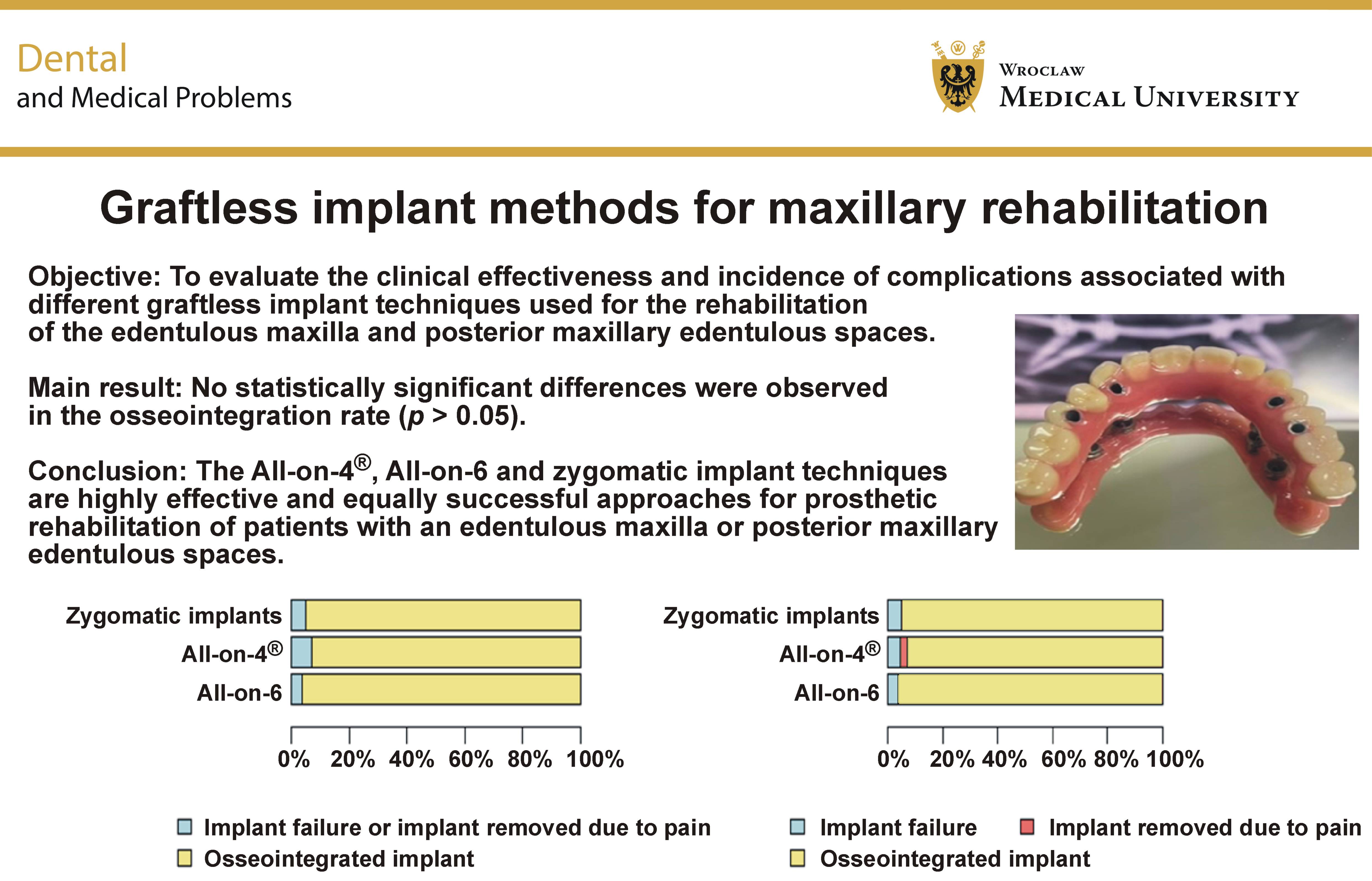
Results. The most frequently selected implantation technique for maxillary rehabilitation was the All-on-6 method, which was associated with a complication rate of 3.70%. The All-on-4® method was the least frequently used and demonstrated a failure rate of 7.05%. A lack of osseointegration was observed in 4.81% of the zygomatic implants. The statistical analysis did not reveal any statistically significant differences in osseointegration rates among the evaluated methods.
Conclusions. The All-on-4®, All-on-6 and zygomatic implant techniques are effective alternatives to conventional approaches for the rehabilitation of the edentulous, atrophic maxilla or posterior maxillary edentulous spaces. All three techniques are associated with comparable complication rates. These methods shorten treatment and rehabilitation time and reduce trauma by minimizing the number of surgical interventions, eliminating the need for guided bone regeneration procedures and interventions into the sinus (sinus lift). Further studies with follow-up periods exceeding 5 years and larger patient cohorts are warranted to confirm these findings.
Keywords: zygomatic implants, All-on-4®, All-on-6, maxillary atrophy
Introduction
Edentulism or posterior maxillary edentulous spaces are serious clinical problems due to both functional limitations and the aesthetic and psychosocial consequences for patients.1 The absence of teeth not only leads to impaired chewing, speech and facial aesthetics, but also results in the gradual atrophy of the alveolar process, further compromising future prosthetic and implant treatment. Alveolar bone resorption results in a reduction in the volume, height and width of the alveolar process beneath the gingiva following tooth loss and prolonged edentulism.2, 3 This phenomenon is considered one of the most common dental problems, occurring in nearly 90% of patients. Bone loss can result from physiological, pathological or iatrogenic processes. The mechanism of bone resorption in the posterior maxilla is also associated with the process of pneumatization of the maxillary sinuses.4 Smoking has been shown to affect bone density throughout the body, including the jaws.5 A greater degree of alveolar bone loss has been reported in females than in males.6
In recent decades, implant-supported prosthetics have become the gold standard for the treatment of patients with edentulism, offering durable and functional solutions while helping to prevent progressive bone resorption. This treatment approach allows for the reconstruction of missing teeth without compromising the structural integrity of adjacent teeth through grinding. Currently, various implant-supported prosthetic rehabilitation options are available, including crowns, bridges and overdentures. Implants are also widely used for prosthetic rehabilitation following extensive maxillofacial resections. They can provide anchorage for facial prostheses, such as ocular, nasal and palatal prostheses, in addition to dental prostheses.7, 8, 9, 10
The traditional approach to managing bone loss often requires regenerative procedures to obtain sufficient bone volume for implant placement, including maxillary sinus lift and alveolar ridge augmentation. However, these treatments involve additional surgical trauma, carry a risk of complications and increased morbidity, prolong the overall treatment time, and increase treatment cost.11, 12 In response to these challenges, an alternative implant technique was developed 25 years ago that enabled immediate full-arch prosthetic rehabilitation using only 4 implants (All-on-4®). Over time, this concept evolved into the All-on-6 approach, in which 6 implants are used.
All-on-4® is an implant treatment method that uses 4 dental implants to support a fixed screw-retained prosthesis,13 typically including second premolars or first molars. The treatment is delivered on the day of surgery. This technique provides a predictable and more affordable solution for individuals who have lost most or all of their teeth. The All-on-4® technique is considered a more efficacious and cost-effective alternative14 to traditional implant treatment, which typically requires 1 implant for each missing tooth.
The All-on-6 procedure is an extension of the All-on-4® concept for patients with sufficient bone volume to accommodate 2 additional implants in the posterior maxilla. This method was developed to increase the stability and predictability of prosthetic restorations. As with the All-on-4® concept, a temporary fixed bridge is usually delivered on the day of surgery and later replaced with a permanent restoration after complete implant healing.
Before both the All-on-4® and All-on-6 procedures, radiological assessment of bone quantity and quality using cone beam computed tomography (CBCT) is required. For both treatment concepts, long-term success depends on patient cooperation, including regular follow-up visits, impeccable oral hygiene, professional maintenance, and avoidance of risk factors such as smoking.
However, in cases of advanced maxillary atrophy15, 16, 17 there may be insufficient bone volume even for All-on-4® using conventional implants. For such patients, zygomatic implants were developed. Zygomatic implants are anchored in the zygomatic bone and may be used in combination with conventional dental implants (2+2 or 1+3) or as 4 zygomatic implants alone. Based on a detailed diagnosis, an experienced surgeon–prosthodontist team selects the most appropriate treatment approach. Regardless of the implant technique used, successful treatment requires extensive surgical experience and a thorough knowledge of head and neck anatomy to avoid and manage possible complications during and after surgery.
Zygomatic implants were first introduced by Brånemark17, 18 in 1988. They had a machined surface and were available in lengths ranging from 35 mm to 52.5 mm.18 The original treatment protocol involved bilateral placement of 2 zygomatic implants in combination with 4 regular implants in the anterior maxilla.18 This protocol was later modified to include 4 zygomatic implants (2 on each side) for patients with insufficient bone in the anterior maxilla.12
Treatment with 4 zygomatic implants is an implant-supported rehabilitation method in which 4 long titanium screws are inserted into the zygomatic bones. If sufficient bone is present in the canine or lateral incisor area, zygomatic implants may be combined with 2 conventional implants to provide additional support for the prosthetic reconstruction.17 The threaded parts of the zygomatic implants are placed in the zygomatic bone, while their prosthetic platforms are located at the maxillary alveolar process in the canine and first molar regions. The decision to use 4 zygomatic implants is made when maximum stability of the prosthetic restoration, both temporary and permanent, is required. Zygomatic implants reduce the overall treatment time and eliminate the need for sinus floor elevation or bone grafting.16, 18, 19, 20
The aim of this study was to comparatively evaluate different graftless implant techniques used for the treatment of the edentulous maxilla and posterior maxillary edentulous spaces, including the All-on-4®, All-on-6 and zygomatic implant techniques, based on both the available literature and retrospective clinical experience. Clinical efficacy, complication rates and long-term implant survival (follow-up: 1–5 years) were analyzed. Immediate and conventional implant placement techniques were also compared. The findings of this study are intended to provide practical guidance for clinicians in selecting the optimal implant technique for patients with an edentulous maxilla or posterior edentulous spaces, taking into account local anatomy, available bone volume, the experience of the surgeon and prosthodontist, and patient preferences.
Material and methods
Ethical considerations
The study was conducted in accordance with the Declaration of Helsinki and received approval from the Bioethics Committee of Medical University of Lublin, Poland (approval No. KE-0254/101/2019).
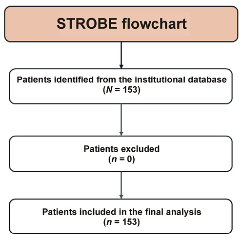
This retrospective observational study was based on the analysis of the medical records of patients who underwent implant treatment at 4 private dental clinics in Poland between 2015 and 2020. The study was performed and reported in accordance with the STROBE (Strengthening the Reporting of Observational Studies in Epidemiology) guidelines. The analysis included cases of completely edentulous maxillae and posterior maxillary edentulous spaces in which implant treatment was performed using the All-on-4®, All-on-6 and zygomatic implant techniques. The aim of the study was to determine which implant procedure was most frequently used for the reconstruction of missing teeth in patients with moderate to severe maxillary alveolar ridge atrophy, which method demonstrated the highest clinical effectiveness, and which treatment was associated with the lowest complication rates during long-term follow-up (1–5 years). Demographic variables, including patient age and sex, were also analyzed. In addition, the implant techniques associated with the highest incidence of complications were identified. Immediate and conventional implant placement protocols were also compared.
Characteristics of the clinical centers
The study included 4 private dental clinics in Poland. A total of 667 implants placed in 153 patients were analyzed.
Inclusion and exclusion criteria
The study included patients with completely edentulous maxillae or posterior maxillary edentulous spaces who provided written consent for implant-supported prosthetic treatment and had complete radiological documentation available (orthopantomogram and/or CBCT). The inclusion criteria comprised eligibility for permanent implant-supported prosthetic rehabilitation of a partially or completely edentulous maxilla in patients with moderate or severe alveolar ridge resorption and maxillary sinus pneumatization, without the need for augmentation procedures or maxillary sinus floor elevation (Figure 1). The exclusion criteria encompassed severe systemic diseases, cancerous conditions, untreated or poorly controlled diabetes mellitus, severe cardiovascular conditions, active autoimmune diseases, immunosuppressive therapy, bisphosphonate therapy, and previous irradiation of the head and neck region. No patients met any of the exclusion criteria. The study participants did not present significant systemic diseases that could affect osseointegration. The medical history of each patient was reviewed. None of the patients had a history of rheumatic disease, diabetes or uncontrolled hypertension, and all had obtained cardiology clearance before treatment. None of the participants were active tobacco smokers at the time of enrollment. All patients included in the study demonstrated good oral hygiene at the time of qualification for the procedure, and no signs of gingivitis or periodontitis were detected.
Additionally, no exclusions were applied because patients with severe systemic diseases or active smoking habits were not considered eligible for implant treatment and, therefore, were not present in the analyzed database.
Distribution of patients by age and sex
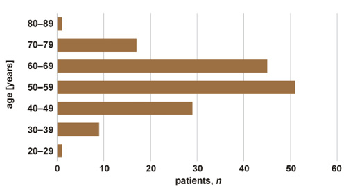
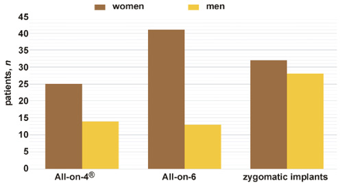
The patients included in the study ranged in age from 20 to 89 years. The largest age group consisted of patients between 50 and 69 years of age (n = 96) (Figure 2). More women than men participated in the study. Women predominated in all implant treatment groups (Figure 3).
Implant classification
Patients who underwent implant-supported prosthetic rehabilitation received one of the 3 types of implants:
• type I – passive implants with a cylindrical implant body (Alfa Dent Classic, Brånemark, Zimmer, Massif Implant);
• type II – active implants with a conical implant body (AB, Adin, Alpha Active, Alpha Bio, Astra, Axis, BEGO, Global D, Implant Direct, iRES, Neodent, Nobel Biocare, Noris, Replace Select, Sterngold, Straumann, Thommen, Top Implants);
• type III – zygomatic implants (Noris Zygoma, Brånemark Zygoma, iRES Zygoma).
Surgical techniques
Implantation procedures were classified as immediate implantation, performed during the same session or immediately after tooth extraction, or conventional (two-stage) implantation, performed after a healing period, usually 2–4 months following tooth extraction.
Surgical protocol
Under local anesthesia, after incision and elevation of a mucoperiosteal flap and preparation of the osseous bed, 4 implants were placed. Two implants were inserted in an axial orientation, whereas the remaining 2 distal implants were placed at an angle of 30–45° in the anteroposterior direction to bypass the maxillary sinus and enhance the longitudinal stability of the planned prosthetic bridge. Once primary stability had been achieved (>35 Ncm), immediate provisional loading was carried out using a temporary prosthetic restoration. The All-on-6 concept represents an extension of the All-on-4® protocol for patients with more favorable anatomical conditions, allowing the placement of 2 additional implants in the posterior region with controlled angulation. The wound was then closed with 4-0 non-resorbable sutures.
Zygomatic implants – extrasinus approach
After elevation of a mucoperiosteal flap and exposure of the surgical field, an initial marking was made on the zygomatic bone using a diameter drill. A groove (approx. 15–20 mm deep) was then created with a thick diamond bur along the lateral wall of the maxilla and directed toward the previously marked point in the root of the zygomatic bone. A 2.5-mm guide drill was introduced into this groove and advanced toward the predetermined intersection point on the zygomatic bone. This procedure allows the implant to penetrate the lateral (buccal) side and emerge from the lateral wall of the maxillary sinus, achieving anchorage in the body of the zygomatic bone. The osteotomy was then expanded using a standard 2.9-mm drill, followed by a 3.5-mm spiral drill. A depth gauge was used to determine the length of the zygomatic implant. The implant was placed in such a manner that its platform emerged at or slightly above the crest of the alveolar process. The wound was closed with 4-0 non-resorbable sutures. Depending on the clinical situation, either 2 or 4 zygomatic implants were inserted. Once primary stability had been achieved (>35 Ncm), an immediately loaded provisional prosthesis was delivered.
Immediate and final prosthetic protocols
All patients underwent rehabilitation according to a standardized two-stage prosthetic protocol. Immediately after implant placement, a screw-retained provisional full-arch prosthesis fabricated from acrylic or composite resin was delivered. The final prostheses veneered with porcelain were placed after 6 months, following confirmation of successful implant osseointegration.
Assessment of complications
Complications were defined as the lack of implant osseointegration within 6 months after placement or the need for implant removal due to chronic pain or infection.
Statistical analysis
For the statistical analysis distinguishing between “implant failure” and “implant removed due to pain”, Fisher’s exact test was used. When the outcomes were analyzed together as a single endpoint, the χ2 test was applied.
Kaplan–Meier curves were compared using the log-rank test. Statistical significance was set at p < 0.05. All statistical analyses were performed using the R software, v. 4.5.1 (R Foundation for Statistical Computing, Vienna, Austria).
Results
A total of 667 implants placed in 153 patients were included in the analysis. The distribution of procedures according to the implantation technique is presented in Table 1.
Zygomatic implants demonstrated a failure rate of 4.81%. In the All-on-4® group, 2.56% of implants were removed due to pain, and the rejection rate was 4.49%, resulting in an overall complication rate of 7.05%. In the All-on-6 group, 3.70% of implants failed to achieve osseointegration.
The evaluated techniques differed with respect to treatment outcomes, as implant removal due to pain occurred only in the All-on-4® group (Table 2). Nevertheless, no statistically significant differences were observed in the percentage of osseointegrated implants (Table 3). Overall, the 3 treatment methods demonstrated comparable clinical performance.
The All-on-6 technique was the most frequently used implantation method, particularly among women (Figure 3).
The conventional implantation protocol was used more frequently in edentulous patients (58%) (Table 4). Overall, 346 implants were placed using the immediate implantation technique, while 321 were placed using the conventional protocol (Table 4).
Complications, defined as failure of osseointegration or the need for implant removal due to pain, occurred in 32 implants. The highest complication rate was observed following conventional procedures (5.92%), whereas immediate implantation was associated with fewer implant failures (3.76%) (Table 4).
Implant survival
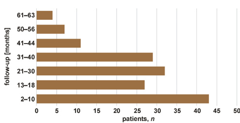
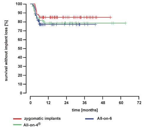
Most implants (85.62%) were followed up for up to 40 months after surgery (Figure 4). During the follow-up period, 28 implant failures and 4 implant removals due to pain were recorded. All remaining implants remained clinically stable throughout the observation period. No additional complications, such as loss of osseointegration, were observed, and the patients reported no pain or functional problems associated with their prosthetic restorations.
For the zygomatic implant, All-on-4® and All-on-6 groups, the median implant survival exceeded the maximum follow-up period. This indicates that more than 50% of the implants in each group remained functional at the end of the observation period. Accordingly, the Kaplan–Meier curves did not reach the 50% survival probability threshold (Figure 5).
Discussion
Nowadays, the number of contraindications to implant placement in the oral cavity has decreased, significantly expanding patient eligibility for implant-supported rehabilitation. Implant dentistry is developing rapidly. Moderate diabetes, head and neck radiotherapy, or chemotherapy are not absolute contraindications to implant treatment.21, 22, 23 Bisphosphonate therapy may cause osteonecrosis and impair implant osseointegration, potentially leading to implant failure.24 However, Ata-Ali et al., based on a systematic review and meta-analysis, concluded that the placement of dental implants in patients receiving bisphosphonate therapy does not reduce implant success rates.25 The dental and zygomatic implants used in this comparative study were biocompatible implants composed of titanium and enriched with zirconium oxide, yttria and hyaluronic acid (iRES implants). Previous studies have demonstrated that zirconia and titanium implants exhibit excellent osseointegration and mechanical resistance.26 Titanium used in dental implants integrates with bone to form a functional unit known as bone-to-implant contact (BIC).27 However, adverse reactions to titanium implants have been reported in numerous clinical studies.28 Recent research indicates differences in the response to titanium implants between peri-implant soft tissues and periodontal tissues.28 Currently, a distinction is made between cylindrical implants with passive thread designs and conical implants with aggressive threads. Conical implants are characterized by self-tapping properties and generally provide high primary stability. Pereira et al. demonstrated that macrogeometry of dental implants does not influence their survival probability; however, it may alter stress distribution within the implant–abutment complex.29 All modifications to implant design and implantation techniques have made any dentate ridge suitable for full implant-supported rehabilitation.
The main aim of this study was to determine whether the tested implantation methods are effective for the reconstruction of the edentulous maxilla. A major challenge in this region is the presence of the maxillary sinus, which increases in volume following tooth loss due to sinus pneumatization. The results of the present study confirmed the effectiveness of modern implant-based solutions for the treatment of edentulous maxilla. The lowest effectiveness was observed with the All-on-4® technique, with a complication rate of 7.05%, including failure of osseointegration or pain requiring implant removal. The zygomatic implant and All-on-6 techniques demonstrated very high effectiveness, with implant survival rates of 95.19% and 96.30%, respectively. Both immediate and conventional implantation showed low rates of failed osseointegration. Despite a maximum follow-up period of 5 years, most implants were observed for up to 40 months after surgery. Only 14.38% of implants were followed for more than 3 years and 4 months.
The All-on-4® concept, developed by Paulo Maló, was introduced as an alternative to time-consuming and costly augmentation procedures.30 Maló et al., in their 2019 study, reported a cumulative implant survival rate of 94.7%.30 Uesugi et al. noted a survival rate of 97.4% for implants placed in the maxilla using the All-on-4® technique.31 Browaeys et al. described a survival rate of 100% among 80 implants.32, 33 In our study, a lower percentage of osseointegrated implants was observed, with 92.95% achieving successful osseointegration during the 5-year observation period. Four implants were removed due to pain, and 7 implants failed because of a lack of osseointegration. The relatively lower percentage of osseointegrated implants may be explained by the limited number of implants included in the analysis (N = 156). It is worth emphasizing that the removal of 4 implants in the All-on-4® group resulted from sphenopalatine neuralgia (Sluder’s neuralgia) diagnosed in a single female patient.
Zhang et al. did not demonstrate significant differences between patients treated using the All-on-4® and All-on-6 techniques.34 Similarly, Tallarico et al. found no statistically significant differences in implant survival, technical and biological complications, marginal bone loss, or periodontal parameters between the 2 groups.35 In contrast, La Monaca et al. reported cumulative implant survival rates of 89.7% for the All-on-4® group and 99.0% for the All-on-6 group after a mean follow-up of 6.46 ±2.236 years (range: 1–10 years).36 In our study, 96.30% of implants achieved osseointegration in the All-on-6 group. The All-on-4® technique demonstrated a 3.35% lower success rate than the All-on-6 technique; however, statistical analysis showed no significant differences in the percentage of osseointegrated implants. The slight superiority of the All-on-6 technique may be attributed to the greater number of implants, which provides increased prosthethic stability. In the All-on-4® concept, the loss of a single implant may pose a significant challenge because maintaining the existing prosthetic restoration is less feasible than in the All-on-6 approach. Moreover, a greater number of implants may improve load distribution and reduce stresses within the prosthetic restoration. Pandey et al. demonstrated that the All-on-6 technique resulted in lower maximum principal stress values in the cortical bone and implants, indicating more favorable biomechanical behavior.37
The third method evaluated in the present study was the use of zygomatic implants for the rehabilitation of the severely atrophic maxilla. Zygomatic implants constitute an important therapeutic option for patients with advanced alveolar ridge atrophy in whom the use of conventional implants without prior regenerative procedures is impossible. The main advantage of this technique is the absence of complex bone augmentation procedures or sinus lift. However, with the intrasinus approach, the zygomatic implant penetrates the Schneiderian membrane and passes through the maxillary sinus cavity, which may represent a limitation of this technique. Although this approach provides high primary stability due to quadricortical anchorage, it may also be associated with a greater risk of sinus complications, including sinusitis or subcutaneous abscesses. In response to these limitations, the extrasinus technique was developed, allowing implant placement without entering the sinus lumen. This technique creates a narrow access to the lateral wall of the sinus (zygomatic antrostomy zone), and enables a more predictable and controlled trajectory toward the zygomatic bone without compromising the Schneiderian membrane.
In our study, 187 zygomatic implants were placed using the extrasinus technique, and 9 failures were recorded, resulting in a survival rate of 95.19%. It should be emphasized, however, that only 60 patients underwent implant-prosthetic treatment using zygomatic implants, and the results might have differed in a larger study population. For comparison, Coppedê et al. reported a survival rate of 98.9% for extrasinus zygomatic implants.38 Chrcanovic et al., in an analysis including 4,556 zygomatic implants, reported a cumulative implant survival rate of 95.21% after 12 years.39 Other studies have demonstrated average survival rates of 96.2%40 and 96.1%16 for zygomatic implants.
In summary, conventional implantation predominated in the present study (58% of cases), which was likely related to the age distribution of the study population, as 92.75% of the patients were between 50 and 69 years of age. The complication rate for immediate implantation was 2.16% lower than that observed for conventional implantation. The practice of immediate implant placement combined with prompt provisionalization is becoming increasingly common.41 According to Tsuda et al., flapless extraction followed by immediate implant placement combined with bone augmentation, connective tissue grafting and the placement of an immediate provisional crown resulted in the lowest variability in treatment outcomes.42 These findings suggest that conventional implantation does not necessarily guarantee greater treatment safety and that local factors, including bone quality, surgeon experience, diagnostic capabilities, and patient compliance play a substantial role in the treatment success.
It should be noted that the retrospective design of the study limited the possibility of complete standardization of procedures and control of confounding variables. Additionally, despite the relatively large patient sample (N = 153), the number of patients differed among the implantation techniques, which may have affected the comparability of the results. Future studies investigating implant-supported rehabilitation of the edentulous maxilla should include follow-up periods exceeding 5 years, involve larger patient cohorts and assess postoperative patient-reported outcomes according to the implantation technique used. It would also be beneficial to ensure comparable sample sizes among the evaluated treatment groups.
Conclusions
The present study demonstrated that the 3 compared methods for the rehabilitation of the edentulous maxilla or posterior maxillary edentulous spaces showed no statistically significant differences in the percentage of successfully osseointegrated implants. Immediate implantation was not inferior to conventional implantation and may therefore be recommended due to its shorter treatment time and greater patient comfort. The selection of the appropriate implant technique should be individualized, taking into account the patient’s anatomical conditions and general health status, available technology, and the experience of the surgical team.
Ethics approval and consent to participate
The study was conducted in accordance with the Declaration of Helsinki and approved by the Bioethics Committee of Medical University of Lublin, Poland (approval No. KE-0254/101/2019).
Data availability
The datasets generated and/or analyzed during the current study are available from the corresponding author on reasonable request.
Consent for publication
Not applicable.
Use of AI and AI-assisted technologies
Not applicable.
- The study compares 3 graftless implant techniques for maxillary rehabilitation: All-on-4®, All-on-6 and zygomatic implants.
- The clinical effectiveness and complications associated with different graftless implant techniques were evaluated, and conventional implant placement was compared with immediate implant placement for maxillary rehabilitation.
- The findings demonstrated no significant differences in osseointegration rates for All-on-4®, All-on-6 and zygomatic implant techniques.