Abstract
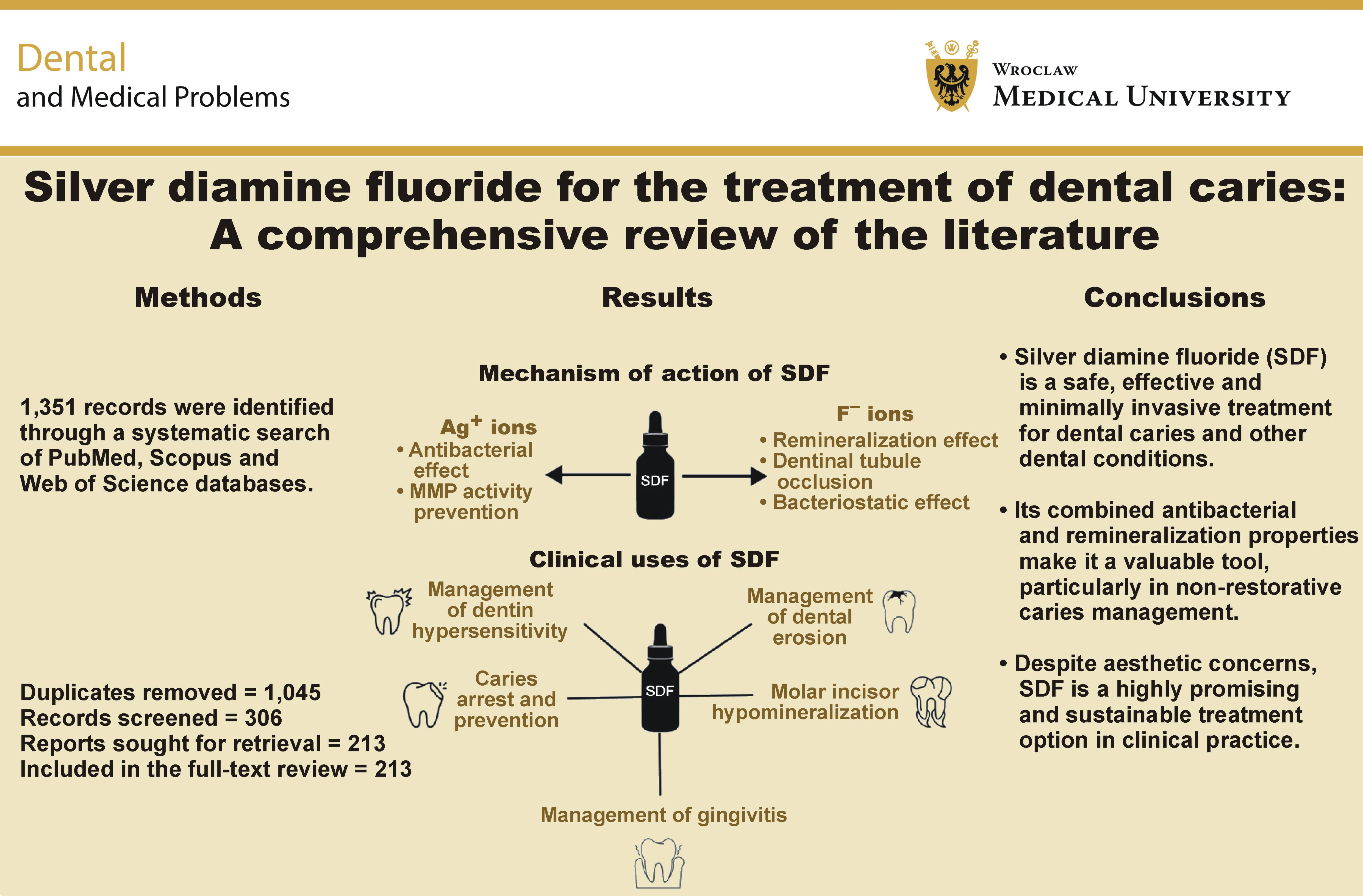
Silver diamine fluoride (SDF) is an aqueous solution of silver (Ag+) and fluoride (F−) ions stabilized by ammonia. It is regarded by the World Health Organization (WHO) as a safe, efficacious and cost-effective pharmacological agent for the minimally invasive management of dental caries. The Ag+ ions in SDF mainly exert an antibacterial effect, while the F− ions remineralize dental tissues. Recently, SDF has also been approved by the US Food and Drug Administration (FDA) for desensitization. The main advantages of SDF over other treatment options are its shorter application time, ease of application and no need for anesthesia. Furthermore, it is suitable for individuals with physical or mental disabilities, non-ambulatory patients, and elderly patients, as it can also be applied by dental nurses, hygienists and healthcare workers. Various studies have confirmed the efficacy of SDF in arresting and preventing coronal and root caries, desensitizing teeth, and disinfecting root canals, highlighting its potential to reduce the global healthcare and economic burden associated with untreated dental caries. The aim of this review is to provide an updated overview of SDF properties and its use in the minimally invasive management of dental caries.
Keywords: dental caries, minimally invasive dentistry, remineralization, silver diamine fluoride
Introduction
Dental caries is a global health problem that affects billions of people worldwide.1, 2, 3 Despite significant advancements in preventive dental care, untreated dental caries is still regarded as one of the most prevalent yet preventable diseases worldwide.4 According to the Global Burden of Disease Study 2017, over 532 million children and 2.3 billion adults were affected by dental caries in 2017.5 Dental caries remains a childhood disease affecting a subset of children in both developed and developing countries.6 Similarly, root caries is a prevalent condition that affects over one-third of the global elderly population.7
Traditionally, dental caries is managed using a surgical approach that involves removing the damaged tooth structure and replacing it with a suitable restorative material. However, this is a destructive process that inevitably results in the removal of healthy tooth structure. The so-called surgical approach is a short-term solution and has become financially unsustainable globally.8 Hence, the trend is shifting toward a minimally invasive approach in which attempts are made to remineralize dental tissues using various antibacterial agents such as chlorhexidine (CHX), calcium phosphate, casein phosphopeptide–amorphous calcium phosphate, and fluoridated compounds such as sodium fluoride (NaF) varnish.9, 10, 11, 12, 13, 14, 15 Among these, silver diamine fluoride (SDF) has recently gained popularity as a potent, efficacious and cost-effective antibacterial and remineralizing agent.
Silver diamine fluoride has been clinically used to arrest cavitated dentin lesions and prevent new cavities.16, 17 In addition, clinicians have used it to manage early childhood caries, root caries, and pit and fissure caries, as well as for desensitization.18 Silver diamine fluoride has also been utilized as a root canal disinfectant and has been suggested as a treatment for dental erosion and a dye to detect carious lesions.19 In 2021, the World Health Organization (WHO) added SDF to its Model List of Essential Medicines for the management of dental caries in children and adults.20
Silver diamine fluoride has become the center of attention for clinicians and researchers. A recent bibliometric analysis has shown that the research interest regarding the use and clinical efficacy of SDF has increased exponentially from 2016 to 2021.21 Therefore, this review aimed to provide comprehensive, updated information regarding the multipronged clinical use of SDF as a non-invasive and cost-effective agent to combat the global burden of dental caries and other preventable dental issues, such as dentin hypersensitivity, molar hypomineralization and gum disease. The review also discusses the adverse effects and toxicity-related issues of SDF and provides an overview of the various strategies used to overcome its inherent tooth-staining potential.
Material and methods
The SANRA (Scale for the Assessment of Narrative Review Articles) reporting guidelines were used in this study to enhance methodological transparency and scientific rigor.22 This involved identifying a clear research objective, documenting the literature search strategy and conducting a thematic synthesis of findings from the included studies.
As this was a narrative review, this study did not follow a methodology of a systematic review. This may have introduced selection bias, resulting in limitations related to the availability and quality of the published studies. A comprehensive search was conducted in 3 databases, namely PubMed, Scopus and Web of Science, up to August 2025. The review focused on studies that provided information on the composition, mechanism of action, clinical applications, and adverse effects of SDF. Only full-text research articles of all types or review articles related to the clinical efficacy, chemistry or safety of SDF were included. Editorials were excluded, and case reports and non-English articles were cited only when no other evidence was available. Furthermore, an additional search was performed for articles that discussed patients’, parents’ and healthcare professionals’ views regarding the clinical acceptability of SDF owing to its tooth-staining effect. The keywords used for the database search are provided in Table 1.
Results
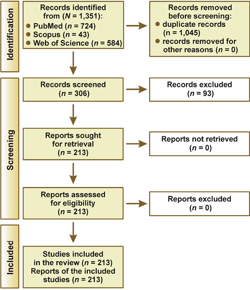
Using the predefined search strategy, a total of 1,351 articles were retrieved from the selected databases. Screening for duplicate records resulted in the removal of 1,045 papers. The remaining 306 articles underwent title and abstract screening, leaving 213 papers for full-text review. A thematic narrative synthesis of the included studies was performed based on the themes of chemical composition, clinical efficacy and uses, adverse effects, and aesthetic concerns related to SDF. A PRISMA (Preferred Reporting Items for Systematic reviews and Meta-Analyses) flow diagram presenting the process of study selection, screening and full-text retrieval is presented in Figure 1.
Silver compounds in dentistry
Silver compounds have been used in medicine and dentistry since the 1800s, owing to their antibacterial and caries-preventive properties. In dentistry, the use of silver compounds dates back to the 1840s, when silver nitrate (AgNO3) was used for a variety of dental restorative and preventive purposes.23 Some of the silver compounds used in dentistry are discussed in the following sections.
Silver nitrate
Silver nitrate was the first silver compound to be used in dentistry.23 Metallic silver (Ag0) is relatively inert chemically. However, silver particles in an aqueous solution of AgNO3 exist in ionic form and exert a bactericidal effect. Silver ions (Ag+) are highly reactive species that have a high affinity for binding to bacterial proteins, thereby causing structural changes in the bacterial cell walls and membranes.24 Klein et al. evaluated AgNO3 and other preventive materials for their effects on artificial caries.25 The results of their study showed that AgNO3 could significantly reduce the progression of caries compared with no treatment.25 However, the use of AgNO3 gradually declined, mainly due to its potential to cause tooth staining and the introduction of fluoride compounds with improved properties.26, 27
Ammoniacal silver nitrate
In an effort to enhance the stability and antibacterial efficacy of AgNO3, Percy Howe combined AgNO3 with ammonia to form ammoniacal silver nitrate, also known as Howe’s solution (HS) – AgNH3NO3.23 The silver ions in HS were shown to be able to penetrate the affected enamel and dentin and exert their antibacterial effect. As a result, HS became popular as a cavity-sterilizing agent and was also advocated for the treatment of endodontic problems, even though its efficacy had not been clinically proven at that time.26
In a clinical study involving ground sections of human and dog teeth, Zander and Smith showed that HS penetrated dentin tissue, despite the presence of a calcified barrier.28 In addition, less pulpal damage was observed when ammoniacal silver nitrate was applied directly to the exposed pulp than when it was applied to a carious lesion.23
Silver fluoride
Silver fluoride (AgF) was introduced in order to combine the antibacterial effect of silver ions with the remineralization potential of fluoride ions. Various in vitro studies have documented the ability of AgF to inhibit the growth of Streptococcus mutans in biofilms, inhibit the metabolic activity of dental plaque, and reduce the depth of carious lesions.25, 29, 30
An in vivo study conducted by Craig et al. demonstrated the ability of AgF to inhibit occlusal and proximal caries in primary molars.31 Green indicated that AgF, with or without the addition of stannous fluoride (SnF2), was able to inhibit the initial progression of carious lesions in permanent teeth.32 Despite its efficacy, the effect of AgF is short-lived due to its unstable chemical nature. Therefore, subsequent studies were aimed at enhancing the stability of AgF. This resulted in the incorporation of ammonia into AgF to form SDF, which possesses superior chemical and light stability.
Table 2 summarizes the historical advancements in the use of silver compounds for caries management in dentistry that led to the development and clinical use of SDF. As this review focuses primarily on SDF, this compound will be discussed in greater detail in a separate section.
Silver diamine fluoride
Silver diamine fluoride is a topical solution containing diamine silver and fluoride ions. The compound has been shown to be more stable and undergo less oxidation than silver fluoride.33 The first use of SDF for caries prevention dates back to the 1960s and 1970s in Japan, where it was used to arrest primary carious lesions.34 The use of SDF remained mostly limited to Japan for many decades until it was recommended for use among schoolchildren in Brazil in the 20th century.23
Over the past decade, interest in the use of SDF for the prevention and management of dental caries has increased.35 In addition, SDF is considered a safe, efficacious, non-invasive, and cost-effective option for caries management, with the potential to support the achievement of the WHO global oral health goals for 2020.35 Subsequently, WHO included SDF as an “essential dental medicine” in the Global strategy and action plan on oral health 2023–2030.36 Various in vitro and clinical studies have shown the efficacy of SDF in preventing and arresting caries and its superiority over other preventive agents.37, 38, 39, 40, 41
Knight et al. conducted a series of in vitro tests between 2005 and 2009 and showed the efficacy of SDF in arresting and preventing caries.42, 43 In 2014, the US Food and Drug Administration (FDA) approved SDF in the USA for preventing dentinal hypersensitivity.34, 44 However, it has also been used off-label as a caries-preventive and caries-arresting agent. Since its approval by the FDA, there has been considerable interest worldwide in the use of SDF for the non-surgical management of dental caries.
Chemical structure and composition
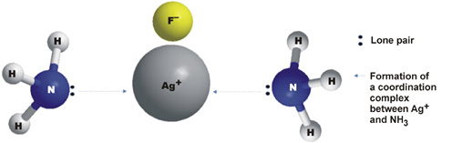
Chemically, SDF is an aqueous solution of AgF stabilized by the addition of ammonia.45, 46, 47 Physically, SDF is a colorless solution with a molecular mass of 160.98 g/mol.48 Overall, SDF has an alkaline pH. However, its alkalinity varies from one product to another. Various studies have shown that the pH of SDF remains stable for up to 180 days.49, 50, 51 Asghar et al. measured the pH of 38% SDF (FAgamin) for up to 90 days.52 Their results showed that the mean pH of SDF was 7.73 ±0.02 after the first hour of measurement, while the mean pH on day 90 was 7.70 ±0.01, indicating minimal time-dependent changes.52 However, once the cap is opened, there is a risk of light-induced reduction of the Ag+ ions in SDF. Hence, care should be taken to prevent direct exposure to light during SDF use and storage.
The chemical structure of SDF is presented in Figure 2, while its composition is outlined in Table 3.
Mechanism of action
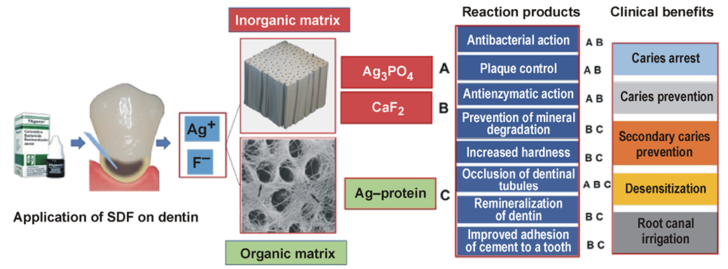
Several mechanisms of action have been proposed for SDF based on the effects of its silver and fluoride ions (Figure 3).
Antibacterial action
Silver ions
As SDF is an aqueous solution, silver ions are continuously released from SDF into the oral environment and can exist in 3 oxidation states: silver ions (Ag+); divalent silver ions (Ag2+); and trivalent silver ions (Ag3+).53 It has been shown that among these 3 states, only Ag+ possesses sufficient stability to exert an antibacterial effect.54 Silver ions in SDF combine with the sulfhydryl groups of bacterial proteins and deoxyribonucleic acid (DNA), resulting in altered hydrogen bonding and inhibition of respiratory processes, DNA unwinding, cell division, and cell wall synthesis.24, 55 In an in vitro study, Knight et al. showed that dentin surfaces treated with SDF had significantly less growth of S. mutans than those in the control group.42 Similarly, Chu et al. observed that the application of SDF to dentin resulted in considerably fewer bacterial colony-forming units (CFU) of S. mutans and Actinomyces naeslundii, two of the most commonly found bacterial species in root dentin caries.56, 57
The silver ions in SDF prevent biofilm formation by reacting with functional and structural bacterial proteins, particularly those containing thiol groups (–SH), resulting in bacterial cell death.46, 58 Silver ions in SDF have also been shown to inhibit collagen degradation and the enzymatic activity of matrix metalloproteinases (MMPs).37 Furthermore, silver ions bind to bacterial cell membranes, leading to membrane leakage and rupture, and can also cause DNA mutations that lead to bacterial cell death.47, 59 Finally, Ag+ can infiltrate the hydroxyapatite (HA) crystals that form on the tooth surface following SDF application and reduce bacterial adhesion.60
Another aspect of the antibacterial efficacy of SDF is the cariostatic reaction between tooth components and SDF. It has been shown that the fluoride ions (F−) released from SDF can penetrate dentin to a depth of 50–100 µm and provide a bacteriostatic effect.18, 61 Similarly, high concentrations of F− in SDF inhibit bacterial biofilm formation by binding to bacterial cellular components and affecting their enzymatic activity related to carbohydrate metabolism.33
Fluoride ions
At high concentrations, fluoride ions have been shown to exert a bacteriostatic effect by binding to bacterial cellular components. The formation of hydrogen fluoride following the application of SDF results in increased bacterial cell membrane permeability coupled with the inhibition of plaque-dependent bacterial metabolism.62 Furthermore, fluoride ions inhibit non-metal enzymes such as phosphatases, thereby preventing acid production by dental plaque.47
A summary of the mechanisms of action of SDF is provided in Table 4.
Effect of silver diamine fluoride on dental tissues
Enamel
The enamel remineralization potential of SDF has been attributed to the presence of a high concentration of fluoride ions and its alkaline pH. Similarly, the antibacterial effect results from the high concentration of Ag+ ions in SDF.63 It has been shown that the application of SDF results in the formation of calcium fluoride (CaF2), fluorohydroxyapatite (FHA), Ag0, and other silver compounds.23, 64 Although CaF2-like globules form due to the high F− ion concentration in SDF, they have been found to be easily washed away by saliva, water or other media in which the samples are stored.63 Similarly, FHA formation occurs due to the reaction of HA with F− ions in SDF.
Li et al. evaluated and compared the penetration of Ag+ ions into primary enamel and dentin tissues.65 Their results showed that Ag+ ions penetrated vertically into the enamel surface, followed the direction of the enamel rods, and then penetrated demineralized dentin. The extent of silver penetration was found to be dependent on the degree of demineralization. It has been shown that there was considerable variability in the depth of penetration of Ag+ ions into the tooth structure, with a mean penetration depth of 744.7 ±448.7 µm.65 In contrast, as reported in a literature review by Shah et al., Suzuki et al. showed that silver ions were mainly deposited on the enamel surface and could penetrate up to 20 µm, while fluoride ions could diffuse up to 25 µm beneath the enamel.18, 66 Similar results were reported by Akyildiz et al., who showed that SDF could penetrate up to 25 µm into enamel.67 This considerable difference in the reported penetration depths may be due to the differences in the types of dental tissues used for analysis, such as primary and permanent dental tissues.
Dentin
Previous studies have shown that the application of SDF on dentin results in a significant reduction in demineralization. Furthermore, higher concentrations of Ca2+ and PO43− ions have been observed in the outer 25 µm of carious dentin treated with SDF compared to demineralized control specimens.57 This indicated that SDF was able to promote remineralization in the previously demineralized dentin specimens. Mei et al. also showed that SDF could inhibit MMPs, an effect attributed to the high pH of SDF, thereby preventing degradation of the dentinal organic matrix.68 The authors of the study observed the formation of dense granules on the dentin surface following SDF application, which were ascribed to extrafibrillar mineral deposition. It has also been shown that the hardness of the dentinal surface following SDF application was higher than that of the underlying tissue.37 The alkaline pH of SDF tends to provide a favorable environment for cross-linking between minerals and collagenous proteins, thereby preventing the degradation of the exposed fibers.44
In addition, fluoride ions have been shown to protect the dentinal collagen structure in 2 main ways.68 First, fluoride ions promote remineralization through the formation of FHA. The newly formed apatite crystals cover and protect the underlying collagen fibers from damage. Mei et al. used an immunolabelling technique and observed that SDF-treated dentin had more intact type I collagen than water-treated lesions.39 The second proposed mechanism of protection of the dentinal organic matrix is the inhibition of MMPs. Mei et al. compared AgNO3 and AgF and demonstrated that AgF was a much stronger inhibitor of MMP-2, MMP-8 and MMP-9.68 Similarly, Kato et al. showed that 200 ppm of fluoride could completely inhibit the active and inactive forms of MMP-2 and MMP-9.69 It has been proposed that the high electronegativity of F− ions prevents the binding of zinc and calcium ions to MMPs,thereby preventing their activation.69
Clinical uses
Caries arrest and prevention
Dental caries in primary teeth
Silver diamine fluoride is most commonly used as a fluoride-based agent for caries arrest and prevention.33 Zhi et al. showed that the annual application of 38% SDF or the use of a flowable, high-fluoride-releasing glass ionomer cement (GIC) had comparable caries-arresting potential. However, the rate of caries arrest with SDF increased when the agent was applied twice a year.40
In 2002, a 30-month clinical trial conducted by Chu et al. in Chinese preschool children showed that the annual application of 38% SDF was more effective in arresting dentin caries in primary anterior teeth than 5% NaF varnish applied every 3 months.16 Caries excavation before the application of SDF provided no significant benefits.16 A meta-analysis by Rosenblatt et al. reported prevented fractions of 96.1% for caries arrest and 70.3% for caries prevention with SDF, compared with 21.3% and 55.7%, respectively, with NaF varnish.46
Llodra et al. conducted a 36-month clinical trial to assess the caries-arresting potential of SDF in primary and permanent molars.70 The results showed that the application of SDF every 6 months was effective in arresting caries in both primary and permanent teeth.70 Yee et al. demonstrated that a single application of 38% SDF was effective in arresting caries in the primary teeth of schoolchildren.38
Braga et al. compared the effectiveness of SDF with other non-invasive measures: the cross-tooth brushing technique (CTT) and sealing with GIC for arresting dental caries in erupting permanent molars.71 After 3 and 4 months of treatment, 10% SDF demonstrated significantly higher caries-arresting potential than CTT and GIC sealants.71 Similarly, dos Santos et al. compared the caries-arresting potential of 30% SDF with GIC used as an interim restorative treatment (IRT) in schoolchildren aged 5–6 years.72 The results showed that SDF was 1.73% more effective in arresting caries than IRT.72
In 2020, a randomized clinical trial (RCT) by Mabangkhru et al. investigated the caries-arresting efficacy of 38% SDF and 5% NaF in Thai children aged 3–5 years.73 Both treatment options were applied every 6 months. At the end of 1 year, the caries arrest rate with 38% SDF was 35.7%, compared with 20.9% for 5% NaF (p < 0.001). Furthermore, the appearance of the children’s teeth following SDF treatment did not affect parental acceptability.73
In 2022, Satyarup et al. conducted a randomized controlled trial to compare the efficacy of 38% SDF (Group 1) with the atraumatic restorative technique (ART) (Group 2) in schoolchildren. The results showed that after 9 months, the mean survival rate (± standard error (SE)) of the teeth in Group 1 was significantly higher (76.7 ±0.21) than that in Group 2 (62.2 ±0.28). Hence, SDF was shown to be more effective at arresting caries than ART in schoolchildren.74
Dental caries in adults
Tan et al. compared the efficacy of 4 different treatment protocols for preventing root caries among older adults who had at least 5 teeth with exposed sound root surfaces.75 The following treatment groups were used in the study: placebo and oral hygiene instructions (OHI); OHI + 1% CHX application every 3 months; OHI + 5% NaF application every 3 months; and OHI + annual application of 38% SDF. Compared with the placebo and OHI groups, the prevented fractions for root caries were 71%, 64% and 57% for SDF, NaF and CHX varnish, respectively. The prevented fraction for root caries with the combination of annual SDF application and OHI was significantly higher than those of the other treatment groups.75
Similarly, Zhang et al. demonstrated the caries-arresting potential of different treatment options in community-dwelling older adults.76 The treatment groups included annual OHI (Group 1), OHI + annual SDF application (Group 2), and OHI + SDF + oral health education program twice a year (Group 3). After a 24-month follow-up, the mean numbers of arrested root caries surfaces in Groups 1, 2 and 3 were 0.04, 0.28 and 0.33, respectively, with Group 3 demonstrating significantly greater caries-arresting potential than the other 2 groups.76
In 2016, Li et al. investigated the efficacy of 38% SDF in arresting root caries among community-dwelling adults in Hong Kong, China.77 The following 3 groups were tested: soda water (control); annual SDF application; and annual SDF application immediately followed by the application of potassium iodide (KI). Tooth color changes were measured using PANTONE color plates. After a 30-month follow-up, the caries arrest rates in Groups 1, 2 and 3 were 45%, 90% and 93%, respectively (χ2 test; p < 0.01). It was concluded that the annual application of SDF, regardless of adjunctive KI application, was effective in arresting root caries in adults.77 Interestingly, it was also shown that the application of KI following SDF treatment was ineffective in mitigating long-term SDF-mediated tooth staining. Additionally, Mitchell et al. showed that the probability of arresting an active root caries lesion with SDF was 91.6% after 18 months of treatment.78
Management of tooth sensitivity
Silver diamine fluoride has been shown to be effective in minimizing dentinal hypersensitivity. It has been demonstrated that the application of SDF results in the formation of compounds such as silver phosphate (Ag3PO4) and CaF2, which form a squamous surface layer, occlude the exposed dentinal tubules and prevent hypersensitivity.34 In 2014, SDF was cleared for use by the FDA for the management of dentin hypersensitivity.
In a multi-center study, Castillo et al. compared the efficacy of 38% SDF with that of a placebo (sterile water).79 In their study, 129 subjects with at least 1 tooth sensitive to air-drying were recruited from 2 different sites, and pain levels before and after treatment were measured using a 100-mm visual analog scale (VAS). At both sites, SDF was shown to significantly reduce dentin sensitivity (−35.8 ±27.7 and −23.4 ±21, respectively) after 7 days, compared with VAS scores of 0.4 ±16.2 and −5.5 ±18.1 for the control group at both sites. The authors concluded that SDF was a clinically effective option for managing tooth sensitivity.79
In another study, Craig et al. evaluated the efficacy of an SDF/KI-based commercial product compared with that of an oxalic acid-based product in reducing dentinal sensitivity.80 Tooth sensitivity was measured in 19 study subjects at baseline and 7 days after treatment using a VAS. It was shown that the SDF/KI-based product could significantly reduce dentin hypersensitivity compared with the oxalic acid-based product 1 week after treatment (p = 0.00134). The SDF/KI-based product was recommended as a potential option for managing dentin hypersensitivity.80
Molar incisor hypomineralization
Dental hypomineralization is a congenital defect that mainly affects the permanent first molars and incisors.81 This condition is characterized by the appearance of well-demarcated, chalky white or brown opacities. Histologically, these opacities appear porous and are located in the deeper parts of the enamel. Children with this condition experience severe tooth hypersensitivity, as their teeth are more prone to wear and dentinal exposure. The application of SDF to these teeth has been shown to effectively reduce sensitivity and arrest the lesions.
An RCT by Ballikaya et al. compared the effects of SDF and silver-modified atraumatic restorative treatment (SMART) sealants for the treatment of initial carious lesions in molars affected by molar incisor hypomineralization (MIH).82 The results showed that SDF and SMART had comparable short-term effectiveness in preventing dental caries in MIH-affected molars.82
Similarly, a study by Saad et al. compared the effectiveness of SMART with that of conventional restorations and fluoride varnish application in moderately hypomineralized and severely hypomineralized molars.83 Their results demonstrated that both SDF and fluoride varnish had a significant and comparable effect on the reduction of hypersensitivity in hypomineralized molars.83
Endodontic irrigant and inter-appointment medication
Silver diamine fluoride has also been investigated as a root canal disinfectant and irrigant. In 2010, Hiraishi et al. studied the efficacy of 3.8% SDF and 5.25% sodium hypochlorite (NaOCl) in penetrating dentin and neutralizing Enterococcus faecalis biofilms.84 It was shown that 3.8% SDF and 5.25% NaOCl were equally effective at disrupting the bacterial biofilm compared with NaCl. The authors also observed silver deposits on 66.5% of the radicular dentin, which penetrated up to 40 µm at 72 h after SDF treatment.84
In another ex vivo study, Mathew et al. compared the antibacterial action of 3.8% SDF and 2% CHX against E. faecalis for endodontic purposes using single-rooted teeth.85 Their results showed an absence of CFU in specimens treated with SDF and CHX 24 h after treatment, compared with a CFU count of 3.8 × 10⁷ for the negative control (0.9% sterile saline). The authors concluded that SDF could be used as an endodontic antibacterial agent. However, one limitation of this study was the short duration of antibacterial testing.85
In contrast, Al-Madi et al. demonstrated that NaOCl was more effective in disrupting bacterial biofilm than SDF or CHX.86 The study investigated the antibacterial potential of 3.8% SDF as a root canal irrigant compared with 2% CHX, using NaOCl and saline as positive and negative controls, respectively. Single-rooted teeth without anatomical or structural deformities were treated with SDF and CHX, and confocal laser scanning microscopy (CLSM) was used to evaluate the live-to-dead ratio of E. faecalis biofilms. After a 10-minute exposure to the treatment solutions, NaOCl had the highest live-to-dead percentage among all treatment groups. Furthermore, SDF had a significantly higher live-to-dead ratio than CHX and saline.86
Management of dental erosion
Dental erosion is the chemical loss of dental tissues due to non-bacterial acids.87 Dental erosion can lead to the exposure of dentinal tubules, resulting in hypersensitivity and the development of carious lesions. Silver diamine fluoride has been shown to be an excellent antierosive agent owning to its remineralization potential. The compound reacts with tooth minerals to form Ag0 and silver chloride on the tooth surface, which may contribute to its antierosive effect.39, 88 A study demonstrated the formation of silver-containing HA on enamel following SDF treatment.63 Similarly, another laboratory-based study showed that SDF could effectively reduce erosion of enamel and dentin.19
Treatment of gingivitis
Owing to its high Ag+ ion concentration, SDF possesses excellent antibacterial properties. Hence, it can also be used for the treatment of gingival problems. In 2020, Rams et al. evaluated the antibacterial efficacy of 19% SDF and 38% SDF against various periodontal pathogens.89 Their results showed that SDF exhibited significant in vitro antibacterial efficacy against putative periodontal pathogens cultured from severe lesions.89 Similarly, a case report by Lim and Yang demonstrated that SDF treatment was effective in the management of gingival hyperplasia due to its anti-inflammatory properties.90
A summary of the clinical uses of SDF is presented in Table 5.
Safety
Although SDF contains high concentrations of Ag+ and F− ions, 1 drop of 38% SDF has been shown to contain 2.24 mg of F−, compared with 11.3 mg of F− in a typical dose of 0.25 mL of 5% NaF. Hence, the annual or biannual application of SDF does not pose a safety or toxicity risk.91
During the process of obtaining the FDA approval, the median lethal dose (LD50) for 38% SDF was found to be 520 mg/kg by the oral route and 380 mg/kg by the subcutaneous route. One drop of SDF has an approximate volume of 25 µL and contains 9.5 mg of the active ingredient, which is sufficient for treating 5 teeth. If SDF is administered to a child weighing only 10 kg, the administered dose would be 0.95 mg/kg of body weight. Therefore, the safety margin of SDF for a child weighing 10 kg would be calculated as 380 mg/kg LD50 or 0.95 mg/kg dose, corresponding to an approx. 400-fold safety margin.27 In fact, the administered dose is much lower. Vasquez et al. showed that the maximum total dose required to apply 38% SDF to 3 teeth was 2.37 mg.92 Therefore, approx. 400 applications of 38% SDF, equivalent to 1 g of silver, would be required to result in silver poisoning.47
Adverse effects
No long-term adverse effects of SDF have been reported in the literature.47 According to the results of 9 RCTs in which SDF was applied to multiple teeth for caries arrest and prevention, the only side effect observed was the appearance of mild, painful, white lesions on the oral mucosa, which disappeared within 48 h.27, 40 In a randomized controlled trial by Milgrom et al., the most commonly reported adverse effects following SDF application were mild diarrhea and headache.93 However, these symptoms resolved within 48 h of SDF treatment. Similarly, the gingival response to SDF has been shown to be minimal.93
In a RCT by Zheng et al., it was shown that there were no short- (1-day) or long-term (1-year) adverse effects of SDF application to primary anterior teeth, such as swelling, irritation or bleaching.94 A RCT by Castillo et al. reported mild erythema in some survey participants during the first 24 h following SDF application.78 However, Horst et al. recommend the application of a coating agent on the mucosa opposite the teeth to which SDF will be applied.27 Due to the high concentration of silver ions present in SDF, it must not be used in patients who have an allergy to silver.47
Aesthetic concerns
Parental aesthetic concerns regarding SDF treatment
Despite its excellent efficacy, ease of application and cost-effectiveness, the use of SDF remains limited by its potential to cause black staining of teeth and to stain clothing and countertops.27, 33, 34, 46, 95 While this staining is an indication of the successful anticaries effect of SDF, it is also an aesthetic concern for many patients.96
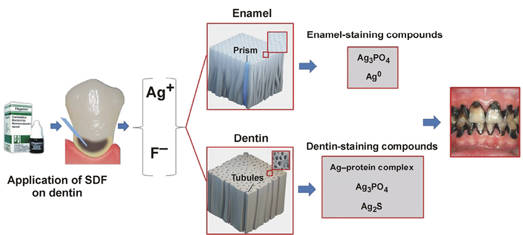
The exact cause of SDF-mediated tooth staining is not fully understood. However, according to Lou et al., silver ions react at the surface of enamel to form Ag3PO4, CaF2 and Ag0.97 The resulting Ag3PO4 is brownish in color and gradually turns black, staining the teeth. Similarly, in the case of dentin, silver ions react with proteins present within dentin to form a silver protein complex, in addition to Ag3PO4 and CaF2, which further contributes to the staining mechanism.98 Tooth staining occurs 2 min after the application of SDF, and a visually discernible color change occurs within 5 min.99
A graphical summary of the proposed mechanism of SDF-mediated tooth staining is shown in Figure 4.
A survey by Crystal et al. was conducted to evaluate parental acceptance of SDF staining.100 Only 29.7% of the parents found SDF-mediated tooth staining acceptable on anterior teeth. Furthermore, parental acceptability was likely to be higher for posterior teeth than for anterior teeth.100 These findings were summarized by Gordon, who noted that parental acceptance of SDF staining was higher when children’s teeth were located in less visible areas of the mouth.101 It has also been shown that parents tend to be more accepting of SDF use on primary teeth than on permanent teeth.102
Alshammari et al. conducted a cross-sectional study in Saudi Arabia in which a total of 222 parents were asked for their opinions regarding SDF staining after viewing before-and-after photographs.103 The results showed that parents considered SDF-mediated staining of anterior teeth unacceptable, with 90% of them strongly refusing treatment. Similarly, in the case of posterior teeth, 28.4% of parents refused the treatment, while 68.5% of them strongly refused SDF treatment due to aesthetic concerns.103 Likewise, a cross-sectional study conducted by Bagher et al. investigating parental acceptance of SDF use on their children’s primary and permanent teeth showed that only 2 (1.9%) of the 104 parents found SDF treatment acceptable, while 46 (44.2%) considered the treatment strongly unacceptable.104
On the other hand, Magno et al. concluded that the parental acceptance of SDF treatment was not affected by its staining potential.105 Instead, the findings of the study showed that clinicians were more concerned about the staining effect of SDF than parents.105 Similar results were obtained by Mabangkhru et al.,73 while Duangthip et al. reported that the overall parental acceptance of the staining potential of SDF was 71%.106
Dental professionals’ aesthetic concerns regarding SDF treatment
Various studies have documented dental professionals’ concerns about the tooth-staining potential of SDF. In a survey of US Pediatric Dentistry Residency Program directors, 91.8% of the respondents believed that parental concerns regarding SDF staining were the main impediment to its widespread clinical use.107
Similarly, a survey of registered dental hygienists conducted by Chhokar et al. revealed that 56% of the respondents believed that patients or their guardians would object to SDF-mediated staining of carious teeth.108 In 2019, another survey of US pediatric dentists demonstrated that only 33% of the respondents agreed or strongly agreed with the application of SDF to primary teeth in the aesthetic zone. Interestingly, only 14.3% of dentists agreed or strongly agreed with SDF application to permanent teeth in the aesthetic zone.109
Methods to reduce silver-mediated tooth staining associated with SDF
Various techniques have been employed to reduce the staining of teeth caused by SDF.
Potassium iodide
Knight et al. incorporated KI into SDF and demonstrated its effectiveness in reducing tooth staining.43 Potassium iodide reacts with silver ions to form silver iodide (AgI), a creamy white mixture that deposits on the surface of the tooth and cosmetically masks the staining effect. However, other studies have shown that the effect of KI is only temporary, and black staining of the teeth still occurs over time.27, 110 Miller et al. evaluated the staining associated with SDF and SDF + KI after the placement of a GIC restoration.111 The results showed that there was no statistically significant difference in the staining between the control and experimental groups.111 Similarly, Zhao et al. compared discoloration around restoration margins in teeth that were treated with either SDF or SDF followed by KI and then restored with GIC.112 The results of their study showed that despite the reduction in staining in the SDF + KI group, perceptible staining still occurred around the restoration margins.112 Furthermore, a radomized clinical trial by Li et al. revealed that the immediate application of KI to the teeth following SDF application could not eliminate long-term black staining.77
Recently, Detsomboonrat et al. investigated the effect of incorporating different concentrations of KI into SDF on its tooth-staining potential.113 The results of their study showed that the inclusion of KI into SDF resulted in a concentration-dependent reduction in tooth staining. However, the color change in the specimens treated with SDF + KI was minimal over the following 14 days. The study did not describe long-term effects of the incorporation of KI into SDF on its ability to minimize tooth staining.113
Glutathione biomolecule
Sayed et al. added 20% glutathione (G) to SDF to reduce its staining potential.110 Glutathione is a biomolecule that coats silver particles and prevents them from forming clusters. As a result, the particles do not agglomerate, thereby reducing the black staining of teeth. It was observed that the overall color change in specimens treated with 20% G + SDF, with or without light exposure, was greater than that in the control group (SDF) and the SDF + KI group. Similarly, G-modified SDF was more effective in preventing staining in enamel than in dentin specimens. The authors of the study evaluated color changes over 14 days only, which is insufficient to establish the long-term staining potential of a silver-based solution.110
Similarly, Gupte et al. compared the staining potential of enamel specimens treated with 38% SDF, SDF application followed by 10% KI, and SDF containing 20% (w/v) G at 4 different timepoints: 1, 7 and 14 days, as well as 1 month after treatment.114 Their results showed that while KI could effectively reduce SDF-mediated staining, G-modified SDF lacked the ability to prevent long-term enamel staining. In another study, Kamble et al. compared the tooth discoloration potential of 38% SDF following the incorporation of KI and the G biomolecule.115 Their results showed that while KI could effectively reduce SDF-mediated staining in primary molar specimens, glutathione could also be used as an alternative.115
Capping agents
Capping agents have also been used to overcome the staining potential of SDF. Their ability to encapsulate Ag+ ions prevents the formation of silver compounds that cause tooth staining. In a laboratory study, Asghar et al. incorporated 4 different capping agents in 30% SDF, namely tannic acid (TA), G, carboxymethyl chitosan (CM), and gallic acid (GA) at 3 different concentrations: 5%; 10%; and 15% (w/v). The study results showed that capping agents could reduce SDF-mediated staining of dentin specimens. However, the stain-reducing effect was observed to be more dependent on the type of capping agent than on its concentration. It was shown that TA and G biomolecules produced the greatest reduction in dentin staining following SDF treatment among all capping agents tested. Hence, the incorporation of capping agents could be an effective method to overcome SDF staining and enhance its clinical use.116
Silver nanoparticles
Another approach to reducing SDF-associated staining involves the use of nanosilver-based formulations. Silver nanoparticles (AgNPs) exhibit enhanced, size-dependent antimicrobial activity compared with conventionally used silver products and SDF. In addition, AgNPs have also been shown to be safe for use inside the human body at low concentrations.117 Osorio et al. demonstrated the role of silver ions in reducing collagen degradation and inhibiting the enzymatic activity of MMPs.118 Similarly, Espíndola-Castro et al. showed that nanosilver fluoride resulted in significantly less staining compared with SDF.119
Comparable results were obtained by Sayed et al., who demonstrated that tooth staining caused by the application of nanosilver was significantly reduced compared with that caused by SDF and SDF + KI. However, one problem associated with AgNPs is their inherent tendency to aggregate after reduction, leading to reduced antibacterial efficacy and the development of tooth stains over time. Hence, further research is required to identify modifications or alternatives to SDF that minimize color change.120
The aggregation of AgNPs in an ionic environment, such as the oral cavity, tends to occur rapidly.121 This can be prevented by coating AgNPs to stabilize them electrostatically.122 Yin et al. used polyethylene glycol (PEG), a synthetic polyether, as a capping and stabilizing agent to prepare fluoride-containing AgNPs.123 The results of their study showed that these AgNPs not only possessed good antibacterial properties but also minimized tooth staining compared with 38% SDF. However, information regarding the remineralization efficacy of these AgNPs and their concentration-dependent effects on tooth staining was not available.123
Currently, no alternative to or modification of SDF can eliminate the black staining of teeth associated with its use. The clinical application of SDF could be considerably enhanced if its staining effect were eliminated. This can be achieved with the use of capping agents. Capping agents are amphiphilic molecules comprising a polar head and a non-polar hydrocarbon-based tail. The tail interacts with the surrounding medium, while the head reacts with metal atoms.124 The addition of capping agents to metallic solutions tends to result in the coating of metal ions, thereby preventing them from forming reduction products that subsequently cause tooth staining.
Conclusions
Silver diamine fluoride has diverse dental applications that make this topical treatment option an excellent, cost-effective, non-invasive therapeutic agent for managing carious lesions, treating dentin hypersensitivity and enamel hypomineralization, performing endodontic irrigation and disinfection, and providing periodontal treatment.
Despite its clinical efficacy, the use of SDF is limited by its potential to cause silver-mediated staining of carious tooth structure, which raises aesthetic concerns. Various approaches to addressing this problem, including the use of capping agents, KI application after SDF treatment, and alternatives to SDF such as AgNPs, have been used with varying degrees of success. However, to date, the staining problem has not been fully resolved. Hence, there is a need to further explore SDF modifications or non-staining alternatives with similar or improved efficacy.
Ethics approval and consent to participate
Not applicable.
Data availability
Not applicable.
Consent for publication
Not applicable.
Use of AI and AI-assisted technologies
Not applicable.