Abstract
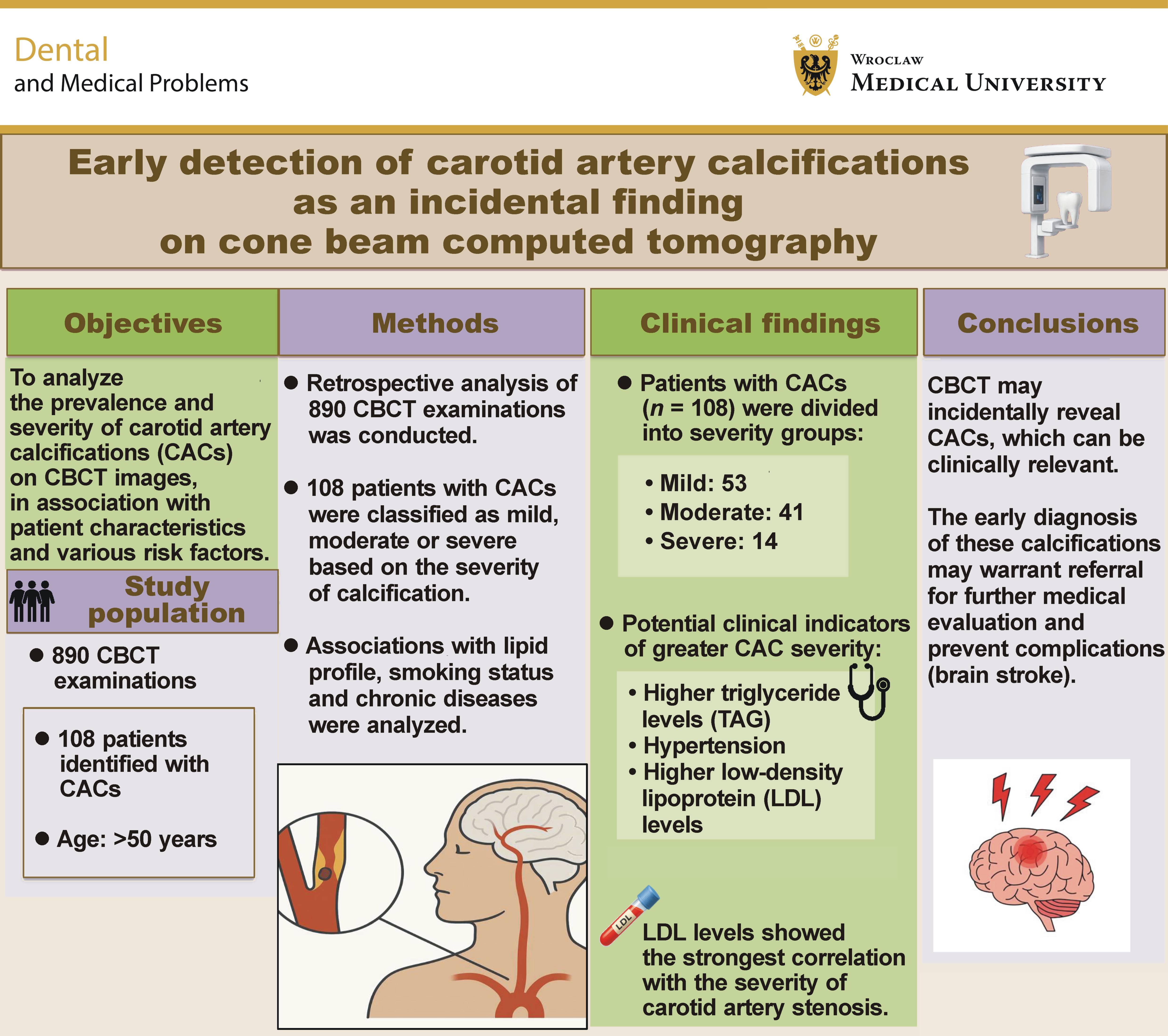
Background. Carotid artery calcifications (CACs) are markers of atherosclerosis and an increased risk of stroke. Therefore, their incidental detection on dental cone beam computed tomography (CBCT) scans can be clinically relevant. Early detection allows timely intervention, potentially reducing the risk of serious cerebral and cardiovascular complications.
Objectives. The aim of the study was to retrospectively analyze the prevalence and severity of CACs on CBCT images and their association with patient characteristics and various risk factors.
Material and methods. This retrospective study evaluated 890 CBCT examinations. Of these, 108 patients exhibiting sclerotic changes in the internal carotid artery (ICA) were included and classified as mild, moderate or severe based on the severity of calcification. The study excluded individuals younger than 50 years, as well as cases in which similar radiopacities could not be clearly differentiated. The associations between various clinical variables, including lipid profile parameters, smoking status, a history of chronic diseases, and the severity of calcifications were statistically analyzed.
Results. A total of 108 patients were categorized based on the severity of calcification as mild (n = 53), moderate (n = 41) or severe (n = 14). The findings indicated significant associations between CAC severity and age (p < 0.001), low-density lipoprotein (LDL) levels (p < 0.001), triglyceride levels (TAG) (p = 0.002), and hypertension (p = 0.027). Low-density lipoprotein levels demonstrated the strongest correlation with the severity of stenosis (p = 0.102).
Conclusions. Cone beam computed tomography may incidentally reveal CACs, highlighting the potential need for further medical evaluation. Larger studies are required to confirm the clinical significance of these findings.
Keywords: cone-beam computed tomography, carotid artery atherosclerosis, vascular incidental findings, calcifications
Introduction
Atherosclerosis is a chronic disease characterized by the buildup of lipid-rich plaques in the arterial walls, leading to intimal fibrosis, necrosis, vessel narrowing, and potential stenosis. It can result in vessel stiffening, hemodynamic disturbances, and acute occlusion due to plaque rupture, which may cause serious secondary conditions such as heart attacks and strokes.1 Atherosclerosis is the primary contributor to cardiovascular and cerebrovascular diseases, which remain the leading causes of mortality and morbidity worldwide, affecting large and medium-sized arteries. It tends to develop at arterial bends or bifurcations, where there is decreased endothelial shear stress or disrupted blood flow. The impact of atherosclerosis varies depending on its location, such as the coronary arteries, where it may lead to coronary artery syndrome, or the carotid and intracranial arteries, where it may result in stroke or transient ischemic attack (TIA).2
Vascular calcifications are present in 80–90% of atheromas and are recognized as both a sign of aging and an active, self-regulating process involving complex cellular and enzymatic mechanisms. Calcification can occur in the medial and intimal layers of blood vessels, including the intracranial and cervical carotid arteries, and contribute to the formation of atheromatous plaques. These plaques can narrow or block blood vessels, potentially resulting in serious health issues such as carotid artery stenosis (CAS), ischemic stroke, vision loss, and an overall increase in morbidity and mortality.3 Carotid artery calcifications (CACs), seen as radiopacities in the cervical region, are known as extracranial carotid calcifications, while those along the internal carotid artery (ICA) within the cranium are referred to as intracranial carotid calcifications. Various methods, such as computed tomography (CT), magnetic resonance imaging (MRI) and angiography, are used to detect and assess these calcifications. The occurrence and frequency of CACs differ according to sex, age, genetic predisposition, and lifestyle-related risk factors and comorbidities.4
Carotid artery stenosis is a frequently encountered condition in routine medical practice. It is a significant cause of acute ischemic stroke, accounting for around 20% of cases. Carotid artery stenosis can be categorized as either symptomatic or asymptomatic, with this distinction greatly influencing the treatment approach. Symptomatic disease may present as acute neurological ischemia or amaurosis fugax, requiring immediate vascular or emergency evaluation. Conversely, asymptomatic disease may be identified after detecting a carotid bruit or incidentally during imaging.5 Panoramic radiographs commonly used in dental practice can incidentally reveal CACs as irregular, nodular opacities near the mandibular angle at the C3–C4 vertebral levels.6 Due to the widespread use of panoramic radiography in routine dental care, dental professionals are well-positioned to detect signs of CAC. This situates them as potential frontline contributors to cardiovascular risk screening, enabling timely identification and appropriate referral for further medical assessment.7, 8 Radiographic examination plays an important role in dentistry, and the detection of carotid artery atheromas on panoramic radiographs may facilitate the prevention of cerebrovascular events, although general practitioners may have limited diagnostic expertise in this area. Advances in cone beam computed tomography (CBCT) allow for detailed three-dimensional (3D) imaging, improving diagnostic accuracy and dental treatment planning. Cone beam computed tomography, with its wide cone-shaped X-ray beam, enables evaluation of the carotid artery bifurcation at the epiglottis level, in relation to the C3 and C4 vertebrae and the upper thyroid cartilage.9
Cone beam computed tomography is becoming increasingly significant due to its accurate multiplanar imaging, which is crucial for thorough structural assessments. The technique enables adjustment of the field of view (FOV) according to the specific requirements of the examination. Smaller FOVs concentrate on the maxillofacial complex, while larger volumes can capture the sinuses and other craniofacial structures. Progress in CBCT technology, improved image quality, heightened awareness of medicolegal issues, and increasing expertise among practitioners have resulted in more incidental findings during 3D evaluations. However, standardized protocols for managing these unexpected outcomes remain limited.10 Unlike traditional two-dimensional (2D) radiography, CBCT provides volumetric data, allowing visualization of anatomical structures in all dimensions. As a result, clinicians can identify pathologies that fall outside the primary region of interest, including those affecting anatomically significant areas.11 Beyond its role in diagnostic imaging, CBCT is increasingly applied to clinical decision-making, such as for the evaluation of sagittal root position for immediate implant placement in the anterior maxilla. The versatility of CBCT is also evident in emerging diagnostic applications, such as the use of pulp chamber volume to estimate chronological age in teeth with open apices.12, 13
An incidental radiographic finding refers to any discovery made by diagnostic imaging techniques such as CT or CBCT that is unrelated to the original reason for conducting the assessment. When examining CBCT scans, it is crucial for medical professionals to analyze the entire image volume instead of focusing solely on the area of interest. Thorough and methodical assessment enables the identification of incidental findings of potential clinical significance.14 In recent years, artificial intelligence (AI) has gained attention as a tool for detecting CACs in dental imaging. A systematic review by Arzani et al. highlighted the growing potential of AI-based methods for identifying CACs on dental radiographs.15 Yoo et al. developed CACSNet, a deep learning model capable of automatically classifying and segmenting CACs on panoramic radiographs.16 While these advances are promising, most focus on 2D imaging, and volumetric CBCT-based assessment with vascular correlation remains limited.15, 16
Missed lesions are the leading cause of malpractice in radiology, raising concerns for clinicians due to the frequent occurrence of incidental findings in CBCT scans. These findings may involve either skeletal structures or soft tissues; however, soft tissues may not be clearly visible due to CBCT’s limited contrast resolution. Therefore, clinicians who use CBCT must have a comprehensive understanding of head and neck anatomy, along with an awareness of the occurrence of pathological abnormalities and false-positive results in CBCT examinations.17 The aim of the present study was to retrospectively evaluate the role of CBCT in the early detection of CACs as incidental findings.
Material and methods
Study sample selection
The protocol of the study was approved by the Ethics Committee of the University Hospital and the Faculty of Medicine in Pilsen, Charles University (reference No. 93/25).
Cone beam computed tomography examinations of 890 patients performed between January 2023 and December 2024 at the Stomatology Department of the University Hospital Pilsen (Czech Republic) were retrospectively reviewed. Of these, 108 CBCT scans obtained from patients aged over 50 years with identified CACs were included in the study. All scans were acquired using the maxillofacial protocol mode, either with a full-volume or large FOV setting, to ensure comprehensive imaging of the anatomical area of interest.
Exclusion criteria included patients under 50 years of age, scans with significant motion artifacts or metallic fixation that could obscure the area being evaluated, and cases with other potential sources of calcification. For each included case, the patient’s age and sex were recorded. When available, data on lipid profile parameters (low-density lipoprotein (LDL), high-density lipoprotein (HDL) and triglycerides (TAG)) was also retrieved from routine clinical records obtained within 6 months before or 3 months after the CBCT scan. Laboratory values were linked to imaging findings and compiled into a de-identified dataset for analysis. Smoking status, history of cardiovascular disease, coagulation disorders, hypertension, diabetes, and the use of anticoagulant therapy were also recorded.
An experienced radiologist evaluated the CBCT scans and categorized the patients into 3 groups based on the severity of atherosclerotic changes: mild; moderate; and severe. The associations between various demographic and clinical variables and the severity of calcification were statistically analyzed. Furthermore, we investigated the relationship between patient characteristics and the degree of CAS in patients with severe calcifications, as assessed by CT angiography.
Imaging procedure
The examinations were performed using the KaVo OP 3D Vision device (KaVo Dental, Biberach, Germany). The FOV of the scans ranged from 150 mm × 150 mm to 160 mm × 100 mm. Of the 108 patients, 68 underwent CBCT with an FOV of 150 mm × 150 mm, 5 with an FOV of 160 mm × 130 mm, and 35 with an FOV of 160 mm × 100 mm. The voxel size ranged from 0.6 mm to 0.3 mm, with a scan time of 10–18 s depending on the selected resolution. The radiation dose was determined based on the slice thickness – for 0.6 mm, the reported dose was 80–100 mGy/cm2, whereas for 0.3 mm, it was approx. 300 Gy/cm2. Radiation dose values were obtained from the metrics reported by the device. Effective dose and dose–area product (DAP) values were not available for retrospective extraction and thus could not be consistently reported.
The most commonly used slice thickness was 0.3 mm. Both CBCT and CT examinations were radiologically analyzed using syngo.via for CT software (Siemens Healthineers, Erlangen, Germany). A total of 108 CBCT images from patients referred for various dental indications, primarily implant planning or third molar assessment, were analyzed. Although the primary indication for imaging was purely dental, the scan volume incidentally included the cervical spine (C3–C4) and extracranial ICA region, allowing the detection of CACs. The scans were evaluated by an experienced radiologist with 9 years of professional experience.
Assessment of findings
A total of 108 CBCT examinations in which CACs were identified in the extracranial segment of the ICA region were selected for analysis. Calcifications were confirmed as carotid in origin only if they were visible in at least 3 consecutive axial slices and aligned with the known anatomical course of the ICA (lateral to the pharyngeal wall, adjacent to the C3–C4 vertebral level, and medial to the styloid process). Calcifications in the tonsillar region and other sources of radiopacity (e.g., tonsilloliths, calcified triticeous cartilage, stylohyoid ligament ossification, and lymph node calcifications) were excluded based on their distinct morphology and anatomical position on multiplanar CBCT reconstructions. Further evaluation was conducted by an experienced radiologist using syngo.via for CT software.
The classification was based on a predefined visual scale introduced by Fisher et al. to estimate the severity of atherosclerosis.18 The grading was performed on axial reconstructions at the level of the extracranial segment of the ICA (segment C1), using bony and cartilaginous landmarks for localization. The severity categories were defined as follows:
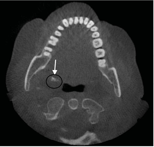
– mild: sclerotic changes are slight, thin or discontinuous, without evidence of plaque thickening;
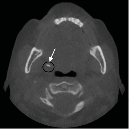
– moderate: sclerotic changes are wider and may be continuous, following the course of the ICA;
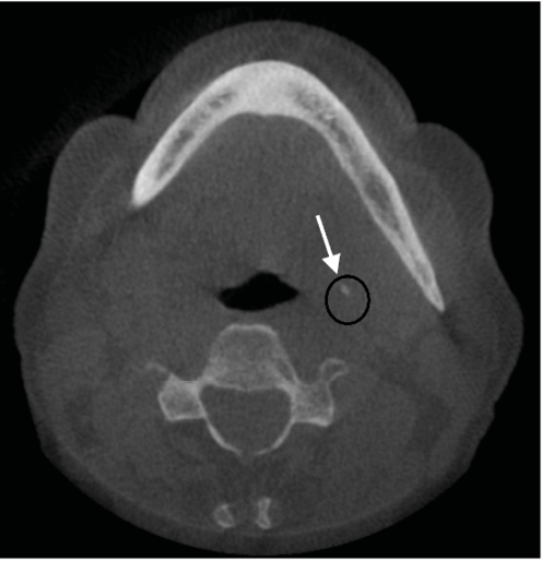
– severe: sclerotic changes are thick, irregular and continuous, consistent with a densely calcified atheromatous plaque.
Statistical analysis
All statistical analyses were performed using Python programming language with appropriate statistical packages, including SciPy, NumPy and Pandas. This study employed a two-tier analytical approach reflecting the hierarchical nature of the data collection: a primary analysis of all patients with CBCT-detected calcifications (n = 108); and a secondary analysis of the subset with severe calcifications confirmed by CT angiography (n = 14).
Primary analysis: CAC severity
This analysis included 108 patients, identified among 890 CBCT examinations, who had detectable CACs and were categorized as having mild, moderate or severe calcifications. Parametric and non-parametric tests were applied for the analysis of associations between patient characteristics and CAC severity, according to the distribution of data:
– continuous variables (age, LDL cholesterol, HDL cholesterol, and TAG): one-way analysis of variance (ANOVA) was used to compare mean values across the 3 calcification severity groups (mild, moderate and severe) for normally distributed continuous variables. Post hoc analyses were performed where appropriate to identify specific between-group differences;
– categorical variables (sex, smoking status, diabetes, hypertension, coagulation disorders, and cardiovascular disease): χ2 test of independence was employed for categorical variables with adequate expected cell frequencies. Fisher’s exact test was used when expected cell frequencies were insufficient, ensuring robust analysis despite small cell counts in some categories.
Secondary analysis: CT angiography stenosis subset
This analysis focused exclusively on the 14 patients with severe calcifications who underwent confirmatory CT angiography. Given the limited sample size, which provided approx. 30% power to detect large effect sizes, non-parametric tests were employed to avoid distributional assumptions. These findings should be interpreted as exploratory and hypothesis-generating rather than confirmatory, as the small sample size limits the ability to detect statistically significant associations even when meaningful trends may exist. Given the small sample size, the following non-parametric tests were applied for the analysis of associations between patient characteristics and the percentage of CAS measured by CT angiography, regardless of data distribution:
– continuous variables (age, LDL cholesterol, HDL cholesterol, and TAG): Spearman’s rank correlation coefficient (rho). This non-parametric correlation method was chosen because it does not assume linearity or normality of data distribution;
– categorical variables (sex, smoking status, diabetes status, hypertension, antihypertensive therapy, cardiovascular disease, coagulation disorders, anticoagulant use, and antidiabetic medication use): Mann–Whitney U test was used to compare percentages of stenosis between two-group categorical variables;
– comparative analysis: Spearman’s correlation was used to examine the relationship between CT angiography and ultrasound measurements. Pearson’s correlation coefficient (r) was additionally reported for comparison.
Interpretation of statistical findings
For the primary analysis (n = 108), we conducted 10 statistical comparisons: age; sex; smoking status; diabetes; hypertension; coagulation disorders; cardiovascular disease; LDL cholesterol; HDL cholesterol; and TAG. To control for type I error inflation due to multiple testing, a Bonferroni correction was applied (adjusted α = 0.05/10 = 0.005). Variables with p-values <0.005 were considered statistically significant after correction. We report both unadjusted and Bonferroni-adjusted significance levels in the results. For the secondary analysis (n = 14), results are presented as exploratory findings with unadjusted p-values, acknowledging the limited statistical power. For the secondary analysis, a p-value <0.05 (unadjusted) was considered statistically significant. Results are presented as mean ± standard deviation (M ±SD) for continuous variables and as frequencies with percentages for categorical variables. All findings from the CT angiography subset should be interpreted as hypothesis-generating rather than confirmatory due to the limited sample size.
Results
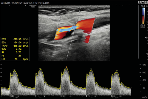
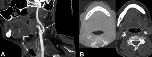
Of the 890 CBCT examinations performed between January 2023 and December 2024, CACs were identified in 108 patients (12.1%). These calcifications were classified as mild (n = 53, 49.1%), moderate (n = 41, 38.0%) or severe (n = 14, 13.0%) (Figure 1, Figure 2, Figure 3). All 14 patients with severe CACs underwent confirmatory vascular imaging: 3 initially underwent Doppler ultrasonography (Figure 4) followed by CT angiography (Figure 5), while 11 proceeded directly to CT angiography. The study population (N = 108) included 53.7% of women and 46.3% of men, all of whom were over 50 years of age.
Analysis of CAC severity in relation to patient characteristics
Analysis of the 108 patients revealed significant associations between calcification severity and several cardiovascular risk factors (Table 1). Age showed the strongest association (p < 0.001), with mean age increasing progressively across the mild (60.8 ±7.4 years), moderate (65.2 ±7.5 years) and severe (73.9 ±5.1 years) groups. Lipid abnormalities were also strongly correlated with severity. High LDL cholesterol was significantly associated with more severe disease (p < 0.001), with 87.0% of patients with high LDL levels presenting with moderate or severe calcifications compared with 24.2% of patients with low LDL levels. Elevated TAG similarly showed a significant association (p < 0.001 for TAG category; p = 0.002 for TAG as a continuous variable), with severe calcifications observed in 26.1% of patients with high TAG vs. 3.2% of those with normal levels. Hypertension was associated with disease severity (p = 0.027), and severe condition was observed in 22.2% of hypertensive patients and 6.5% of normotensive patients.
After Bonferroni correction for multiple testing (adjusted α = 0.005), age, LDL cholesterol and TAG remained statistically significant. Hypertension no longer reached statistical significance after correction, suggesting that this correlation should be interpreted with caution. No significant associations were found with sex, smoking, diabetes, coagulation disorders, or a history of cardiovascular disease. High-density lipoprotein cholesterol approached significance at p = 0.051.
Association between patient characteristics and the degree of carotid artery stenosis on CT angiography
This exploratory analysis examined the 14 patients with severe calcifications who underwent confirmatory CT angiography. Given the limited sample size, these findings should be interpreted cautiously as hypothesis-generating rather than definitive. We investigated the relationship between patient characteristics and the degree of stenosis as measured by CT angiography. This subset had a mean age of 73.9 years and was predominantly female (n = 10, 71.4%) (Table 2,Table 3).
Categorical variables vs. stenosis
As shown in Table 2, none of the examined categorical variables demonstrated statistically significant associations with the percentage of stenosis measured on CT angiography. The relationship between coagulation disorder status and the percentage of stenosis approached but did not reach statistical significance (U = 23.00, p = 0.052). Patients without coagulation disorders exhibited higher mean stenosis values (73.75%) compared to those with coagulation disorders (57.50%); however, this difference was not statistically significant and should be interpreted with caution given the small sample size. Among the other categorical variables, smokers had higher mean percentages of stenosis (75.00%) than non-smokers (72.08%), and patients with diabetes showed higher stenosis values (75.00%) compared to non-diabetic individuals (70.00%). However, these differences did not reach statistical significance (p = 0.226 and p = 0.430, respectively).
Continuous variables vs. stenosis
Table 3 presents the correlations between continuous variables and the percentage of stenosis. Low-density lipoprotein levels showed the strongest correlation with stenosis severity (rho = 0.4549, p = 0.102), indicating a moderate positive association, although this did not reach statistical significance. Higher LDL levels were associated with greater percentages of stenosis, which aligns with established pathophysiology of atherosclerosis. Age showed a weak negative correlation (rho = −0.1904, p = 0.514), while HDL cholesterol demonstrated a very weak negative correlation (rho = −0.0871, p = 0.767). Triglyceride levels exhibited a weak positive correlation with the percentage of stenosis (rho = 0.1962, p = 0.501). We also examined the association between CT angiography and ultrasound measurements, finding a moderate positive correlation (rho = 0.5000, p = 0.667) that did not reach statistical significance, likely due to the limited sample size.
None of the examined continuous variables demonstrated statistically significant correlations with the percentage of stenosis. Given the small sample size (n = 14) and lack of statistical significance, these numerical associations should not be interpreted as clinically meaningful trends but rather as preliminary observations requiring validation in adequately powered studies. In summary, the analysis of the CT angiography subset (n = 14) did not identify any statistically significant associations between patient characteristics and the severity of stenosis. The limited sample size precludes definitive conclusions, and all observations from this subset should be considered exploratory and hypothesis-generating only.
Discussion
The carotid arteries deliver oxygen-rich blood to the head, neck and brain, bifurcating into the external and internal carotid arteries typically at the C3–C4 vertebral level. Carotid atherosclerosis accounts for approx. 20% of all ischemic strokes and TIAs. According to most studies, prevalence of CACs in dental images ranges from 2.00% to 31.57%. Thus, early detection of carotid calcifications may help identify previously undiagnosed coronary artery disease and facilitate referral for further evaluation.19 Atherosclerosis is influenced by various factors, such as diabetes mellitus, obesity, hypertension, smoking, alcohol intake, and chronic renal disorders. Atherosclerosis can lead to strokes when the carotid arteries, which supply blood to the brain, are compromised. It can also cause myocardial infarctions when the coronary arteries, which supply blood to the heart, are affected. These conditions contribute substantially to global mortality.20
Sialolithiasis, tonsilloliths and calcification of lymph nodes and blood vessels may manifest in the cervical area. The need to distinguish these calcifications not only from each other but also from different anatomical structures and structures capable of calcification, such as the stylohyoid ligament, triticeous cartilage and thyroid cartilage, makes the diagnosis of atheromas more complex. While the oral surgeon may find it challenging to differentiate atheromatous calcifications on CBCT from other anatomical and pathological structures, the ability to detect carotid atheromas using CBCT presents an opportunity for early treatment, thereby potentially reducing morbidity and mortality.9 Cone beam computed tomography scans have been increasingly used in recent years for various applications in the dental field. Their value lies in the practitioner’s ability to discover clinically significant incidental findings beyond the area of interest for which CBCT was indicated.14
The objective of this study was to retrospectively assess the prevalence and severity of CACs identified incidentally on CBCT. A study comparing carotid ultrasonography and CBCT revealed that CBCT may identify and quantify minimal calcification volumes, as little as 1 mm3, indicating its high sensitivity in selected cases. Nevertheless, CT remains the preferred imaging modality for a more thorough assessment of CACs, especially in regions outside the typical coverage of CBCT.21 Several imaging modalities have been used to evaluate CACs, including modified posteroanterior radiographs of the neck, where calcifications manifest lateral to the spine, along with CT, which exhibits high sensitivity for identifying intracranial calcifications. Additional techniques include panoramic radiographs, magnetic resonance (MR) angiography, Doppler ultrasonography, and CBCT. In contrast to Doppler ultrasonography and MR angiography, CBCT provides high-resolution 3D imaging of calcifications while potentially reducing costs and radiation exposure compared with conventional CT in selected dental indications.22
The present study highlights the critical role of CBCT in the early detection of CACs as incidental findings during routine dental imaging. A total of 890 patients were evaluated, and CACs of mild, moderate or severe grade were identified in 108 patients along the course of the ICA. Among these patients, severe calcifications were detected in 14 individuals, underscoring the potential of CBCT as a valuable diagnostic tool beyond its primary dental applications. Our findings revealed significant associations between severe CACs and established cardiovascular risk factors, including advanced age, elevated LDL levels, increased TAG levels, and hypertension. These results are consistent with existing literature, indicating that dyslipidemia and blood pressure abnormalities significantly contribute to atherosclerotic lesions in the carotid arteries. Moreover, patients with severe CACs underwent further vascular assessment with CT angiography, which allowed for the evaluation of the presence and degree of CAS. However, due to the small sample size and the fact that CT angiography was performed only in patients with severe CACs, the present study cannot establish a direct correlation between the extent of calcification on CBCT and the degree of CAS.
Such findings emphasize the need to refer patients with suspected CACs for further comprehensive medical assessment, as they may be at elevated risk for cerebrovascular events such as stroke. The results of this study are notable for combining a detailed evaluation of CACs with an assessment of a broad range of associated risk factors. The identification of significant associations between severe calcification and risk factors such as elevated LDL, TAG, hypertension, and advanced age highlights the multifactorial nature of carotid artery disease. By considering these varied parameters together, our study provides broader insight into the possible systemic implications of incidental calcifications identified on CBCT scans.
Our study aligns with previous research showing a clear association between abnormal lipid profiles and CACs. A study by Taengsakul et al. found that high levels of LDL and TAG were closely linked to the development of carotid artery narrowing in patients receiving radiotherapy.23 Likewise, Smith et al. reported that lipid disorders significantly contributed to plaque buildup and vascular stenosis in this population.24 These findings reinforce our conclusion that lipid imbalances play an important role in the progression of carotid calcification. Monitoring lipid levels in patients with CBCT-detected calcifications may therefore enable earlier identification of cardiovascular risk and prompt clinical management.23, 24 Brar et al. provided additional evidence supporting the clinical relevance of CACs seen on dental imaging.6 Their findings demonstrated a strong correlation between calcifications detected on panoramic radiographs and the presence of conditions such as hypertension, hyperlipidemia, diabetes mellitus, stroke, and heart disease.6
Cone beam computed tomography provides 3D information, allowing precise localization of calcifications and volumetric assessment. In addition, by applying a structured severity-based classification and correlating selected cases with vascular imaging, this study highlights aspects that have not been systematically addressed in earlier dental imaging research. These features support the novelty of our approach and underline the role of CBCT as a useful adjunct for the incidental detection of potentially significant findings. Damaskos et al. examined 391 CBCT scans and reported both extracranial and intracranial carotid calcifications, showing associations with systemic risk factors.25 Their findings support the clinical relevance of CBCT in vascular assessment and are in line with the correlations observed in our study.25
Similarly, the CBCT-based staging approach for external carotid calcifications proposed by Kadyan et al. illustrates the value of structured classifications.19 Our work differs by applying a severity-based assessment to internal CACs, with additional correlation with vascular imaging, thereby extending the potential diagnostic contribution of CBCT.19 Furthermore, our retrospective design is consistent with other CBCT-based studies that have examined anatomical and pathological associations, such as the link between Schneiderian membrane thickness and the severity of periodontitis.26
The incidental finding of CACs on CBCT scans provides a distinctive opportunity for the early identification of individuals who may be at increased risk for cardiovascular disease, particularly among asymptomatic patients who might otherwise remain undiagnosed. This highlights a valuable role for dental professionals and radiologists, who should recognize these findings and refer patients for further medical evaluation when necessary. While CBCT is not a substitute for dedicated vascular imaging, its widespread use in dental clinics makes it a practical, non-invasive tool for identifying patients who may benefit from further assessment. As CBCT becomes more common in clinical practice, the implementation of appropriate training and protocols for the recognition of incidental CACs may contribute to earlier diagnosis and the improvement of prevention strategies.
Limitations
This study provides important observations; however, several methodological limitations should be acknowledged. The study is limited by its retrospective design and the relatively small CT angiography subset, which may affect generalizability. However, the use of standardized assessment criteria and a large initial CBCT sample provides meaningful insights. Further prospective studies are needed to confirm these findings.
Conclusions
Cone beam computed tomography has become a valuable tool not only for dental diagnosis but also for the incidental detection of clinically relevant findings such as CACs. This study demonstrates that CACs may be identified incidentally on CBCT examinations and that their presence may warrant further medical evaluation. Given the retrospective design and the limited sample, these findings should be interpreted cautiously and confirmed in larger studies.
Cone beam computed tomography provides 3D visualization, which may facilitate more accurate detection of CACs than conventional dental radiographs. Early recognition of such findings may prompt appropriate medical referral and potentially prevent serious complications.
Ethics approval and consent to participate
The protocol of the study was approved by the Ethics Committee of the University Hospital and the Faculty of Medicine in Pilsen, Charles University (reference No. 93/25).
Data availability
The datasets generated and/or analyzed during the current study are available from the corresponding author on reasonable request.
Consent for publication
Not applicable.
Use of AI and AI-assisted technologies
AI-assisted tool (ChatGPT; OpenAI, San Francisco, USA) was used to refine the language. All scientific content and references were written, reviewed and verified by the authors.