Abstract
Background. Dental implants are a common and effective solution for replacing missing teeth. However, the effects of implant placement on adjacent teeth, particularly at the molecular level, remain incompletely understood. Osteogenic genes play a crucial role in bone formation and remodeling, and their expression in the roots of adjacent teeth may be influenced by the presence of dental implants.
Objectives. The aim of the study was to investigate the effect of implant placement on the expression of osteogenic genes, specifically osteopontin (OPN) and osteocalcin (OC), in the root of the adjacent tooth.
Material and methods. An experimental study was conducted from January to June 2024 using 20 Wistar rats. Implants were placed adjacent to one of the teeth in each rat, while the contralateral tooth served as the control. Tissue samples were collected from the root of the tooth adjacent to the implant (implant group) and from the corresponding contralateral tooth (control group). RNA was extracted and analyzed using real-time polymerase chain reaction (PCR) to assess the expression levels of the OPN and OC genes. Western blot was performed to evaluate protein expression.
Results. The expression of OC was lower in the teeth adjacent to the implants compared with the control group; however, this difference was not statistically significant (p = 0.082). No significant correlation was found between the expression of OC or OPN and the sex of the rats (p > 0.05).
Conclusions. Implant placement has a significant influence on the expression of osteogenic genes in the root of the adjacent tooth, particularly by increasing the expression of OC. These findings suggest that dental implants may influence the molecular environment of adjacent teeth, potentially affecting long-term implant success and the health of adjacent teeth. Understanding these molecular changes can help optimize implant procedures and manage potential complications, ultimately improving patient outcomes.
Keywords: dental implants, gene expression, osteocalcin, osteogenic genes, osteopontin
Introduction
Dental implant placement stimulates ossification in the toothless area and helps preserve the structure of the jaw and face. In other words, after implant placement, several biological reactions occur during bone healing, and their course depends on multiple factors that may determine the success of implant-supported rehabilitation.1 One of the problems associated with dental implant treatment is long healing period of the bone surrounding the implant. Microscopic and macroscopic studies on implant structure have shown that implant characteristics may contribute to accelerating bone adhesion.2 Investigations have demonstrated that the composition and topography of the implant surface have a direct effect on the osteogenic, osteoinductive and osteoconductive activity of osteoblasts. These cellular interactions depend on the coating and binding of adhesive blood proteins to the implant surface, which subsequently serves as a platform for participation and signaling of specific cell receptors. This signaling induces the spatial and temporal expression of growth factors, including bone morphogenetic proteins (BMPs) and transforming growth factor beta (TGF-β). The expression of BMPs initiates a biological cascade of cellular events, including chemotaxis, proliferation and differentiation, ultimately leading to new bone formation.3, 4 The structure of the implant also affects the osseointegration, i.e., the gradual integration of the implant with the surrounding bone tissue.5, 6 Two genes involved in bone morphology and mechanical properties are osteocalcin (OC) and osteopontin (OPN).7, 8 Osteocalcin is an important protein containing γ-carboxyglutamate domains. It plays a role in calcium binding to hydroxyapatite and in bone formation. This protein is produced by osteoblasts.7, 9 Osteopontin gene has a sequence rich in aspartic and glutamic acids that contribute to calcium binding and is considered an important factor in the mineralization of bone-forming cells. Osteopontin also functions as a cytokine involved in the regulation of interferon gamma (IFN-γ) and interleukin (IL)-12 signaling.8 The results of previous studies have shown that the modification of implant surfaces may positively affect initial bone repair and lead to the formation of lamellar bone due to initial bone remodeling. Furthermore, the surface characteristics of biomaterials are important determinants of cellular responses at the implant–bone interface and may influence both the quantity and quality of newly formed bone tissue.10 In addition, the chemical composition of implants may affect cellular functions and the expression of genes involved in bone formation. Cell survival and proliferation are strongly influenced by the surrounding microenvironment, which may consequently affect treatment outcomes.11 Understanding the behavior of osteogenic genes may provide insight into their expression patterns following implant placement.
Rodent models have been extensively used to investigate interventions targeting both bone and oral tissues.12 For instance, Yazarloo et al. performed a systematic review of orthodontically induced white spot lesions in Wistar rats and reported that preventive and therapeutic interventions, such as 5% sodium fluoride varnish and carbon dioxide (CO2) laser irradiation, effectively inhibit enamel demineralization and promote remineralization.13 In a separate toxicological study, Shamel et al. evaluated the effects of chronic intragastric administration of titanium dioxide (TiO2) nanoparticles in albino rats, reporting significant epithelial atrophy and apoptotic changes in the lingual mucosa that were at least partially reversed after a four-week recovery period.14 Rady et al. further showed that first- and second-generation H1 antihistamines induced histological and gene expression alterations involving inducible nitric oxide synthase (iNOS), caspase-3 and α-smooth muscle actin (α-SMA) in the parotid glands of albino rats, highlighting dose- and generation-dependent effects on salivary tissue homeostasis.15 Collectively, these studies underscore the sensitivity of oral and maxillofacial structures to both local and systemic interventions and support the usage of real-time polymerase chain reaction (PCR) to detect slight changes in gene expression. Given the importance of the described issue, the present study was conducted to investigate the impact of implant placement on the expression of osteogenic genes in the root of the adjacent tooth.
Material and methods
Materials and compounds
A real-time PCR kit (Roche, Penzberg, Germany) was used to perform PCR analysis, and ketamine and xylazine (Pfizer Inc., New York, USA) were used to anesthetize the rats. The required buffers for RNA solubilization and purification were obtained from Sigma-Aldrich (Darmstadt, Germany). Gel electrophoresis compounds were acquired from Thermo Fisher Scientific (Waltham, USA).
Ethics approval
The study was conducted in accordance with the ARRIVE (Animal Research: Reporting of In Vivo Experiments) 2.0 guidelines. All experimental procedures were approved by the Ethics Committee of the Clinic of Dentistry and Maxillofacial Surgery (Academy of Košice, Pavol Jozef Šafárik University, Košice, Slovakia) (approval No. REK01/2023) and performed in compliance with institutional and national guidelines for the care and use of laboratory animals.
Inclusion and exclusion criteria
Adult Wistar rats weighing 200–250 g were included in the study. Animals showing signs of illness, abnormal behavior or a weight loss >10% during the acclimatization period were excluded from the study.
Randomization
Animals were assigned to the experimental groups using a computer-generated randomization sequence to minimize selection bias.
Blinding
Investigators performing the molecular analyses were blinded to group allocation during sample processing and data collection.
Sample size calculation
The sample size was determined based on a previous study in the field of implant research and osteogenic gene expression analysis.16 A total of 20 Wistar rats (10 per group) was considered sufficient to detect significant differences in gene expression levels with adequate statistical power, while accounting for potential sample loss during the experimental procedures.
Study sample
After obtaining the required institutional approval, 20 Wistar rats weighing 200–250 g were selected and examined. The animals were fed standard rat chow and provided with water ad libitum throughout the experiment. The animal facility was maintained under 12-h light/12-h dark cycle at a temperature of 23 ±2°C. The laboratory animals were maintained according to the guidelines of the National Institutes of Health (NIH).
Implants
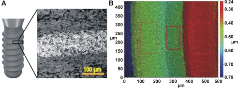
The surface roughness parameter (Sa – arithmetical mean height) was analyzed at ×20 magnification using three-dimensional (3D) optical microscopy (VR-6000; Keyence, Itasca, USA). Micro-rough-surfaced titanium implants (2 mm in diameter × 3 mm in length), manufactured from commercially pure grade IV titanium, were obtained from Southern Implants Ltd. (Irene, South Africa). The surface roughness parameters were: Sa = 849.81 ±11.34 nm; and root mean square height (Sq) = 1.12 ±0.09 µm. The micro-rough implant surface was produced using aluminum oxide blasting, consistent with the technique used for commercially available dental implants (Figure 1).
Grouping and sampling
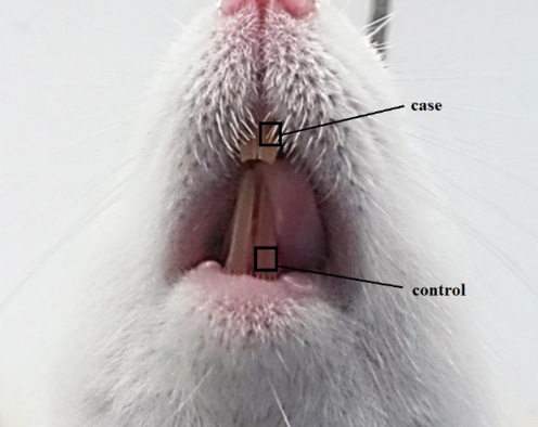
After the extraction of the upper left central incisor, an implant was placed immediately. The upper left central incisor adjacent to the implant was considered the experimental tooth (implant group), whereas the corresponding lower left central incisor in animals that did not undergo implant placement was considered the control tooth (control group) (Figure 2).
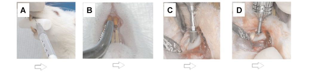
Briefly, implants were placed in the extraction sockets of the upper left central incisors. The osteotomy was prepared using a 1.2-mm pilot drill and a 1.7-mm-diameter final drill (Southern Implants Ltd.). All osteotomy procedures were performed with copious saline irrigation. Micro-rough-surfaced implants were placed into the prepared sockets with a torque of approx. 15 Ncm until the implant shoulder was positioned at the level of the alveolar bone crest. The wound was then sutured with slowly resorbable sutures. Antibiotic cover (enrofloxacin 2.3 mg/kg) was administered by intraperitoneal injection immediately after surgery and once daily for 3 postoperative days (Figure 3).
The control and intervention groups were euthanized 1 week after the procedure. In accordance with the ethical requirements for tissue collection, during the procedure the animals were anesthetized by intraperitoneal injection of ketamine (100 mg/kg) and xylazine (5 mg/kg),17 and the target tissue adjacent to the implant was collected and immediately immersed in TRIzol™ reagent. The material obtained from the dental tissue adjacent to the implant was subjected to real-time PCR analysis to assess the expression of the target genes.
RNA extraction from dental root cells
RNA extraction was performed using a kit (Pars Tous Biotech, Mashhad, Iran). Briefly, to extract RNA from cells, the collected cells were first centrifuged. The resulting cell pellet was washed 3 times with phosphate-buffered saline (PBS), and the cells were sedimented. Then, 350 µL of buffer RLT was added to the pellet, and the sample was centrifuged at 8,000 g for 15 s. After centrifugation, 700 µL of buffer RW1 was added to the column, and the column was centrifuged at 8,000 g for 15 s. After RNA extraction, its quantity and quality were checked by ultraviolet (UV) spectrophotometry (Pico200; Picodrop Limited, Saffron Walden, UK) and agarose gel electrophoresis.
Synthesis of cDNA
The design and preparation of primers for the specific genes (OPN, OC) were analyzed using Primer-BLAST software available in the National Center for Biotechnology Information (NCBI) database (https://www.ncbi.nlm.nih.gov/tools/primer-blast). The GAPDH gene was considered an internal control. The primers were lyophilized and synthesized in Copenhagen, Denmark. In order to prepare the primers for real-time PCR, they were diluted. The sequence of the designed primers for 3 investigated genes is presented in Table 1.
After the master mix reaction and cDNA sample preparation, the PCR product was electrophoresed on a 2% agarose gel at a voltage of 100 V to ensure optimal amplification of the desired fragment and to visualize the product band. Electrophoresis was performed according to the manufacturer’s instructions. The PCR product was mixed with 2 µL of sample buffer and poured into the gel wells.
Western blotting
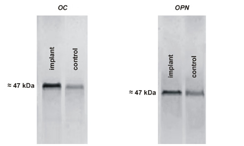
The expression of OC and OPN at the protein level was determined by Western blotting based on the binding of specific antibodies to the OC and OPN proteins, using the method of transferring the stain from sodium dodecyl sulfate–polyacrylamide gel electrophoresis (SDS-PAGE) gel to the nitrocellulose membrane. Protein bands were calculated using ImageJ software (https://imagej.net/ij). Specific antibodies conjugated against histidine were used. The steps were carried out according to previously described methods.
Assessment of the expression of OC and OPN using real-time PCR
A total of 8.8 μL of the prepared mixture and 1 μL of the desired cDNA were added to each real-time PCR microtube. For the positive control, 1 μL of cDNA was transferred to the corresponding microtube. To check for the presence of contamination during the procedure, 8.8 µL of the reaction mixture was added to the negative control microtube along with 1 µL of sterile distilled water. After preparing each microtube, they were placed in the real-time PCR instrument following centrifugation, and the thermal cycling program was carried out according to the manufacturer’s instructions.
Statistical analysis
Statistical analysis was performed using the IBM SPSS Statistics for Windows software, v. 26.0 (IBM Corp., Armonk, USA). The comparison of gene expression among the groups was analyzed using one-way analysis of variance (ANOVA) and multivariate analysis of covariance. The obtained values were expressed as mean ± standard deviation (M ±SD), and p-values <0.05 were considered statistically significant.
Results
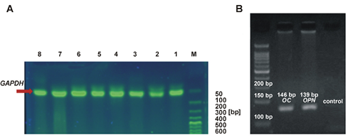
The quality of the extracted RNA was evaluated on an agarose gel. The presence of GAPDH bands indicated the desired quality of the extracted RNA (Figure 4A). To determine the expression of OC and OPN, mRNA was first converted to cDNA using the TRIzol™ method and a cDNA kit. The desired gene fragments can be seen on the agarose gel electrophoresis (Figure 4B).
The expression of OC and OPN was assessed on a 1.5% agarose gel stained with ethidium bromide. The OC gene (146 base pairs) and the OPN gene (139 base pairs) are shown in Figure 4B. Western blot analysis shows clear expression of OC and OPN in the teeth of the implant group. According to Figure 5, the bands corresponding to OC and OPN can be seen in both the control and implant groups.
OC and OPN gene expression
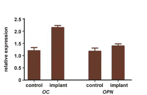
The expression of the GAPDH gene is constant, and a comparison of the treated sample with the control and internal control allows the detection of a decrease or increase in gene expression. The general findings related to gene expression showed that OC expression was 2.17 in the control group and 1.23 in the implant group; the difference was not statistically significant (p = 0.082). The expression of OC in the root of the implanted central teeth was higher than that in the control group; the difference was not statistically significant (p = 0.082).
The expression of OPN was 1.47 in the control group and 1.21 in the implant group. The expression level of the OPN gene was about 1.18 times higher in the implanted samples, but this increase was not statistically significant (p > 0.05) (Figure 6).
In order to investigate the possibility of sex influencing the expression of genes, the association between gene expression in the roots of central teeth and the sex of the rats was investigated. The findings showed that the expression of OC and OPN was not related to the sex of the rats (p > 0.05) (Table 2).
Discussion
Osteogenic genes such as OC and OPN play an important role in bone formation. These genes affect pathways related to proliferation and regeneration.17, 18 The findings of the present study showed that the expression of the OC gene in the implanted central teeth was lower than that in the control teeth; however, the difference was not statistically significant (p = 0.082). Also, the expression level of the OPN gene was lower in the implanted central teeth compared with the controls, but this difference did not reach statistical significance. In a previous study, a significant increase in OC expression after implant placement was reported. In addition, micro-rough implants increased the expression of alkaline phosphatase (ALP), receptor activator of nuclear factor kappa-B ligand (RANKL) and dentin matrix protein 1 (DMP1).19 Another study also reported an increase in the expression of bone-forming genes after implant placement in osteoblast cells.20
Osteopontin is synthesized and secreted by different types of body tissues, including cardiac fibroblasts, osteoblasts, osteocytes, odontoblasts, bone marrow cells, hypertrophied chondrocytes, dendritic cells, macrophages, smooth muscle cells, and skeletal muscle myoblasts. Choi et al. showed that OPN is an important factor in bone regeneration.21, 22
One of the causes of the smaller changes in OC expression is its role in bone formation. The findings show that OC regulates bone quality by modulating the alignment of biological apatite with collagen fibrils. In fact, an increase in OC expression may improve bone structure.23 In addition to the mentioned factors, OC and OPN are related to RUNX2 and DLX5 in osteoblastic differentiation through different cellular pathways.24, 25
Several transcription factors contribute to this process, among which RUNX2 and DLX5 play an important role.26, 27, 28 RUNX2 is essential for osteoblast differentiation and skeletal morphogenesis.19 DLX5 plays a role in bone development and fracture healing.28
Considering the relationship between these proteins, implant placement can affect the levels of OC and OPN through different pathways.
The findings of this study should be considered in the context of the broader molecular landscape of implant osseointegration. While our focus was on OC and OPN, several other biomarkers could provide additional insights into the biological responses to dental implants. Alkaline phosphatase is a key early marker of osteoblastic activity and bone formation, and its expression is typically upregulated during the initial phases of implant healing. Inflammatory cytokines such as IL-1β, IL-6 and tumor necrosis factor-alpha (TNF-α) play crucial roles in the inflammatory response following implant placement and can influence osteogenic differentiation. Matrix metalloproteinases (MMPs), particularly MMP-2 and MMP-9, are involved in extracellular matrix remodeling and bone tissue integration. Additionally, bone turnover markers such as C-terminal telopeptide (CTX) and N-terminal telopeptide (NTX) could provide information about bone resorption activity around implants. Several studies have investigated the effects of implant surface characteristics on various biomarkers. Rough implant surfaces have been shown to enhance osteoblast adhesion, proliferation and differentiation, leading to improved bone–implant contact and osseointegration.29, 30, 31, 32 The surface topography and chemical composition of implants can modulate the expression of multiple genes involved in bone metabolism, including ALP, osteonectin and insulin-like growth factor 1 (IGF-1).33 Future studies should consider a more comprehensive panel of biomarkers to better understand the multifaceted biological responses to dental implant placement and to provide a more complete picture of the molecular mechanisms underlying implant osseointegration.
Some studies have found sex to be associated with bone formation.34 In the present study, in order to investigate the possibility of sex influencing the expression of genes, the association between gene expression in the roots of central teeth and the sex of the rats was examined. The findings showed that the expression of OC and OPN genes was not related to the sex of the rats. In this study, rough-surfaced titanium implants were used. The type of titanium implant has been established as appropriate for this purpose.
The long-term success of dental implants depends on several factors, including osseointegration. In addition, the chemical biocompatibility of the material and the microgeometry of the implant surface are important for implant success and long-term bone grafting. They also influence the cellular response, causing increased bone formation and thus improving implant stability.35 In addition, implant morphology can affect bone metabolism: rougher surfaces stimulate the growth and attachment of bone cells and increase mineralization.36 The development of bone–implant interfaces depends on the direct interaction of the bone matrix and osteoblasts with biomaterials. Osteoblast adhesion is essential for bone–biomaterial interaction.37, 38 Studies have shown that implants with rough surfaces have better bone-to-implant contact (BIC) than those with smooth surfaces. Surfaces with moderate roughness (Sa between 1.0 µm and 2.0 µm) provide a better bone response than smoother surfaces. Among conical implants, the input torque is influenced by the design and implant length.39
Some researchers have found that implant design and placement techniques are effective in promoting bone formation and faster healing. It has been reported that shear force during implant placement affects the proliferation of bone cells.40, 41 On the one hand, considerable research has been conducted to improve the microscopic and macroscopic structure of implant surfaces in order to accelerate and increase bone adhesion to the implant surface, which has resulted in positive outcomes.2 On the other hand, by using biological agents such as BMPs and growth factors in the vicinity of the implant surface or in the defects created around it, researchers have attempted to increase the rate and speed of bone formation. In this regard, many studies have been conducted on plasma growth factors that are obtained from platelets, which have resulted in different findings.42 Therefore, the use of structures effective in ossification around implants may increase the efficiency of treatment and the expression of osteogenic genes.
Although OC and OPN are well-established markers of osteoblastic activity and matrix maturation, bone remodeling and implant osseointegration are orchestrated by a broader network of anabolic and catabolic factors. Key enzymes and cytokines, such as ALP, ILs (IL-1β, IL-6), TNF-α, MMPs (MMP-2, MMP-9), and resorption markers like CTX or NTX, play pivotal roles in the initiation, progression and resolution of bone turnover around implants. By limiting our analysis to OC and OPN, we may have overlooked important pro-inflammatory or resorptive signals that could modulate implant stability and adjacent tooth root remodeling. Future studies should therefore employ a multiplex approach (e.g., Luminex, proteomic arrays) or targeted quantitative PCR panels to capture both osteogenic and osteoclast biomarkers. This more comprehensive profiling will yield a more comprehensive picture of the molecular microenvironment governing peri-implant bone healing and long-term integration.
Limitations
This study has several limitations that should be acknowledged. Firstly, the use of rat models, while appropriate for initial experimental studies, may not fully represent human biological responses due to species-specific differences in bone metabolism and gene expression patterns. Secondly, the relatively short follow-up period of 1 week may not reflect long-term molecular changes and gene expression dynamics that occur during the osseointegration process. Thirdly, the analysis was limited to 2 osteogenic biomarkers (OC and OPN), which may introduce selection bias and provide an incomplete picture of the complex molecular responses occurring around dental implants. Many other important biomarkers were not evaluated, including ALP as an early osteoblastic differentiation marker, inflammatory cytokines such as ILs (IL-1β, IL-6, IL-10) and TNF-α, which play crucial roles in the inflammatory phase of osseointegration, as well as MMPs involved in tissue remodeling, and bone resorption markers such as CTX and NTX. The inclusion of these additional biomarkers would have provided a more comprehensive and realistic assessment of the biological processes involved. Future studies should consider a broader panel of biomarkers to better understand the multifaceted molecular responses to dental implant placement.
Conclusions
In this controlled animal study, placement of micro-rough-surfaced titanium implants in the maxilla was associated with a significant upregulation of OC mRNA and protein expression in the roots of adjacent teeth as early as 1 week after implantation. The expression of OPN also showed an upward trend, although the change did not reach statistical significance. No sex-related differences in OC or OPN expression were observed. These findings suggest that peri-implant osteogenic activity extends beyond the implant site and modulates molecular markers in neighboring tooth structures. Nevertheless, the short follow-up period of 1 week, limited biomarker panel, and absence of histological or biomechanical assessments limit the generalizability of the findings. Future studies with longer observation periods, broader panels of anabolic and catabolic markers (e.g., ALP, RANKL/osteoprotegerin (OPG), cytokines, MMPs, and CTX/NTX), and complementary imaging or push-out tests are needed to characterize the temporal and functional aspects of adjacent tooth remodeling. Such studies may provide further insight into the development of implant surfaces and adjunctive therapies aimed at optimizing both implant osseointegration and preservation of neighboring dental tissues.
Ethics approval and consent to participate
All experimental procedures were approved by the Ethics Committee of the Clinic of Dentistry and Maxillofacial Surgery (Academy of Košice, Pavol Jozef Šafárik University, Košice, Slovakia) (approval No. REK01/2023) and performed in compliance with institutional and national guidelines for the care and use of laboratory animals.
Data availability
All data generated or analyzed during this study is included in the published article.
Consent for publication
Not applicable.
Use of AI and AI-assisted technologies
Not applicable.