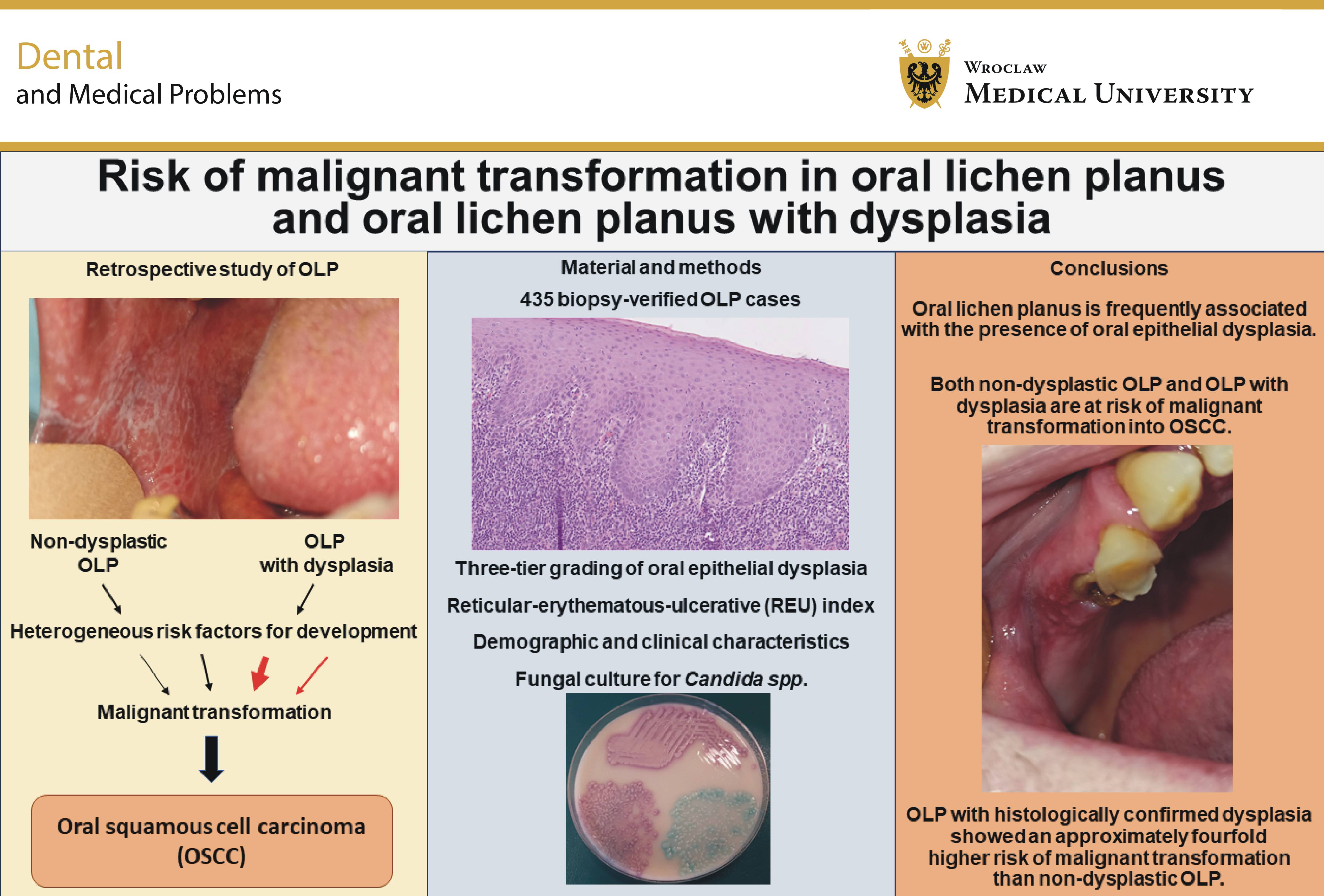
Abstract
Background. Chronic activity of the subepithelial lymphocytic infiltrate may induce epithelial changes associated with malignant transformation (MT) in oral lichen planus (OLP) cases.
Objectives. This study compared the risk of developing oral squamous cell carcinoma (OSCC) in a large cohort of patients with OLP and OLP with epithelial dysplasia (OLD). Special attention was paid to carcinoma location and clinical factors, such as desquamative gingivitis and reticular, erosive or ulcerative status.
Material and methods. This retrospective study included 435 histologically verified OLP/OLD cases treated between 2004 and 2024. To assess risk factors, medical history data on medication, smoking and comorbidities was collected. Clinical and pathological aspects were correlated. Statistical analyses were performed using SigmaXL software.
Results. Of the cases included in the study, 59.8% were classified as OLP (260/435) and 40.2% as OLD (175/435). A case was considered a true MT only if it followed the OLP/OLD biopsy results. According to this criterion, 30 OSCCs were diagnosed in 21 patients. Oral squamous cell carcinoma developed in 2.3% of OLP cases (6/260) and 8.6% of OLD cases (15/175), with a statistically significant difference between the groups (p = 0.0027). The most common site for OSCC was the gingiva/alveolar mucosa, accounting for 33.3% (10/30) of tumors, followed by the tongue (20.0%, 6/30). Two alveolar carcinomas were located at sites of peri-implantitis associated with desquamative gingivitis. In 2 patients, synchronous OSCCs were diagnosed in the maxilla and mandible.
Conclusions. In this study, 8.6% of patients with OLD developed OSCC. Although OLP without dysplasia still carries a risk of OSCC, this risk was approx. 4 times lower, at 2.3%. Cases with desquamative gingivitis require close monitoring due to their higher risk of carcinoma development. We recommend careful observation of desquamative gingivitis around dental implants or in areas of ongoing dental inflammation. Surgical revision with biopsy should be considered if a resorptive process around an implant is suspected.
Keywords: oral lichen planus, oral squamous cell carcinoma, oral potentially malignant disorder, desquamative gingivitis, oral epithelial dysplasia
Introduction
Oral lichen planus (OLP) is one of the most common non-infectious inflammatory diseases of the oral mucosa, affecting approx. 0.5–2.5% of the population.1, 2, 3 The condition was first described by Sir Erasmus Wilson in 1869.3 Oral lichen planus predominantly affects individuals over the age of 40, with a higher prevalence among women.4
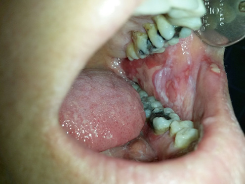
Clinically, OLP is characterized by white hyperkeratotic reticular (lacy) or linear patches, as well as atrophic and erosive lesions.1, 2, 3 The clinical presentation of OLP is illustrated in Figure 1. A specific manifestation commonly observed in OLP is desquamative gingivitis, whereas plaque-like lesions may also be seen on the dorsal surface of the tongue.4, 5, 6 While the etiology of OLP remains largely idiopathic, OLP-like lesions may sometimes be associated with adverse effects of medications or local irritation caused by dental materials.2, 4, 7, 8, 9, 10, 11, 12 The clinical course of OLP is typically characterized by periods of remission and relapse.1 Furthermore, OLP is frequently associated with comorbidities, particularly immune-mediated liver and thyroid disorders.13, 14, 15
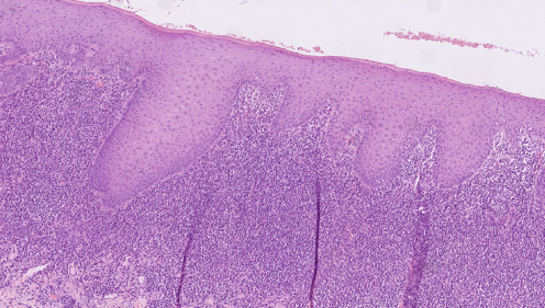
The histopathological criteria for OLP include parakeratosis, acanthosis and a well-defined subepithelial lymphocytic infiltrate.2, 6, 16, 17, 18 This infiltrate consists primarily of CD8+ cytotoxic T lymphocytes, which may contribute to impaired epithelial regeneration by targeting basal cells.4, 19 Oral lichen planus is associated with systemic immune responses.20 Specific antibodies against OLP have not been identified, but patients may have elevated antinuclear antibody titers.4 Salivary immunoglobulin A may contribute to the local immune response.20, 21, 22 The diagnosis of OLP is established through clinical evaluation and histopathological correlation, and augmented by direct immunofluorescence that typically demonstrates shaggy fibrinogen deposition along the basement membrane zone (Figure 2).23, 24, 25
The reticular-erythematous-ulcerative (REU) index and the visual analog scale (VAS) are established tools used to comprehensively assess the progression of lesions and subjective complaints during the monitoring process.26, 27 The REU index was introduced by Piboonniyom in 2005.26 Numerous studies have indicated a correlation between hyperkeratotic lesions and an asymptomatic disease course, whereas erythematous and ulcerative lesions are often associated with pain and burning sensation.1, 2, 3
Biopsy of an OLP lesion is a critical procedure not only for establishing an accurate initial diagnosis but also for identifying the presence of oral epithelial dysplasia (OED).2, 23, 28, 29 When OED coexists with OLP, the condition is termed OLP with dysplasia (OLD).2 Immunohistochemistry serves as a vital tool in enhancing the interpretation of histological findings. The most frequently used markers include p53 and Ki-67 proteins.28, 30, 31 The p53 protein plays a significant role in cell cycle regulation,31 whereas Ki-67 positivity is associated with increased cellular proliferation.32 Notably, many dysplastic epithelial nuclei may exhibit a mutated form of p53.1, 33, 34 In cases of OLD, a differentiated type of oral intraepithelial neoplasia (dOIN) is often observed. This pathology was defined in 2010 as a high-risk lesion with a considerable likelihood of malignant transformation (MT) into oral squamous cell carcinoma (OSCC) within 5 years.35
The first case of an MT of OLP to OSCC was recorded over a century ago. Since 1978, OLP has been recognized as a precancerous condition. It was included among oral potentially malignant disorders (OPMDs) in the 3rd and 4th editions of the World Health Organization (WHO) classification.1, 28 According to the definition of an OPMD provided in the 5th edition of the WHO classification, published in 2022, it is important to accurately correlate the clinical behavior of chronic lesions with the criteria for OED and to assess human papillomavirus (HPV) status in biopsy samples.28 Malignant transformation associated with OLP is characterized by increased proliferation of atypical epithelial cells alongside the accumulation of changes induced by carcinogens or chronic inflammatory processes.36, 37 Research that fails to differentiate between OLP, OLD and other related lesions may overestimate the risk of carcinoma attributable specifically to OLP. Systematic reviews have indicated that the rate of MT in OLP ranges from 0% to 3.5%.38 However, when cases lacking a minimum 6-month interval between the diagnosis of OLP and OSCC are excluded, this rate decreases significantly to approx. 0.44%.36 Furthermore, erosive forms of OLP are considered to carry a greater risk. Other significant contributing factors include the infection with Candida albicans and the presence of OED in biopsy specimens.28
Accurate differentiation between these disorders is essential, particularly when distinguishing OLP and OLD from the more serious condition of proliferative verrucous leukoplakia (PVL).28 These conditions share several characteristics, including multifocal hyperkeratotic lesions, a lymphocytic subepithelial infiltrate and frequent involvement of the gingiva.28 However, PVL is distinctly characterized by the presence of verrucous lesions, aggressive proliferation and a high propensity for early recurrence following excision, as specified in the diagnostic criteria for this condition.39 Notably, the rates of MT associated with PVL range from 50% to 100%.38
Topical treatment of OLP primarily involves corticosteroids such as dexamethasone acetate or clobetasol propionate.40, 41 In cases where a lesion is associated with severe clinical symptoms, surgical removal is performed, and the tissue is submitted for histopathological examination. Comprehensive treatment also addresses subjective complaints and includes the management of secondary oral candidiasis that may arise following corticosteroid use. Supportive management may include a combination of dexpanthenol, lidocaine hydrochloride, laser therapy, and antifungal medications.6, 42, 43, 44, 45, 46
The issue of MT in OLP remains controversial. The available data is often limited to malignancy rates without consideration of the clinical aspects of OLP. This study aims to conduct a comprehensive comparison of factors associated with MT in differentiated OLP and OLD case cohorts. Additionally, we aim to examine the correlation between risk factors and clinical or demographic characteristics, such as the REU index, mycological culture results and smoking status.
Material and methods
This retrospective study examined 435 histologically verified cases of OLP and OLD that were diagnosed and treated between 2004 and 2024 at the Department of Stomatology-Oral Medicine (University Hospital Pilsen, Czech Republic). The mean duration of clinical follow-up was 5.4 years.
Inclusion criteria
Only OLP and OLD cases verified both clinically and histopathologically were included in the study. For the calculation of MT, only OSCCs that developed within clinically documented OLP or OLD lesions were included.
Exclusion criteria
Clinical cases of OLP without histopathological verification were excluded, as were cases meeting the Cerero-Lapiedra criteria for PVL.39 Five patients previously treated for OLD were excluded due to a diagnosis of PVL. Oral squamous cell carcinoma cases with no proven association with previously verified OLP or OLD sites were also excluded.
Histopathological, clinical and microbiological assessment
The histopathological diagnosis of all cases of OLP and OLD was based on routine biopsy specimens stained with hematoxylin and eosin. All histological examinations were performed at a single institution, the tertiary care hospital. Oral epithelial dysplasia was graded using a three-tier system (OED I–III) according to the proportion of the epithelial thickness exhibiting dysplastic features. To clarify findings concerning epithelial atypia and dysplasia, immunohistochemical assessment of mutated p53 protein expression was conducted and classified as negative, positive in single nuclei, or positive in short or long lines of nuclei. The positivity of mutant p53 protein in a long line of nuclei was considered a risk-associated sign. The final outcome was the occurrence of MT of OLP/OLD lesions into OSCC.
Medical histories were obtained to collect data on comorbidities, particularly lichen ruber planus and lichen atrophicus, as well as medication use and allergies. The clinical manifestations of OLP and OLD were assessed using the REU index (range: 1–115, divided into 10 reticular, 45 erythematous and 60 erosive OLP areas), with cases categorized as either erosive or non-erosive and gingival involvement recorded. Mucosal swabs for fungal culture were obtained from all patients to diagnose primary oral candidiasis at the beginning of treatment or secondary corticosteroid-related fungal infection. The results were assessed by the microbiology laboratory using the colony-forming unit (CFU) system for both C. albicans and non-albicans Candida species (range: 0–150). A VAS was used to measure subjective complaints such as pain and burning sensation in all cases (range: 0–10). The VAS was assessed regularly throughout follow-up. The collected data was analyzed using SigmaXL software (v. 10; SigmaXL Inc., Kitchener, Canada).
Results
This retrospective study comprised 435 OLP/OLD cases, including 300 females and 135 males. The female-to-male ratio was 2.2:1 (69% female and 31% male). The average age of the participants at the time of first contact was 56.5 years. The mean duration of treatment was 5.4 years. Within the study cohort, 19.7% (86/435) of patients reported smoking habits. Additionally, thyroid disease was observed in 19.3% (84/435) of cases, with one-third of these diagnoses made following clinical referral.
Surgical interventions included either diagnostic or therapeutic excision of the most clinically significant lesion in each case. Furthermore, topical antifungal agents, including nystatin suspension and miconazole gel, were used in 20.7% (90/435) of cases with elevated Candida culture results.
The cohort was categorized according to the presence of OED in biopsy specimens, resulting in 2 distinct groups: OLP and OLD. Patients with OLP represented 59.8% of the cohort, comprising 260 cases out of 435, while OLD accounted for 40.2%, with 175 cases. The OED criteria were present in one-third of the epithelium in 80.0% (140/175) of OLD cases, in two-thirds in 14.3% (25/175), and in the entire epithelial thickness in 5.7% (10/175). Regarding direct immunofluorescence testing, positive results were identified in 64.3% of OLP cases (144/224) and in 37.4% of OLD cases (52/139). The demographic and clinical characteristics, including sex distribution, age at onset, smoking status, subjective complaints, and REU index, were comparable between the 2 groups. Furthermore, dysplastic cases were more frequently associated with erosive lesions and exhibited p53 positivity in long lines of basal cell nuclei. The correlation between OLD and the presence of p53-positive nuclei arranged in long lines within the basal cell layer was statistically significant (p < 0.0001; two-sample t-test; SigmaXL). Table 1 presents a comparison of demographic and clinicopathological characteristics between OLP and OLD.
An OSCC was classified as an MT only when it was diagnosed more than 6 months after a biopsy-confirmed diagnosis of OLP or OLD. According to these criteria, 30 OSCCs were diagnosed in 21 patients. Four additional cases of OLD were excluded from the MT calculations because OPMD and OSCC were diagnosed on concurrent biopsies or because OLD was identified after a previous diagnosis of malignancy.
In the cohort of patients with OLP, the rate of MT into OSCC was 2.3% (6/260). In contrast, the rate of MT in OLD was significantly higher, at 8.6% (15/175), with the difference being statistically significant (p = 0.0027; two-sample t-test). If the minimum 6-month interval were not applied, the rate of MT in OLD would increase to 10.9% (19/175) and potentially to 13.3% (24/180) if PVL-positive cases were included.
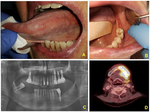
The gingiva/alveolar mucosa was the most frequently affected site of OSCC, accounting for 33.3% (10/30) of cases. A case of OLP with MT in the alveolar region is shown in Figure 3.
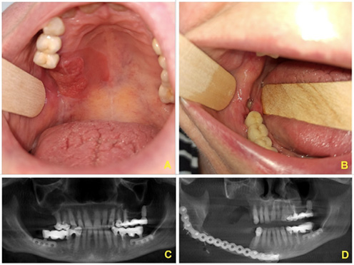
The tongue and labial mucosa were the next most frequently affected sites, accounting for 20.0% each (6/30), followed by the buccal mucosa at 16.7% (5/30 cases). Additionally, 2 cases of alveolar carcinoma were identified in patients with peri-implantitis accompanied by desquamative gingivitis. Furthermore, 2 individuals presented with synchronous OSCCs involving both the maxilla and mandible. Seven patients developed 2 carcinomas, while 1 patient developed 3 carcinomas. A case of OLP exhibiting synchronous MT in the maxillary and mandibular regions is presented in Figure 4.
Table 2 presents a comparison of the 6 OLP and 15 OLD cases that underwent MT into OSCC. The sex ratio among patients with MT was comparable to that of the overall cohort, with 66.7% of patients with MT being female. Notably, the mean REU score was 15, compared with 10 for the entire cohort. Furthermore, risk factors appeared to be more prevalent among OLD cases with an erosive form, which accounted for 73.3% (11/15), and among OLD cases with desquamative gingivitis, which accounted for 53.3% (8/15).
Discussion
The etiology of OLP remains largely unclear despite its prevalence exceeding 2% in the general population.1, 2, 3 Current understanding suggests that an inappropriate immune response to an unidentified antigen may impair epithelial regeneration, a process mediated in part by cytotoxic CD8+ T lymphocytes.2, 4, 19
Typically, individuals with OLP are older than 40 years, with a slight female predominance, reported at a ratio of 1.4:1.4 Our study showed a more pronounced female predominance, with female-to-male ratios of 2.33:1 for OLP cases and 2.13:1 for OLD cases.
Oral lichen planus is associated with thyroidopathy in approx. 8% of cases.14, 15 In our study, this comorbidity was present in nearly one-fifth (19.3%) of patients. Notably, many cases of thyroidopathy were identified following our recommendation for further evaluation after the diagnosis of OLP or OLD,14, 15 highlighting the importance of comprehensive patient assessment in these instances.
Corticosteroids are frequently used for the topical treatment of OLP; however, they may contribute to secondary Candida growth in certain cases.40, 41, 42 In our study, topical antifungal treatment was necessary in 20.7% of the cases analyzed.
Malignant transformation should be understood as a gradual process characterized by an increase in epithelial cell atypia rather than as a single event. Therefore, it is crucial to perform histological verification of OLP to detect the presence of OED. This verification enables appropriate adjustments to monitoring protocols and informs decisions regarding potential re-excision, both of which are key aspects of effective patient management. According to a systematic review published in 2019, the MT rate for OLP ranges from 0.0% to 3.5% over a period of 5–7 years.38 This range is consistent with our MT rate for non-dysplastic OLP cases, which was 2.3%. Conversely, OLD cases showed a significantly higher MT rate of 8.6%. These calculations were performed after excluding cases with synchronous or preceding OSCC diagnoses and cases associated with PVL.
Several factors have been identified as potential predictors of MT in OLP, including the erosive form of the condition, tongue localization, smoking, and the presence of Candida.18, 28 In our study, 50.0% of MT cases involving OLP and 73.3% of OLD cases with MT presented with erosive manifestations. Additionally, MT cases exhibited a higher mean REU index than the general cohort (15 vs. 10), although there was considerable variation, with the values ranging from 2 to 59. Notably, smoking did not appear to be a direct risk factor, as the prevalence of smokers among patients with MT (23.0%) was comparable to that in the general study population (19.8%).
In our study cohort, one-third of OSCCs (10/30) occurred in the alveolar mucosa or gingiva. Within this subset, 2 cases presented with synchronous tumors involving both the maxilla and mandible, as illustrated in Figure 3 and Figure 4. These specific cases were identified in regions affected by peri-implantitis. Other gingival cases were associated with heightened inflammation at periodontal sites. Chronic inflammation has the potential to disrupt healing pathways, which may contribute to the emergence of epithelial atypia. Furthermore, aggressive periodontal pathogens have been associated with several local or distant malignancies.47, 48, 49, 50, 51 This pathogenetic cofactor is likely responsible for the higher number of cases with MT observed at alveolar sites in the OLP cohort. Management of OLP affecting the gingiva should therefore include comprehensive periodontal care. In instances where there is suspicion of a progressive inflammatory process around dental roots or implants, particularly in conjunction with desquamative gingivitis, surgical revision with biopsy should be considered to establish an accurate diagnosis. Consequently, it is advisable to monitor the alveolar manifestations of OLP or OLD using orthopantomography or cone-beam computed tomography.
It is important to note that a persistent source of bias in research of this nature is the potential for inaccurate data. To minimize this issue, we included only histopathologically confirmed cases with comprehensive medical history and clinical documentation. There is a pressing need for more targeted studies to clarify the interactions between the oral microbiome, genetic alterations and the development of OED. In the near future, the use of neural networks could improve the detection of high-risk cases.52 The issue of MT in alveolar OLP, particularly at dental implant sites, warrants investigation in large multicenter studies, similar to other projects in the field of cancer prevention and the management of malignant disease-related complications.53, 54
Conclusions
The current criteria for OPMDs, published in the 5th edition of the WHO classification, are proving to be effective. Our study found that 8.6% of patients with OLD developed OSCC. Oral lichen planus without dysplasia was associated with a lower risk of MT, at 2.3%. Hence, our findings align with the WHO’s assessments of these pathologies.
It is essential to manage cases of desquamative gingivitis carefully, as they demonstrate the highest risk for carcinoma development. Therefore, we recommend close clinical monitoring of desquamative gingivitis, especially in the vicinity of dental implants or in conjunction with ongoing dental inflammation. If a progressive resorptive process around an implant is suspected, surgical revision with biopsy should be considered.
Ethics approval and consent to participate
The scientific project was approved by the Institutional Review Board and Ethics Committee of the University Hospital Pilsen (Faculty of Medicine in Pilsen, Charles University, Pilsen, Czech Republic) (reference No. 231/24; August 1, 2024). The study was conducted in accordance with the Declaration of Helsinki. Written informed consent was obtained from all participants before their inclusion in the study.
Data availability
Not applicable.
Consent for publication
Not applicable.
Use of AI and AI-assisted technologies
Not applicable.