Abstract
Background. The oral microbiota consists of a diverse range of microorganisms, with Streptococcus spp. and Candida spp. frequently coexisting in oral infections.
Objectives. The aim of the study was to investigate the impact of Er:YAG (erbium-doped yttrium aluminum garnet) laser therapy, utilizing the PS04 fractional beam, on the in vitro growth and biofilm formation of clinical strains of Candida albicans, Candida glabrata and Streptococcus mutans.
Material and methods. Single- and dual-species planktonic cultures and biofilms were exposed to an Er:YAG laser using a fractional PS04 handpiece. The effects of the laser were evaluated immediately after irradiation and 24 h post-irradiation by measuring colony-forming units per milliliter (CFU/mL). Biofilm biomass (single- and dual-species) was quantified using the crystal violet staining method. The study tested 2 sets of laser parameters: group 1 (T1): 1.5 W, 10 Hz, 30 s, 0.4 J/cm2, irradiance: 3.9 W/cm2; and group 2 (T2): 6.15 W, 10 Hz, 30 s, 1.6 J/cm2, irradiance: 16 W/cm2. Non-irradiated samples served as controls. The parameters were selected based on their frequent clinical use for snoring treatment and facial rejuvenation.
Results. Candida albicans exhibited a significantly greater reduction under T2 settings in comparison to T1 (85.3 ±1.2% vs. 43.9 ±4.5%, respectively; p = 0.006) within single-species biofilms. For C. glabrata, a significant reduction was observed under T1 parameters (69.8 ±14.9%). Furthermore, S. mutans demonstrated a significantly higher reduction at T2 settings (97.1 ±0.6%) compared to T1 settings (81.1 ±19.6%), with statistically significant differences noted between S. mutans and both C. albicans and C. glabrata under T1, as well as between S. mutans and C. glabrata under T2. In dual-species biofilms, T2 fluence led to greater reductions in C. glabrata, S. mutans and C. albicans in mixed cultures (p < 0.05).
Conclusions. The Er:YAG laser, when used in conjunction with the PS04 handpiece, demonstrated a substantial reduction in the biofilms of C. albicans and C. glabrata. Higher fluence maintained reductions over a 24-h period, particularly in the case of C. glabrata and S. mutans. This study highlights the antifungal potential of low-fluence laser settings that are commonly used in facial aesthetic procedures and snoring treatment.
Keywords: Candida albicans, candidiasis, oral biofilm, erbium laser, Streptococcus mutans
Introduction
The health of the oral cavity is closely interconnected with systemic health.1, 2, 3 Rather than being merely susceptible to microbial colonization, the oral cavity naturally harbors a diverse and dynamic microbial community. This is due to its continuous exposure to the external environment through food, air and other factors, which contribute to the establishment and maintenance of a complex ecosystem of bacteria, fungi and other microorganisms. These microbes can disrupt oral homeostasis, leading to dental caries, periapical infections and candidiasis. In some cases, microbial dysbiosis may contribute to systemic diseases, including cardiovascular disorders, pneumonia and stroke.4 Among the primary pathogens involved, Streptococcus mutans is a key etiological agent in dental caries and a contributor to systemic conditions such as infective endocarditis.5 Additionally, fungal infections, primarily caused by Candida albicans, can disrupt the balance of oral microbiota and pose significant challenges due to their opportunistic nature and resistance.6
A critical factor in the development of oral candidiasis is its nature as a multi-species infection, as opposed to being caused by a single fungal species. While C. albicans is the predominant species responsible for infections, it exists alongside diverse strains and other Candida species, which further complicates infection management. In severe cases, Candida spp. may invade the bloodstream, leading to systemic, life-threatening conditions such as candidemia.6 Conventional treatment for oral candidiasis includes antifungal agents such as nystatin, amphotericin B, or systemic therapies like ketoconazole, fluconazole and 5-fluorocytosine. However, systemic antifungal treatments frequently cause adverse effects, including gastrointestinal disturbances, nephrotoxicity and hepatotoxicity, highlighting the need for innovative strategies.7 Emerging alternatives, such as probiotics, show promise in preventing oral candidiasis and alleviating symptoms, yet they remain less effective for treating established infections.8, 9, 10, 11, 12
Laser therapy has emerged as a promising and increasingly popular approach in disease management over the years.13, 14, 15, 16, 17, 18, 19, 20, 21, 22, 23, 24, 25 The antimicrobial action of the laser is particularly noteworthy and widely emphasized.26, 27, 28, 29, 30 Among various types of lasers, the Er:YAG (erbium-doped yttrium aluminum garnet) laser has found wide application in multiple fields of dentistry.31, 32, 33, 34, 35, 36 Studies have demonstrated the effectiveness of the Er:YAG laser in managing microbial biofilm during endodontic treatment37, 38, 39, 40, 41 and reducing biofilm on dental implants affected by peri-implantitis.42, 43 Additionally, Er:YAG laser bleaching has been shown to reduce biofilm formation on enamel surfaces in comparison to conventional bleaching methods without laser application.44 These findings, along with others, highlight the beneficial effects of Er:YAG laser therapy in inhibiting or reducing biofilm formation in the oral cavity.45, 46, 47, 48 Beyond its efficacy in destroying bacterial biofilms, the Er:YAG laser has also shown potential in reducing Candida biofilm formation, further expanding its potential applications in dental and oral health management.22, 49, 50
The present study aims to evaluate the effects of the Er:YAG laser on planktonic single-species, dual-species, and biofilm composed of C. albicans, Candida glabrata and S. mutans strains. Additionally, it explores innovative approaches to eradicate these pathogens using low-fluence laser parameters, specifically designed for the clinical management of snoring and frequently employed in facial aesthetic procedures.
Material and methods
The research was carried out on clinical strains of C. albicans, C. glabrata and S. mutans at the Department of Microbiology, Wroclaw Medical University, Poland, under the approval of the Institutional Research Ethics Committee (approval No. KB-429/2024). A power meter was utilized, and the output was calibrated prior to the commencement of the study.
Culture condition
Clinical cultures of C. albicans, C. glabrata and S. mutans were obtained from patients with candidiasis and caries lesions and stored at −80°C in Trypticasein Soy Broth (BioMaxima, Lublin, Poland). The strains were frozen in replicates and multiplied before experiments (S. mutans on Brain Heart Infusion (BHI) LAB-AGAR™; Candida spp. on Sabouraud Dextrose LAB-AGAR™ (BioMaxima)).
Two types of samples were prepared for experimentation:
• planktonic solutions: suspensions of microorganisms (0.5 McFarland standard) were combined with BHI broth (BioMaxima) containing 5% sucrose, forming single-species (C. albicans, C. glabrata, S. mutans) or dual-species combinations (C. albicans + S. mutans; C. glabrata + S. mutans; C. albicans + C. glabrata). The treatments were conducted in dark Eppendorf tubes (SARSTEDT AG & Co. KG, Nümbrecht, Germany);
• biofilm: single- and dual-species biofilms were formed on 96-well polystyrene plates (SPL Life Sciences Co., Ltd., Pocheon, Korea). For single-species biofilms, 100 µL of microbial suspensions (0.5 McFarland standard) and 150 µL of BHI broth were added per well. For dual-species biofilms, 100 µL of each microorganism suspension and 50 µL of BHI broth were used, totaling 250 µL per well. The plates were incubated at 37°C for 24 h under aerobic or CO2-enhanced conditions (S. mutans).
Laser irradiation
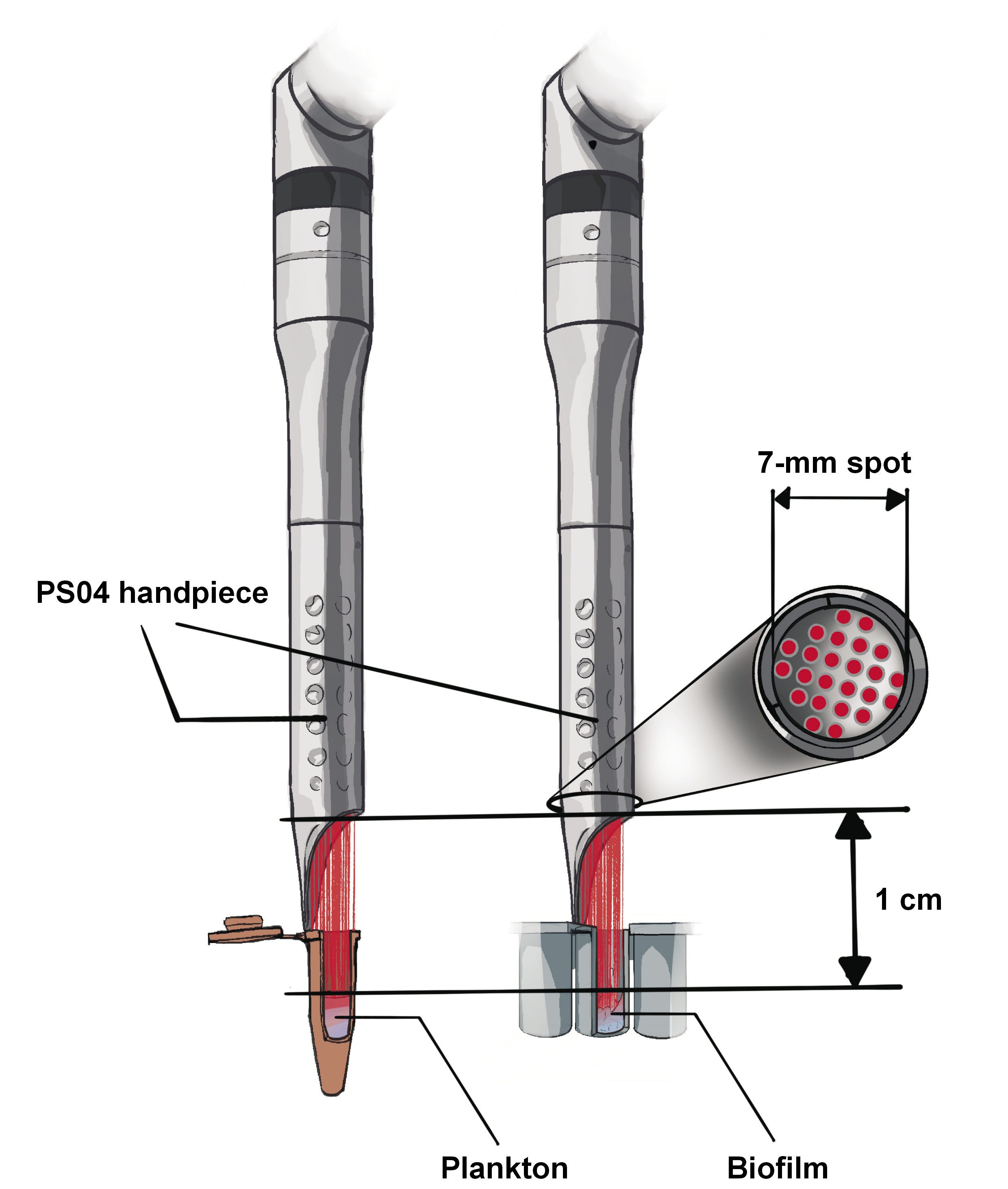
The experiment used an Er:YAG laser (LightWalker AT-S; Fotona, Ljubljana, Slovenia) equipped with a fractional PS04 handpiece (Fotona). The laser was operated in a non-contact mode with a 600-μs long pulse duration, maintaining a 10-mm distance from the surface of strain suspension in the Eppendorf tube (plankton) or 96-well polystyrene plates (biofilm) (Figure 1). The delivery system employed an articulated arm. In both test groups, the same fractional handpiece was used, with 2 variations in parameters. One set was designed for facial rejuvenation, while the other was specifically configured for the clinical treatment of snoring. The control group was biofilm without laser irradiation. The parameters used in the study are outlined in Table 1.
Microorganism quantification
Planktonic cell suspensions
Single- and dual-species planktonic suspensions were exposed to laser light in dark Eppendorf tubes. For the assessment of immediate effects, 100 µL of suspension was serially diluted, and 100 µL from each dilution was plated on BHI agar. The plates were then incubated at 37°C for 24 h under aerobic (yeast) or elevated CO2 conditions (S. mutans and dual-species). Colonies were counted to calculate colony forming units per milliliter (CFU/mL). Control suspensions were subjected to the same protocol without laser exposure.
To assess the effects 24 h post-irradiation, the suspensions underwent incubation at 37°C under aerobic or CO2 conditions, followed by serial dilution, plating and colony counting. Controls were prepared in a similar manner, without irradiation.
Effect of laser on single- and dual-species biofilm (quantification of CFU/mL)
Biofilm samples cultivated for 24 h were rinsed with 0.9% NaCl to remove any unattached cells. Thereafter, the samples were irradiated with a laser, scraped using sterile swabs and shaken in a 0.5% saponin solution. Subsequently, they were processed via serial dilution and plating. The plates were incubated at 37°C for 24 h under aerobic or CO2 conditions, after which CFU/mL values were calculated.
Effect of laser on single- and dual-species biofilm (crystal violet method)
Biofilm samples cultivated for 24 h were rinsed with 0.9% NaCl to remove any unattached cells. Subsequently, the samples were exposed to laser irradiation. Then, they were fixed by drying for 45 min at room temperature. After the drying period, 250 µL of 0.1% crystal violet solution (Chempur®, Piekary Śląskie, Poland) was added to the wells, and the plate was left for 20 min at room temperature. Subsequently, the violet was removed and the wells were washed 3 times in distilled water. Thereafter, they were left to dry for 20 min at room temperature. After adding 200 µL of 95% ethanol (Honeywell, Charlotte, USA), the absorbance was measured at a wavelength (λ) of 540 nm (Asys UVM 340; Biochrom Ltd., Holliston, USA). The control group consisted of a microbial biofilm that had not been exposed to the laser.
Statistical analysis
The Kolmogorov–Smirnov test was performed at a 95% confidence interval (CI) to evaluate the normality of data distribution. Differences in microorganism reduction following laser irradiation were analyzed using multivariate analysis of variance (MANOVA). Post-hoc adjustments were implemented using the Holm–Bonferroni method to control for multiple comparisons. All statistical analyses were carried out using Statistica software, v. 12 (StatSoft, Kraków, Poland), with a significance level set at p < 0.05.
Results
Reduction of microorganisms in single-species planktonic cultures
The results demonstrate that laser application significantly reduced microorganism cell counts in single-species planktonic cultures in comparison to the control group. The most substantial reductions were observed for S. mutans and C. albicans when the PS04 handpiece was used directly after irradiation (DAI) at higher fluence settings (T2). Statistically significant differences were identified between the DAI and 24 h after irradiation (24AI) time points for S. mutans (80.0 ±1.3% vs. 0.0 ±0.0%; p < 0.001) and C. albicans (74.0 ±8.3% vs. 31.7 ±10.2%; p = 0.045) under T2 settings. Furthermore, higher laser fluence (T2) resulted in a significantly greater reduction of C. albicans immediately after irradiation (p < 0.05). Conversely, lower fluence (T1) achieved superior reductions for C. glabrata and S. mutans 24 h post-irradiation compared to T2 (p < 0.05) (Table 2).
Reduction of microorganisms in single-species biofilm cultures
The study assessed the reduction of microbial cell counts in biofilm cultures of C. albicans, C. glabrata and S. mutans, directly after irradiation, with an Er:YAG laser used in conjunction with the PS04 handpiece. The results showed that for C. albicans, T2 parameters achieved a significantly higher reduction compared to T1 (85.3 ±1.2% vs. 43.9 ±4.5%, respectively; p = 0.006), thereby demonstrating enhanced efficacy. In contrast, C. glabrata exhibited a substantial reduction under T1 settings, but no reduction at higher fluence (69.8 ±14.9% vs. 0.0 ±0.0%, respectively; p = 0.022). Streptococcus mutans demonstrated consistently high reductions in both configurations (81.1 ±19.6% for T1 and 97.1 ±0.6% for T2; p > 0.05). Inter-group comparisons indicated significant variation among the species (p < 0.05), with S. mutans showing the highest overall reduction (T2: 97.1 ±0.6%; T1: 81.1 ±19.6%), which was significantly greater compared to C. albicans and C. glabrata under T1 settings, as well as C. glabrata under T2 settings (p < 0.05) (Table 3).
Reduction of microorganisms in dual-species planktonic cultures
The results of microbial cell counts in dual-species planktonic cultures (C. albicans + C. glabrata, C. albicans + S. mutans, and C. glabrata + S. mutans) following Er:YAG laser application using the PS04 handpiece revealed significant variability in reduction rates depending on the species combination, laser settings (T1 vs. T2) and timing (DAI vs. 24AI). Candida albicans in the C. albicans + S. mutans mixture showed a significantly higher reduction with T2 settings at 24AI compared to DAI (83.7 ±5.9% vs. 7.4 ±0.1%, respectively; p = 0.003). A similar trend was observed for S. mutans in the same mixture, which exhibited significant reductions with T1 settings at 24AI compared to DAI (24.9 ±2.3% vs. 0.0 ±0.0%, respectively; p = 0.004). For the C. glabrata + S. mutans biofilm, reductions were significantly greater at 24AI compared to DAI under certain conditions (T2: 39.2 ±10.4% at 24AI vs. 0.0 ±0.0% at DAI; p = 0.033). Moreover, a greater microbial reduction for S. mutans in the C. albicans + S. mutans mixture was observed with T2 settings immediately after laser application, while the opposite was observed 24 h post-irradiation (p < 0.05). Additionally, for the C. glabrata + S. mutans mixture, a greater reduction in S. mutans was found with T1 settings immediately after laser application compared to results measured 24 h after laser irradiation (p < 0.05) (Table 4).
Reduction of microorganisms in dual-species biofilm cultures
Higher laser fluence (T2) exhibited a greater reduction in C. glabrata (C. albicans + C. glabrata and C. glabrata + S. mutans mixtures), S. mutans (C. albicans + S. mutans and C. glabrata + S. mutans mixtures) and C. albicans (C. albicans + C. glabrata mixture) (p < 0.05). No significant increase in reduction was observed for C. albicans in the C. albicans + S. mutans mixture when comparing the 2 irradiation configurations (Table 5).
Laser-induced biomass reduction in single- and dual-species biofilms evaluated using the crystal violet method
The application of the Er:YAG laser in conjunction with the PS04 handpiece demonstrated varying effectiveness in reducing biofilm biomass, depending on laser power settings. For C. albicans, an increase in power from T1 (13.6 ±2.5%) to T2 (14.9 ±1.4%) did not result in a significant reduction (p = 0.571). In contrast, C. glabrata exhibited a substantial decrease in biomass from T1 to T2 (13.4 ±3.1% vs. 51.15 ±8.0%, respectively; p = 0.023), suggesting a greater sensitivity to elevated laser power. Streptococcus mutans showed an increase in biomass reduction from T1 (13.8 ±5.3%) to T2 (24.05 ±1.3%), but this change was not statistically significant (p = 0.099). Multivariate analysis of variance confirmed significant differences between the species (p = 0.009), with the most notable differences observed between C. glabrata and C. albicans (p < 0.009) and between C. glabrata and S. mutans (p < 0.022) (Table 6).
The application of the Er:YAG laser (PS04 handpiece) resulted in varied biomass reduction across different dual-species biofilms, depending on microbial composition and laser power settings. In the C. albicans + S. mutans biofilm, a substantial biomass reduction was observed, decreasing from T1 to T2 (18.8 ±0.8% vs. 11.4 ±0.9%, respectively; p = 0.014), indicating lower efficacy at higher power. Conversely, the C. glabrata + S. mutans biofilm reduction demonstrated a significant decrease from T1 to T2 (23.2 ±5.4% vs. 0.3 ±0.2%, respectively; p = 0.027), suggesting that lower laser power was highly effective against this combination. Moreover, a reduction in biomass was observed for the C. albicans + C. glabrata biofilm when comparing the T1 and T2 settings (31.0 ±13.2% vs. 49.4 ±14.7%, respectively; p = 0.318), implying greater laser efficiency under high-energy conditions (Table 7).
Discussion
The present study is an in-depth examination of the impact of the Er:YAG laser on single-species and dual-species planktonic and biofilm cultures formed by C. albicans, C. glabrata and S. mutans. Additionally, it investigates novel methods for eliminating these pathogens by utilizing low laser parameter settings tailored for the clinical management of facial rejuvenation and snoring. The findings of the current study highlight the potential of the Er:YAG laser with a fractional handpiece as an effective tool for reducing microbial populations in both planktonic and biofilm cultures of C. albicans, C. glabrata and S. mutans. The observed reductions were dependent on laser settings (T1: 0.4 J/cm² vs. T2: 1.6 J/cm²) and microbial species, suggesting that the efficacy of laser treatment can be optimized through the precise customization of parameters to the specific clinical context. Notably, the higher fluence settings (T2) demonstrated superior efficacy in disrupting C. albicans and S. mutans in single-species and dual-species planktonic cultures immediately after irradiation, while the lower fluence settings (T1) were more effective for C. glabrata in single-species planktonic cultures 24 h after irradiation. These results align with earlier studies that emphasize the versatility and efficacy of laser-based approaches in microbial management, particularly in addressing challenges related to biofilm-associated infections in the field of dentistry.23, 48, 51
With respect to the results obtained for planktonic cultures, the application of the Er:YAG laser in conjunction with the PS04 handpiece demonstrated significant reductions in microbial cell counts, particularly for S. mutans and C. albicans. The most notable reductions were observed under T2 settings, with a 80.0% and 74.0% decrease, respectively, immediately after irradiation. These outcomes are consistent with earlier studies, which also found laser treatment to be effective in reducing the growth of these species, suggesting that the intensity of the laser has a significant impact on microbial survival.52, 53, 54 While S. mutans showed no detectable recovery at 24 h post-irradiation, C. albicans exhibited partial resurgence, with a reduction of 31.7%. These findings support earlier research indicating that laser efficacy diminishes over time, particularly for fungal organisms like C. albicans, which may require repeated interventions for long-term control.54 Interestingly, while higher fluence (T2) was more effective against C. albicans immediately after irradiation, lower fluence (T1) yielded better results for C. glabrata and S. mutans at 24AI. These results underscore the importance of optimizing laser parameters for different microorganisms, highlighting that microbial responses to laser treatments can vary significantly depending on the organism type and laser fluence.23, 48, 51
In biofilm cultures, the Er:YAG laser, operated with the PS04 handpiece, demonstrated species-specific efficacy in reducing microbial cell counts. This finding is consistent with the existing literature on biofilm management. For single-species biofilms, S. mutans exhibited the highest reduction rates, with T2 settings achieving nearly complete eradication (97.1 ±0.6%). This result aligns with the findings reported in the study by Grzech-Leśniak et al., which emphasized the susceptibility of S. mutans biofilm to treatments targeting the biofilm matrix using the Nd:YAG (neodymium-doped yttrium aluminum garnet) laser.23 A comparable response was observed for C. albicans, which exhibited a significant reduction under T2 parameters (85.3 ±1.2%), reflecting its vulnerability to interventions that disrupt extracellular polymeric substances (EPS), as discussed by Salehi et al., who highlighted the challenges of disrupting resilient polymicrobial biofilms.55 In dual-species biofilms, T2 fluence generally achieved greater reductions, particularly for C. glabrata and S. mutans in mixed cultures, as well as for C. albicans in combination with C. glabrata. These findings support evidence presented by Salehi et al., which underscored the significance of tailored approaches to address the synergistic interactions between microbial species within biofilms.55 However, no significant improvement with T2 over T1 settings was observed for C. albicans in combination with S. mutans, suggesting that interspecies interactions might alter biofilm susceptibility. These findings emphasize the potential of the Er:YAG laser in the management of biofilms and highlight the importance of understanding microbial dynamics to optimize treatment protocols.48, 51, 56
This study builds upon our previous findings, in which the antimicrobial impact of the Nd:YAG laser on C. albicans and S. mutans was explored using both CFU quantification and Janus green staining.22 The use of Janus green staining allowed for a detailed assessment of metabolic activity and biofilm viability, providing insights beyond the microbial counts achieved through CFU analysis. This method addressed structural and metabolic biofilm responses, which are critical for understanding the underlying mechanisms of laser-induced microbial reduction. In the current study, CFU quantification was used to evaluate the antimicrobial efficacy of the Er:YAG laser. While this method effectively quantifies microbial reduction, it lacks the ability to visualize biofilm structure and viability. Additionally, the study utilized the Nd:YAG laser for photobiomodulation (PBM) applications,22, 23 targeting deeper tissue interactions. In contrast, the present study explores the surface-level effects of the Er:YAG laser using the fractional PS04 handpiece. The utilization of distinct laser systems and their intended applications highlights the adaptability of these technologies for specific clinical scenarios. While the Nd:YAG laser is effective in managing deeper biofilm in periodontal pockets,14, 21, 24, 26, 49 the Er:YAG laser14, 18, 31, 32, 33, 34, 35, 36, 38, 39, 40, 41, 42, 45, 46, 47, 48, 50, 51, 52, 53 has shown promise in disrupting surface biofilm and enhancing aesthetic or surface-level treatments.57, 58, 59, 60, 61 Overall, integrating advanced visualization techniques, clinically relevant substrates and combined laser systems will provide a more comprehensive understanding of biofilm responses to laser treatment and enhance the translational value of these findings for dental and medical applications.
Limitations
Despite the promising findings of this study, it is imperative to acknowledge its limitations. First, the study was conducted in vitro, which may not fully replicate the complex conditions present in vivo, where additional factors such as host immune responses, saliva and tissue interactions could influence the effectiveness of Er:YAG laser treatments on biofilm. The crystal violet staining method, a widely utilized technique for biofilm biomass quantification due to its simplicity and cost-effectiveness, is not without its limitations, which may affect its reliability. A significant drawback is its inability to differentiate between live and dead cells, as it quantifies total biomass without providing insights into cell viability or metabolic activity. This limitation may lead to an incomplete assessment of the functional state of biofilms. A study by Fernandes et al. emphasizes the importance of multiparametric approaches for more comprehensive evaluations.62 Complementary methods such as live/dead staining, metabolic assays, or confocal microscopy could enhance reliability and address potential biases, including material–biofilm interactions. Future studies should consider these approaches to strengthen findings. Additionally, the study’s focus on only 3 species, C. albicans, C. glabrata and S. mutans, limits the generalizability of the results to other pathogens commonly involved in oral infections. Furthermore, although the study demonstrated significant reductions in microbial populations, the long-term efficacy of the laser treatment was not assessed beyond the 24-hour post-irradiation time point. In clinical settings, the potential for biofilm regrowth necessitates the consideration of repeated treatments or combination therapies. The absence of a comprehensive analysis on the potential tissue damage or adverse effects of laser application also represents a limitation, as these factors are crucial in clinical applications. These limitations indicate that while Er:YAG lasers demonstrate potential for biofilm disruption, further research, particularly clinical studies, is needed to fully assess their effectiveness in diverse oral environments.
Conclusions
The present study aimed to evaluate the effects of the Er:YAG laser on biofilms composed of C. albicans, C. glabrata and S. mutans strains, with a particular focus on its antifungal and antibacterial properties. However, the findings are limited by the in vitro nature of the study, which does not fully replicate the physiological conditions of the oral environment. Consequently, the outcomes may not directly translate to clinical dentistry. The results suggest that the efficacy of laser treatment is influenced by several factors, including the fluence settings (0.4 J/cm2 and 1.6 J/cm2), microbial species and the time elapsed post-irradiation. Generally, the antifungal effects were evident in the reduction of fungal strains (C. albicans and C. glabrata) immediately after irradiation, with higher fluence settings showing greater efficacy. In contrast, the antibiofilm effect was characterized by sustained microbial reduction over a 24-hour period, particularly at lower fluence settings, with notable effectiveness against C. glabrata and S. mutans in single-species planktonic cultures. Future research should focus on in vivo studies to assess the laser’s effectiveness under real-world conditions, including the complex interplay of oral microbiota, tissue responses and environmental factors.
Ethics approval and consent to participate
The study was approved by the Institutional Research Ethics Committee of Wroclaw Medical University, Poland (approval No. KB-429/2024).
Data availability
The datasets generated and/or analyzed during the current study are available from the corresponding author on reasonable request.
Consent for publication
Not applicable.
Use of AI and AI-assisted technologies
ChatGPT 4.0 (OpenAI) was used to verify grammar and make minor adjustments for clarity and accuracy.