Abstract
Background. The success of endodontic retreatment relies on the effective elimination of pathogenic microflora from the root canal.
Objectives. The study aimed to investigate the effects of an erbium-doped yttrium-aluminum-garnet (Er:YAG) laser and a 635-nm laser on the healing of asymptomatic chronic periapical lesions (PLs) in endodontically treated teeth and the reduction of postoperative pain.
Material and methods. Forty patients with PLs in mandibular molars were referred for root canal retreatment (RCR). Conventional chemo-mechanical endodontic treatment was conducted in the control group (G1; n = 20). In the test group (G2; n = 20), in addition to conventional chemo-mechanical treatment, Er:YAG laser-activated irrigation (LAI) with 2% NaOCl and 17% EDTA was performed. The laser parameters were as follows: 50 mJ; 25 Hz; 1 W; 300 µs; a tip diameter of 300 µm; fluence of 71.4 J/cm2; and power density of 1,428.6 W/cm2. Subsequently, the canals were filled with thermo-condensed gutta-percha, using the AH Plus sealer. In group G2, additional photobiomodulation (PBM) with a wavelength of 635 nm (400 mW, 5 s per point, a dose per point: 2 J, a dose per square centimeter: 4 J, an applicator diameter of 8 mm) was applied, with 2 application points at the apex level, administered over 4 sessions – on the treatment day, and after 24 h, 48 h and 96 h. Endodontic lesion remission was assessed by measuring the PL size with the use of cone-beam computed tomography (CBCT) at 6 and 12 months postoperatively. Postoperative pain was evaluated using the visual analog scale (VAS) after 1, 2 and 4 days.
Results. The study results demonstrated a statistically significant decrease in the mean PL size at 6 months postoperatively in the test group (mean PL size: 1.55 ±0.51 mm) as compared to the control group (mean PL size: 1.95 ±0.71 mm) (p < 0.05). In the test group, postoperative pain on VAS was significantly lower after the procedure (p < 0.05).
Conclusions. The application of Er:YAG and 635-nm diode lasers improved PL healing and decreased postoperative pain.
Keywords: periapical periodontitis, photobiomodulation, endodontics, erbium laser, Er-YAG
Introduction
The failure rate in endodontic therapy typically ranges from 2% to 16%.1 The success of root canal therapy relies on the effective elimination of pathogenic microflora from the root canal.2, 3 In comparison with primary treatment, the revision of endodontic procedures poses greater challenges due to technical hurdles in canal preparation and the persistent activity of intracanal microorganisms.4 Bacterial infection not only results in chronic apical periodontitis (CAP), but also heightens the potential for postoperative discomfort. The fundamental objective of endodontic treatment is to thoroughly eliminate the pathogenic microorganisms residing within the intricate network of the root canal system, thereby promoting the restoration of optimal oral health and preventing potential reinfection. However, the achievement of effective disinfection can be challenging due to the intricate nature of the root canal system, which encompasses features like isthmuses, oval extensions and lateral canals.1, 2, 4
Teeth affected by apical periodontitis (AP) pose a greater challenge in terms of endodontic treatment as compared to those without lesions during the treatment phase.5 This complexity primarily arises from the persistent bacterial activity within the root canal system. The time required for a lesion to heal varies between 6 months and 2 years, occasionally extending even further.6 Sathorn and Parashos stated that if a lesion remains unresolved for 4 years, spontaneous resolution becomes unlikely.6 Indeed, if a lesion maintains its size or expands after a year of follow-up, or if a lesion appears in a tooth that previously exhibited none, root canal retreatment (RCR) is warranted.7 Radiographically, the complete healing of the cortical bone and a healthy appearance within 6–24 months post-treatment signify the success of the intervention.8 Nevertheless, cases of post-treatment AP can emerge even when the initial treatment was executed correctly, due to various factors, such as the presence of anatomical complexities, an incomplete removal of the infected tissue, undetected accessory canals, or microbial resistance, emphasizing the necessity for vigilant follow-up and potential retreatment to ensure successful outcomes. The occurrence of post-treatment affliction is discerned in 5–15% of cases previously exhibiting pre-treatment AP, regardless of adherence to proper procedural standards.9 The smear layer, which permeates dentinal tubules up to 40 µm along with dentin debris, tends to deactivate root canal medicaments and irrigants while obstructing their penetration into the biofilm.10 Thus, the imperative to eliminate the smear layer and debris cannot be overstated, as far as averting AP is concerned.
Clinicians employ diverse methodologies to ensure precise cleaning and disinfection in endodontic retreatment.2, 11, 12, 13 In this context, various irrigation systems have become integral for achieving improved treatment outcomes, addressing the challenges associated with the failure rates in endodontic retreatment cases.14 Achieving comprehensive root canal decontamination is imperative, requiring the meticulous removal of gutta-percha and the sealer not only from the main canal, but also from anatomically intricate lateral canals or fins. This meticulous approach ensures thorough disinfection and prevents potential microbial reservoirs, contributing to the success of endodontic treatment.15 While the existing studies have primarily focused on the removal of calcium hydroxide (Ca(OH)2) and debris from the root canal and the simulated lateral canals,16 there is still a notable gap in the literature concerning the specific removal of the sealer, using varied irrigation methodologies. Recent advancement has introduced various irrigation systems and techniques.17, 18, 19 RinsEndo, characterized by a hydrodynamic mechanism involving alternating positive and negative pressure, induces macroscopic and microscopic blistering to activate the irrigant, thereby enhancing its efficacy.19 Approaches such as passive ultrasonic irrigation and the EndoVac™ system leverage negative pressure to actively administer and remove irrigation solutions, ensuring thorough cleansing.17, 19 The CanalBrush endodontic instrument, a malleable plastic micro-brush integrated into a dental handpiece operating at 600 rpm, in conjunction with manual irrigation, presents a promising yet inconclusive approach, with varying conclusions across the existing studies.20 Conversely, EndoActivator, a battery-powered handpiece featuring a non-cutting polymer tip, achieves the sonic activation of the intracanal irrigant.20 Widely adopting an oscillating, non-cutting ultrasonic tip for activation, the available literature consistently validates the heightened efficacy of ultrasonically activated irrigation (UAI).19 Both ultrasonic and sonic irrigation methods capitalize on acoustic vibrations to dislodge debris, thereby bolstering the efficacy of irrigation solutions.19, 21 However, a study by van der Sluis et al. indicates the potential for file breakage and the limited penetration in curved canals.18 In turn, laser activation employs laser energy for irrigant activation.21
A distinctive method for activating irrigants in endodontic procedures is laser-activated irrigation (LAI), leveraging pulsed erbium, chromium-doped yttrium-scandium-gallium-garnet (Er,Cr:YSGG) and erbium-doped yttrium-aluminum-garnet (Er:YAG) lasers. These lasers induce optical cavitation within the irrigant, causing the formation of expanding and imploding vapor bubbles at the fiber tip, with smaller secondary bubbles emerging deeper in the canal undergoing acoustic streaming.22, 23 Notably, controlled laboratory studies have reported an impressive 99.5–100% reduction in bacterial load within the infected root canals through the application of LAI.24, 25 Furthermore, rigorous laboratory investigations have demonstrated the heightened efficacy of LAI with the Er:YAG laser over conventional irrigation and UAI, specifically in removing debris from intricate canal structures.23, 26 However, it is essential to note that the outcomes of studies do not uniformly reveal a statistically significant difference between LAI and UAI. Furthermore, photobiomodulation (PBM) emerges as a promising intervention in mitigating postoperative pain following root canal treatment.27, 28 By harnessing the properties of the laser wavelength within the optical window, PBM has demonstrated efficacy in reducing postoperative pain associated with root canal treatment.29 The application of low-level laser therapy (LLLT) adheres to 3 fundamental principles: biostimulation; analgesia; and the modulation of inflammatory processes.30, 31, 32, 33, 34, 35, 36, 37 This therapeutic modality has demonstrated efficacy in mitigating postoperative pain and edema following the extraction of impacted mandibular third molar teeth,38 managing dentine hypersensitivity,39 alleviating postoperative pain after endodontic surgery,40 addressing postoperative pain subsequent to RCR,27, 41 and attenuating postoperative pain following the initial root canal treatment of mandibular molar teeth with symptomatic CAP.42
The principal aim of the present investigation was to examine the synergistic effect resulting from the application of an Er:YAG laser in conjunction with a 635-nm diode laser, specifically assessing their combined influence on the remission of periapical lesions (PLs). Additionally, the study aimed to evaluate the potential mitigation of postoperative pain associated with this combined laser approach. The null hypothesis in the present study was that there would be no differences in the PL size and the pain experienced by patients at 24 h, 48 h and 96 h post-treatment after the application of Er:YAG LAI and PBM as compared to endodontic retreatment using classical methods.
Material and methods
The trial was structured as a randomized and controlled study. Approval was obtained from the Local Ethics Committee at the Regional Medical Chamber (permission No.: 318/KBL), and informed consent was acquired from all the participating subjects in line with the principles of the Declaration of Helsinki.
Subjects
All patients underwent treatment at a dental office, under the care of the same endodontist (I.K-B.). Patients’ randomization was done using the https://www.randomizer.org webpage. The average age of the participants was 45.3 ±9.8 years. The sample size of 20 subjects in each group was determined using the G*Power software from Kiel University, Germany. The calculations were based on our preliminary tests, considering a significance level of 0.05, an effect size (d) of 0.81 and a power of 80%. The sample size was calculated based on our preliminary results with a smaller number of patients, and on a similar study related to this topic.27
The allocation of participants into 2 groups followed a 1:1 ratio and was carried out using a coin toss. The selection criteria for the cases were as follows: teeth treated endodontically, with PLs having a diameter of less than 10 mm; overall good health; a diagnosis of asymptomatic CAP in mandibular molars, with a favorable prognosis for functional reconstruction post-endodontic treatment; and primary endodontic treatment performed a minimum of 4 years prior to the current trial. The exclusion criteria encompassed systemic disorders, pregnancy and breastfeeding, periodontal diseases (pocket depth (PD) of over 3 mm), the use of analgesic agents within 5 days before the procedure, the administration of antibiotics up to 1 month before the procedure, teeth affected by post-trauma complications and those with the materials extending beyond the apical foramen, teeth with broken instruments, teeth with root resorption, and type 3 of canal curvature according to Schneider’s classification. To maintain the integrity of the study, blinding was implemented during the recording of postoperative pain evaluations and cone-beam computed tomography (CBCT) assessments. This ensured that the individuals responsible for gathering and assessing this data remained unaware of treatment conditions or group assignments, minimizing bias and enhancing the reliability of the study outcomes.
Clinical procedure
A total of 40 patients requiring root endodontic retreatment in mandibular molars were randomly divided into 2 groups using a coin toss, based on the treatment proce-dure. The control group (G1; n = 20) received conventional chemo-mechanical endodontic treatment. In the test group (G2; n = 20), conventional treatment was supplemented with the application of 2 lasers: an Er:YAG laser (AdvErL EVO; J. Morita, Ina-machi, Japan) for activating the irrigant; and a 635-nm diode laser (SMARTmPRO; Lasotronix, Piaseczno, Poland) for postoperative PBM. In the control group, EndoActivator (EndoActivator System Kit; Dentsply Sirona, Bensheim, Germany) was employed for the activation of the irrigant (Figure 1).
After administering anesthesia, using 1.8 mL of 4% articaine (Citocartin 200 with 1:200,000 epinephrine; Molteni Dental, Milan, Italy), and removing the filling material with a diamond bur on a high-speed, contra-angle handpiece, a rubber dam was placed. Before commencing the cleaning and shaping procedures for endodontic treatment, each tooth crown was reconstructed with a composite material. After preparing access to the tooth chamber, the tooth cavity was irrigated with 2% sodium hypochlorite (NaOCl). The activation of the irrigant was performed using either EndoActivator (30 s, 15/02 tip) or the Er:YAG laser (20 s, R300T tip) by placing the tips into the tooth chamber following each instrumentation with the file.
The dissolution of the endodontic filling material (gutta-percha) was accomplished through the application of orange oil. Subsequent to achieving canal negotiation by utilizing C-pilot files (VDW, Munich, Germany), and confirming the attainment of the full working length through measurements with the Raypex 6 apex locator (VDW) and radiographic verification featuring an endodontic instrument positioned within the root canal, the canals underwent irrigation with 2% NaOCl. The Er:YAG laser R300T tip was positioned at a depth of 3–5 mm from each tooth canal orifice. In contrast, EndoActivator was placed into the root canal as deeply as possible after canal instrumentation, not exceeding 2 mm before the working length.
Hand instruments were utilized until achieving a canal size of 20/02, after which rotary instrumentation (E3 Azure Endostar files; Poldent, Warsaw, Poland; X-Smart® Plus; Dentsply Sirona), operating at a speed of 300 rpm, with a torque set between 2.1 and 3.0 N·m, was employed until attaining a size of 35/04.
After completing the remaining canal preparation up to a size of 35/04, rinsing was conducted using 10 mL of NaOCl and 2 mL of 17% ethylenediaminetetraacetic acid (EDTA) on each canal, using either EndoActivator or the Er:YAG laser. Final irrigation using an endodontic needle (positioned 2 mm short of the working length) was performed with 2 mL of a saline solution per canal, without activation. After achieving dryness, the canals were then obturated with thermally condensed gutta-percha and the AH Plus sealer (Dentsply Sirona). The teeth were temporarily restored using a composite material. In all cases, single-visit treatment was performed (Figure 2).
Laser application
The 2,940 nm wavelength of the Er:YAG laser was utilized for LAI, with the following parameters: energy – 50 mJ; frequency – 25 Hz; power – 1 W; pulse width – 300 μs; tip diameter – 300 µm; fluence – 71.4 J/cm2; and power density – 1,428.6 W/cm2. The duration of LAI was 20 s.
The PBM of the periapical area was performed immediately after endodontic treatment, using the 635-nm diode laser with the following parameters: power – 400 mW; time – 5 s per point; dose per point – 2 J; dose per square centimeter – 4 J; applicator diameter – 8 mm; spot area – 0.5024 cm2; average power density – 0.8 W/cm2; 2 application points at the buccal and lingual apex levels; 4 sessions – immediately after RCR, and subsequently after 24 h, 48 h and 96 h.
Measurement of periapical lesions
Endodontic lesion remission was evaluated using CBCT (CS 8100 3D; Carestream Dental, Marne-la-Vallée, France). The PL size was measured in various dimensions, and the largest measured dimension of the lesion was considered for calculations. The measurements were taken at baseline (before treatment), as well as at 6 and 12 months postoperatively. The initial size range of lesions was similar for both groups – 3.7–5.9 mm and 3.4–6.0 mm for groups G2 and G1, respectively.
Measurement of postoperative pain
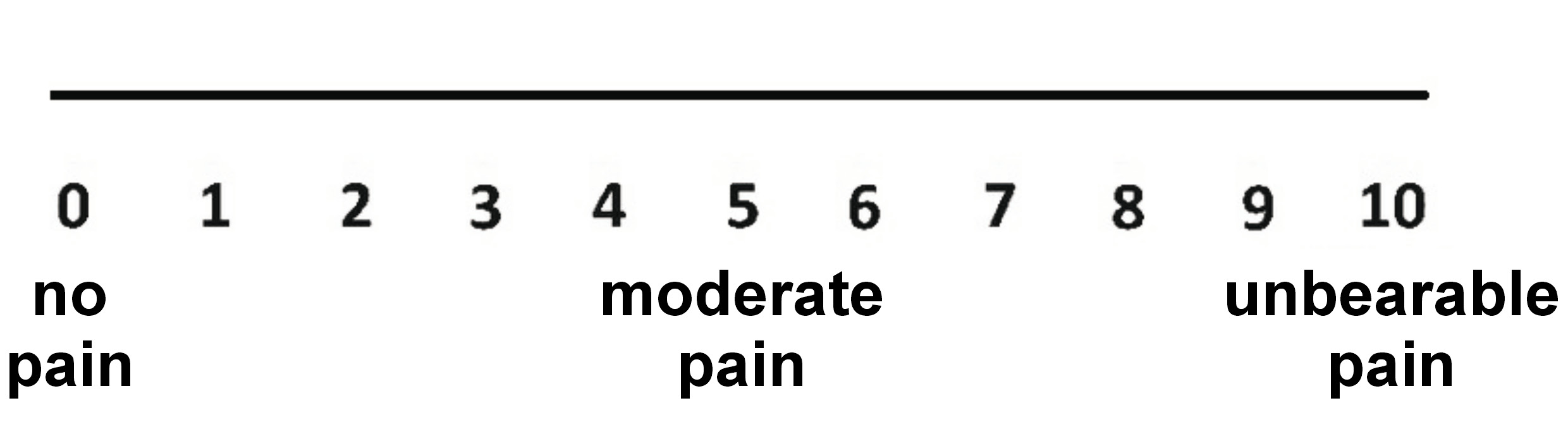
Postoperative pain assessment was conducted utilizing the visual analog scale (VAS) at specific intervals – 1, 2 and 4 days post-procedure. The VAS, a numerical scale ranging from 0 (denoting the absence of pain) to 10 (indicating unbearable pain), served as the instrument for quantifying the pain levels. The patients were administered structured questionnaires to articulate their pain experience on the designated days. The collated responses were gathered during the subsequent follow-up visit scheduled 7 days post-procedure, contributing to a comprehensive evaluation of postoperative pain dynamics (Figure 3).
Statistical analysis
The normality of the data was assessed using the Kolmogorov–Smirnov test. Since the data distribution exhibited normality, statistical analysis for the PL size and the VAS pain level was conducted using Student’s t test. This analysis was performed with IBM SPSS Statistics for Windows, v. 22.0 (IBM Corp., Armonk, USA), with a significance level set at p = 0.05.
Results
Periapical lesion healing following root canal retreatment
The assessment of the PL size, measured in millimeters, after 6 months revealed a significant difference in the reduction of the lesion dimensions between the control subjects (mean PL size: 1.95 ±0.71 mm) and the test group (mean PL size: 1.55 ±0.51 mm) (p < 0.05). However, the results obtained after 1 year demonstrated insignificant improvement, i.e., a greater reduction in the PL size, following the application of lasers (mean PL size: 0.90 ±0.64 mm) in comparison with the control group (mean PL size: 1.11 ±0.66 mm) (p > 0.05) (Table 1, Figure 4).
Pain level following root canal retreatment
The reduction in postoperative pain was assessed in both groups at 1, 2 and 4 days after the procedure. The mean VAS scores for pain were significantly lower in group G2 (3.11 ±1.29 and 2.16 ±0.96) as compared to group G1 (4.10 ±1.86 and 2.95 ±1.39) at the 24-hour and 48-hour marks, respectively (p < 0.05). However, the VAS scores at 4 days (96 h) showed an insignificant difference between the groups (p > 0.05) (Table 2).
Discussion
The null hypothesis in the present study regarding the lack of differences in the PL size change after RCR when applying Er:YAG LAI and PBM was rejected. In terms of postoperative pain reduction, the null hypothesis about the lack of differences between the test and control groups was partially rejected. Our present findings showed a significant reduction in the average PL size at 6 months of Er:YAG laser- and PBM-assisted RCR as compared to conventional secondary root endodontic therapy. Furthermore, the pain reduction in the test group measured after 24 h and 48 h was significantly greater as compared to the control group, while the difference was insignificant at 96 h after treatment. The energy of the erbium laser induces cavitation, triggering an effervescent effect on the irrigant, thereby significantly enhancing its disinfection efficacy – the volume of the activated irrigant can surge up to 1,600 times.21 Moreover, the application of non-ablative photonic energy through PBM modulates biological processes within tissues, affecting the larger biological system. This modulation extends to cellular metabolism, leading to secondary effects that alter cellular behavior.43
The primary aim of the study was to assess the effectiveness of Er:YAG LAI. The photoacoustic effect evoked in the fluids irradiated with the tip placed in a tooth chamber enables precise cleaning of the root canal and removes the smear layer produced by the mechanical shaping of canal dentin with endodontic instruments.44 The research conducted by van der Sluis et al. revealed that LAI involved stimulating irrigants to heighten their penetration into dentinal tubules.18 This stimulation renders the irrigant more reactive, enabling it to flow in a three-dimensional (3D) manner within root canals, thereby augmenting its antibacterial and cleansing effects.18, 23, 45 In the present study, the application of LAI serves the precise and effective removal of old endodontic material from root canals. In other studies, LAI has demonstrated positive outcomes in bolstering the efficiency of the mechanical elimination of the root canal treatment filling and enhancing the disinfection attributes of irrigants.23, 45 In a study by Neelakantan et al., 3 different irrigation protocols utilizing distinct activation methods (ultrasound, and diode and erbium lasers) were evaluated on mature Enterococcus faecalis biofilms.44 The study determined that energizing the irrigants with laser energy emerged as the most effective technique among those considered.44 Furthermore, multiple studies have shown the heightened bactericidal efficacy achieved by combining chemical irrigation with laser irradiation, as opposed to non-irradiated canals, disinfected solely with irrigants.5, 44, 46, 47 This improved effectiveness in root canal cleansing can expedite the PL healing process, as demonstrated in our current investigation.
An additional aspect addressed in our current research pertained to postoperative pain, an important concern in endodontic treatment. The incidence of pain following endodontic treatment has been documented to vary between 3% and 58%.29, 48 Moreover, RCR has been associated with a higher frequency of flare-up pain in comparison with the initial root canal treatment.49 Low-level laser therapy emerges as a non-pharmacological method capable of expediting cellular processes and averting postoperative pain.30 Numerous investigations utilizing VAS to gauge pain intensity have indicated that LLLT effectively diminishes postoperative discomfort after both root canal treatment and RCR.41, 42, 50 Asnaashari et al. conducted a study demonstrating significantly reduced pain scores within 48 h of treatment.41 However, they observed limited pain reduction effects from LLLT/PBM irradiation in the context of endodontic retreatment involving mandibular first and second molars.41 Naseri et al. exhibited a substantially greater pain reduction post-endodontic treatment as compared to a placebo across all study intervals.50 Moreover, the test group necessitated significantly fewer analgesics than the placebo group. They concluded that LLLT effectively supplemented oral analgesics in mitigating postoperative pain.50 Nevertheless, findings from Arslan et al. show that while the LLLT group exhibited significantly reduced postoperative pain in the initial 4 days as compared to the placebo group, no statistically significant differences were discerned between the 2 groups on days 5 and 7.27 This led them to conclude that PBM might alleviate postoperative pain subsequent to RCR in mandibular molars.27 In our study as well, we observed a notable reduction in the pain levels on the 1st and 2nd days following treatment.
The rationale behind the role of the 635-nm wavelength diode laser in augmenting the healing of PLs is rooted in its ability to enhance cell viability through the stimulation of mitochondrial and cell membrane photoreceptors.28 This stimulation prompts the synthesis of adenosine triphosphate (ATP), further fostering osteoblast proliferation.28, 50 This makes it a potential component in novel clinical strategies that leverage laser energy to bolster the healing process.41 In a study by Ng et al., the application of LLLT demonstrated a reduction in pain intensity to some degree, albeit within a limited 4-hour postoperative timeframe.51 Photobiomodulation has exhibited promising outcomes in facilitating periapical tissue healing.28 Low-level laser therapy has also shown effectiveness in advancing the healing of both soft and hard tissues following endodontic microsurgery, leading to enhanced bone volume and density, as verified by CBCT imaging.52 It is important to acknowledge that the current number of studies is constrained, necessitating further research to more definitively establish the efficacy of PBM.53 Notably, the beneficial clinical effects of PBM have been substantiated on a biochemical level as well. Laser therapy has been shown to elevate prostaglandin levels, imparting anti-inflammatory effects, alongside immunoglobulins and lymphokines that elicit the immune system response.54, 55 Additionally, the documented increase in beta-endorphins, pivotal in analgesia, underscores the impact of the therapy.56 With promising clinical results and biochemical correlations, the application of PBM emerges as a valuable supplementary approach in post-RCR pain management.51, 56
Limitations
The primary limitation of this study arises from the restricted number of participants included. The assessment of postoperative pain is inherently subjective, relying on various factors, such as the operator’s experience and adherence to clinical protocols. This subjectivity introduces potential variability in the interpretation and reporting of pain outcomes. Additionally, the absence of consideration for canal curvature in the eligibility criteria represents a notable constraint in the study. The study findings may be influenced by variations in the Er:YAG laser parameters, as well as the shape and type of the tip, especially when using an Er:YAG laser from a different manufacturer. Furthermore, within the test group, we examined the synergistic impact of 2 laser wavelengths (2,940 nm and 635 nm) on the healing process. Future research should involve distinct groups to assess the individual effects of each laser wavelength on PL healing, providing a more comprehensive understanding of the therapeutic contribution of each wavelength.
Conclusions
The present study revealed that the application of the 2,940 nm laser wavelength for irrigant activation and a 635-nm diode laser for PBM enhanced a decrease in the PL size after RCR. Moreover, the application of LAI and PBM resulted in a lower level of postoperative pain after secondary root canal treatment. The synergistic effect of both lasers during RCR allows clinicians to gain precise and adequate cleaning of the root canal, and ameliorate the shaping process by removing the debris produced during mechanical instrumentation. Furthermore, PBM added to endodontic treatment together with the Er:YAG laser reduces pain occurring in the first 2 days after root canal filling. The application of the Er:YAG laser and PBM seems to bring additional benefits in comparison with classical endodontic treatment.
Ethics approval and consent to participate
The trial was structured as a randomized and controlled study. Approval was obtained from the Local Ethics Committee at the Regional Medical Chamber (permission No.: 318/KBL), and informed consent was acquired from all the participating subjects in line with the principles of the Declaration of Helsinki.
Data availability
The datasets supporting the findings of the current study are available from the corresponding author on reasonable request.
Consent for publication
Not applicable.
Use of AI and AI-assisted technologies
Not applicable.